. 2025 Jun 16;32(1):57.
doi: 10.1186/s12929-025-01150-w.

Targeting neuroinflammation: 3-monothiopomalidomide a new drug candidate to mitigate traumatic brain injury and neurodegeneration

Affiliations

Targeting neuroinflammation: 3-monothiopomalidomide a new drug candidate to mitigate traumatic brain injury and neurodegeneration

Shih Chang Hsueh et al. J Biomed Sci. .

Abstract

Background: Traumatic Brain Injury (TBI) is a major risk factor for neurodegenerative disorders such as Parkinson's disease (PD) and Alzheimer's disease (AD), with neuroinflammation playing a critical role in the secondary cell death that exacerbates the initial injury. While targeting neuroinflammation holds significant therapeutic promise, clinical trials of available anti-inflammatory agents have fallen short. 3-Mono-thiopomalidomide (3-MP), a novel immunomodulatory imide drug (IMiD), was designed to curb inflammation without the adverse effects of traditional IMiDs and was evaluated across models involving neuroinflammation.

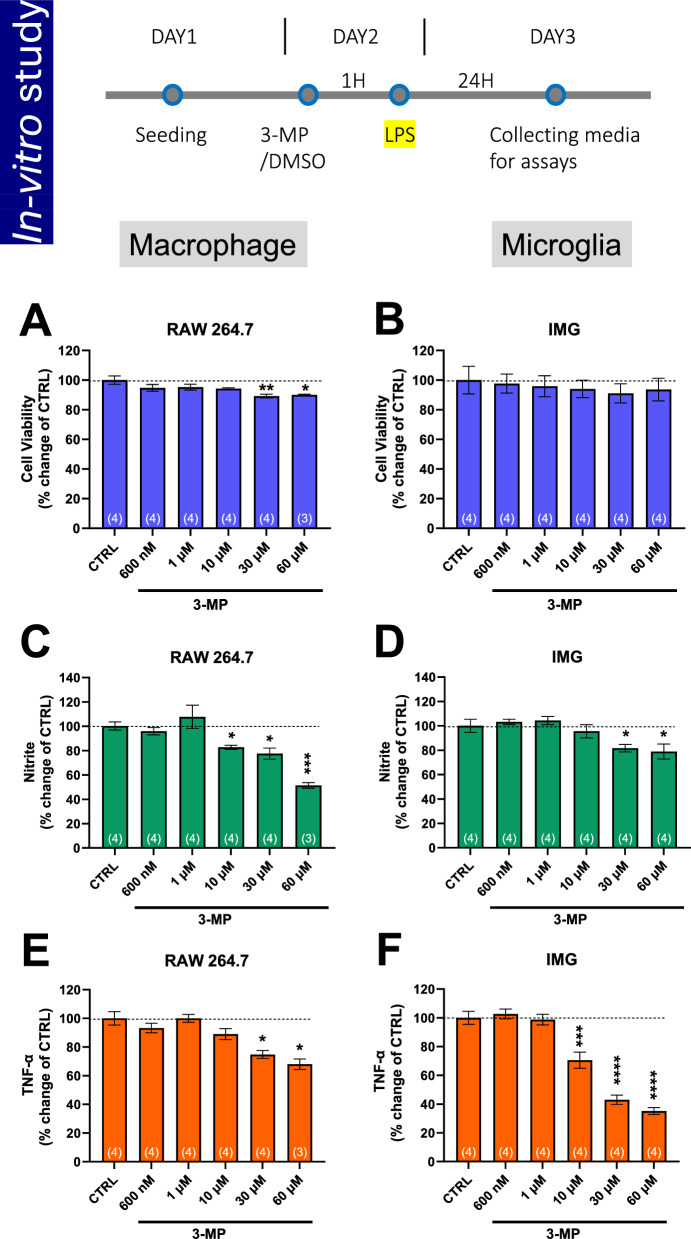
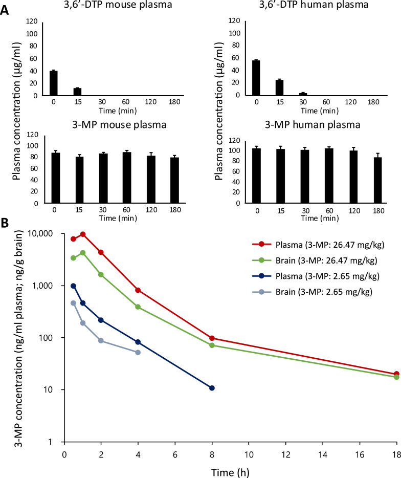
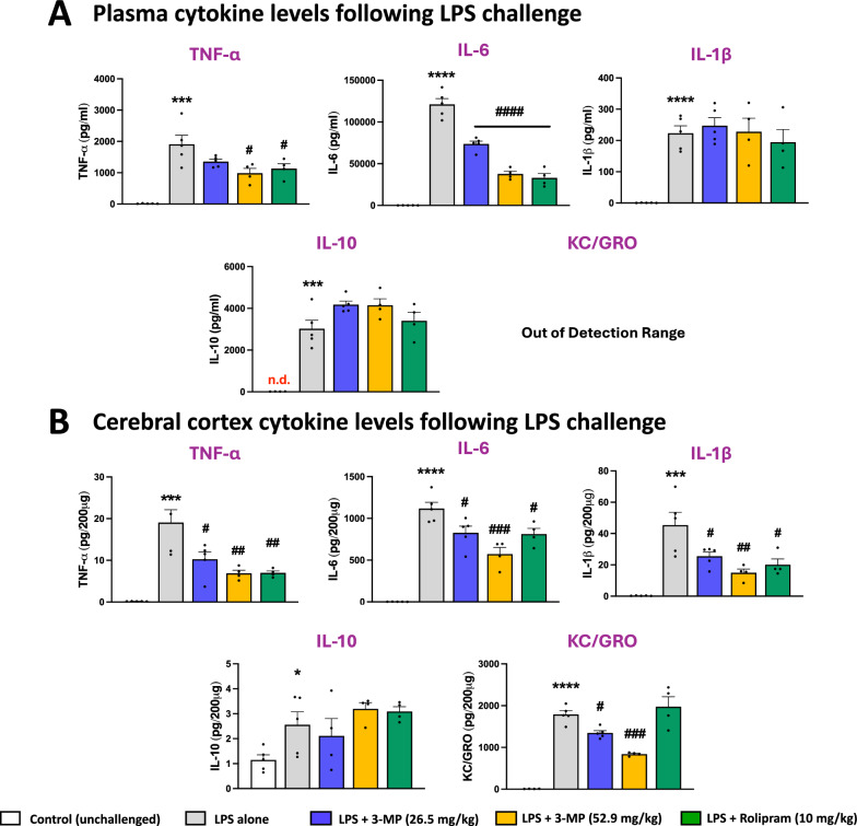
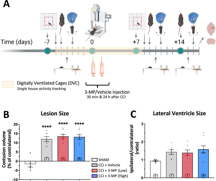
Methods: 3-MP anti-inflammatory activity was evaluated across cellular (RAW 264.7, IMG cells) and mouse studies following lipopolysaccharide (LPS)-challenge (for pro- and anti-inflammatory cytokines/chemokines), and mice subjected to controlled cortical impact (CCI) moderate traumatic brain injury (TBI). 3-MP human cereblon binding, including neosubstrate and molecular modeling evaluation, as well as chicken teratogenicity, ex vivo mouse and human stability studies, and mouse pharmacokinetics were appraised.

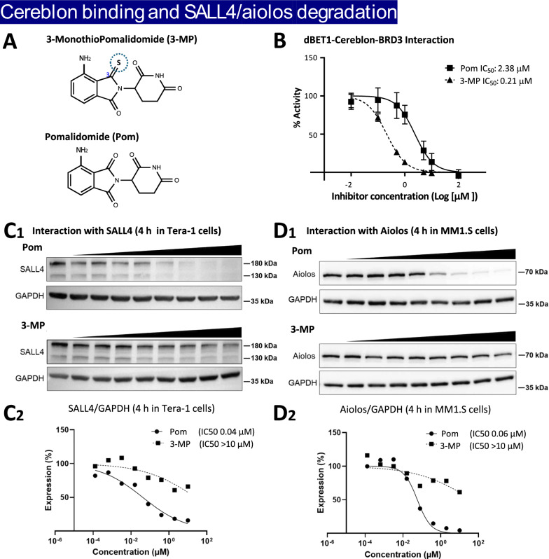
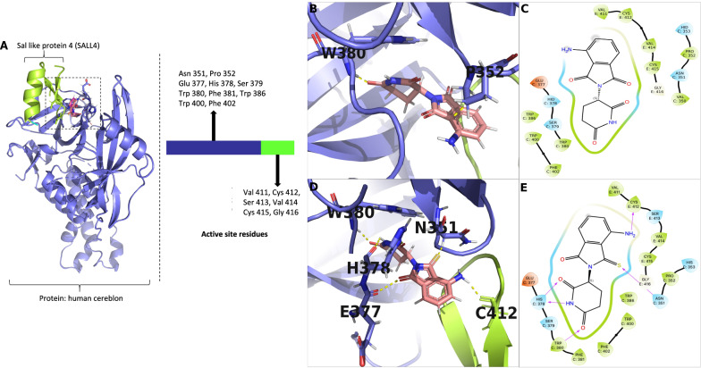
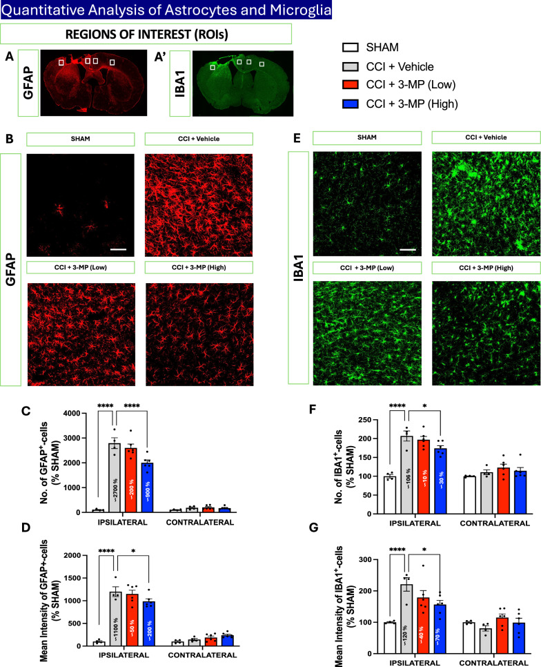
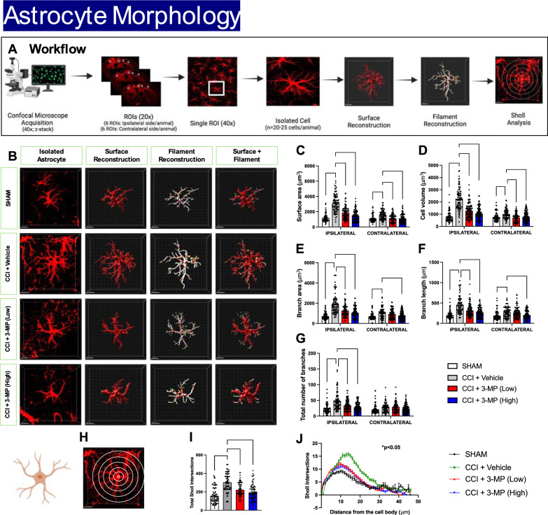
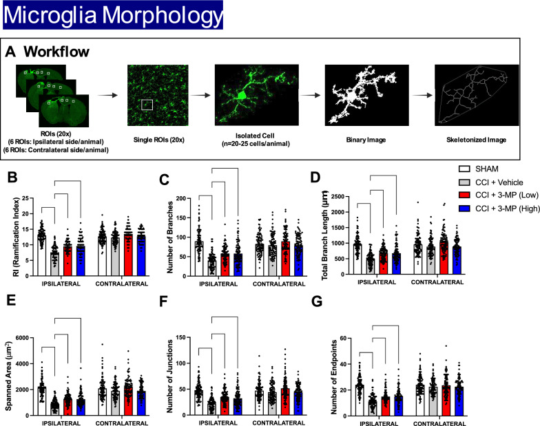
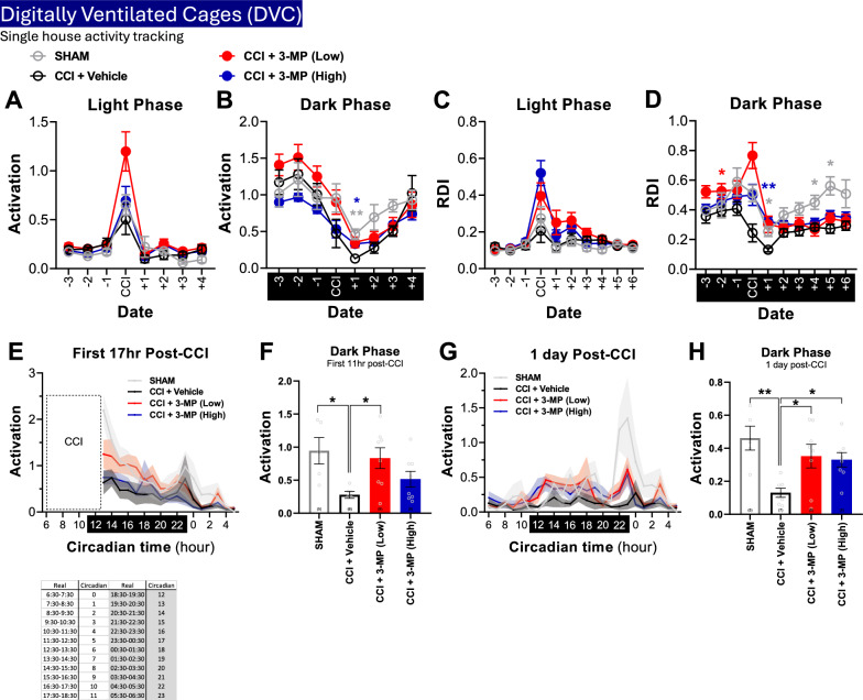
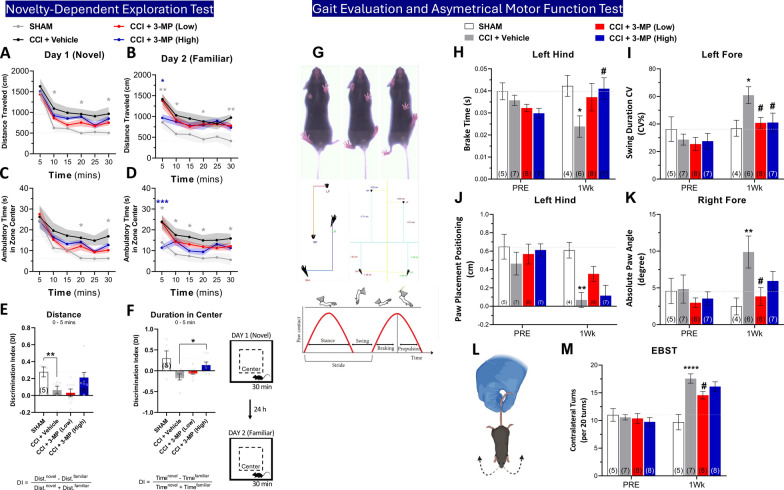
Results: 3-MP binds human cereblon, a key protein in the E3 ubiquitin ligase complex, without triggering downstream cascades leading to thalidomide-like teratogenicity in chicken embryos. 3-MP reduces pro-inflammatory markers in LPS-stimulated mouse macrophage and microglial cell cultures, and lowers pro-inflammatory cytokine/chemokine levels in plasma and brain of mice challenged with systemic LPS without lowering anti-inflammatory IL-10. 3-MP readily enters brain following systemic administration, and achieves a brain/plasma concentration ratio of 0.44-0.47. 3-MP mitigates behavioral impairments and reduces activation of astrocytes and microglia in mice challenged with CCI TBI.

Conclusion: 3-MP represents a promising new class of thalidomide-like IMiDs with potent anti-inflammatory effects that offers potential for treating TBI and possibly other neurodegenerative diseases possessing a prominent neuroinflammatory component.

Keywords: Cereblon; Immunomodulatory imide drugs (IMiDs); Microglia; Neurodegeneration; Neuroinflammation; Pomalidomide; Spalt like transcription factor 4 (SALL4); Teratogenicity; Traumatic brain injury.


Conflict of interest statement

Declarations. Ethics approval and consent to participate: This is a preclinical (non-clinical) research study that does not require ethics approval and consent to participate for human subjects. All preclinical animal studies were undertaken on fully approved Institutional Animal Care and Use Committee protocols (see Materials and Methods). Consent for publication: Not applicable (our manuscript does not contain any individual person’s data in any form). Competing interests: DSK, YKK, SK, MYL, JK and IH are employed by and have stock in Aevis Bio Inc., which has patent coverage on 3-MP. All other authors declare a lack of competing of interest.

Figures

Fig. 1
Fig. 1
3-MP potently binds to human cereblon but does not trigger degradation of the neosubstrates SALL4 and Aiolos. A Chemical structures of 3-monothioPomalidomide (3-MP) and Pomalidomide (Pom). B Concentration-dependent evaluation of the interaction between Immunomodulatory drugs (IMiDs) and cereblon by FRET assay provided an IC50 value for 3-MP of 0.21 μM and for Pom of 2.38 μM. The thalidomide analog-mediated degradation of neosubstrates, SALL (C1C2) and Aiolos (D1D2) were evaluated by Western blotting in Tera-1 cells, and the relative expression level of each neosubstrate was quantified in relation to the housekeeping protein GAPDH (whose level was unchanged by drug incubation). IC50 values to lower SALL4 and Aiolos protein expression values were > 10 μM for 3-MP, and 0.04 μM and 0.06 μM for Pom, respectively
Fig. 2
Fig. 2
Docking showing the structural basis for thalidomide teratogenicity revealed by the Cereblon-DDB1-SALL4-Pomalidomide complex (PDB ID:6UML) into which 3-MP is docked. A 3-D structure of human cereblon (blue) and sal-like protein 4 (SALL4) (green) define the binding site for thalidomide-like compounds and key amino acids involved. B 3-D interactions of pomalidomide with interacting amino acids of Cereblon-DDB1-SALL4-Pomalidomide complex; C 2-D interactions of pomalidomide with interacting amino acids of Cereblon-DDB1-SALL4-Pomalidomide complex. D 3-D interactions of 3-MP with interacting amino acids of Cereblon-DDB1-SALL4-Pomalidomide complex and E 2-D interactions of 3-MP with interacting amino acids of Cereblon-DDB1-SALL4-Pomalidomide complex
Fig. 3
Fig. 3
3-MP mitigates LPS-induced inflammation in cultured RAW 264.7 and IMG cells. Cultured RAW 264.7 and IMG cells were pretreated with either vehicle or 3-MP (0.6 − 60 μM) and challenged with LPS (20 ng/mL) 1 h later. At 24 h following LPS challenge, cellular viability (A, B), nitrite (a stable marker of NO generation) (C, D), and TNF-α levels (E, F) were quantified. 3-MP significantly lowered LPS-induced elevations in nitrite and TNF-α levels. *p < 0.05, **p < 0.01, ***p < 0.001, ****p < 0.0001 vs. the control (LPS + Veh) group
Fig. 4
Fig. 4
Time-dependent ex vivo and in vivo pharmacokinetics of 3-MP. A Ex-vivo disappearance of 3,6′-DTP (100 μg/ml) in mouse (upper left) and human plasma (upper right), and of 3-MP (100 μg/ml) in mouse (lower left) and human plasma (lower right)—expressed as a percent of compound initially added (at time zero) (n = 3). B Plasma and brain 3-MP concentrations in mice following I.P. administration of 3-MP high (26.5 mg/kg) and low (2.65 mg/kg) doses (n = 3 to 4 animals per time point). Of note, the data and Figure ((A) lower right panel) relating to the ex vivo stability of 3-MP in human plasma derives from [64]—study evaluating the stability and pharmacokinetics of 3-MP in rat, with a comparison to human
Fig. 5
Fig. 5
3-MP mitigates LPS-induced elevations in key pro-inflammatory cytokines without altering anti-inflammatory cytokines in plasma (A) and cerebral cortex (B). Systemic administration of LPS (3 mg/kg, I.P.) to mice induced significant increases in pro- (plasma: TNF-α, IL-6, IL-1β, KC/GRO; brain: TNF-α, IL-6, IL-1β, KC/GRO) and anti-inflammatory (plasma and brain: IL-10) cytokines at 4 h. One hour pre-treatment with 3-MP (26.5 and 53 mg/kg, I.P.) significantly mitigated LPS-induced pro-inflammatory cytokines changes (plasma: TNF-α, IL-6; brain: TNF-α, IL-6, IL-1β, KC/GRO) in the absence of alteration in anti-inflammatory IL-10. *p < 0.05, ***p < 0.001, ****p < 0.0001 refers to the effects of LPS compared to the control value (drug vehicle + saline without LPS). #p < 0.05, ##p < 0.01, ###p < 0.001, ####p < 0.0001 refers to the effect of drug treatments vs. drug vehicle + LPS. Rolipram (10 mg/kg, I.P.), a discontinued PDE4 inhibitor with known anti-inflammatory activity [48], was used as a positive control. Values are presented as mean ± S.E.M., of n observations (Control, n = 5; LPS alone, n = 6; 3-MP (Low) + LPS, n = 5; 3-MP (High) + LPS, n = 4; rolipram + LPS (positive control), n = 4)
Fig. 6
Fig. 6
Timeline of mouse model of TBI with 3-MP treatment experimental design and contusion and lateral ventricle size at two weeks. A Mice were first evaluated for their baseline novelty exploration memory, gait function, asymmetrical motor function and motor coordination/balance function by novelty-dependent exploration test, DigiGait analysis, elevated body swing test and beam walking test at one week prior to and at 7 and 14 days after CCI or sham injury. Single house activity tracking was conducted from day 6 prior to CCI to day 7 after CCI TBI by Digital Ventilated Cage (DVC) system. Mice received two injections of 3-MP or Vehicle at 30 min and 24 h following CCI TBI. After two weeks, mice were euthanized, followed by perfusion for assessment of histology and immunohistochemistry. Thereafter, B TBI contusion volume and C lateral ventricle size were quantified from 25 µm Giemsa-stained coronal brain sections (n = 5 to 8 animals/group; ****p < 0.0001 vs. SHAM group w/o CCI TBI)
Fig. 7
Fig. 7
3-MP treatment ameliorates astrocytes and microglia activation following CCI injury. Region of Interests (ROIs) of ipsilateral and contralateral cortex used for the quantitative analysis of GFAP (A) and IBA1 (A′)-positive cells. Representative images of GFAP-positive cells in the ipsilateral cortex (B). Histogram indicates the number (C) and mean intensity (D) of GFAP-positive cells. Representative images of IBA1-positive cells in the ipsilateral cortex (E). Histogram indicates the number (F) and mean intensity (G) of IBA1-positive cells. Values are expressed as a percentage of the SHAM group (n = 5 to 8 animals/group). Black dots within bars represent the values of individual mice; Two-way ANOVA with Dunnett's multiple comparisons test, error bars representing mean ± SEM; *p < 0.05, ****p < 0.0001, SHAM or CCI + 3-MP (Low) or CCI + 3-MP (High) versus CCI + Vehicle. Scale bar = 50 μm
Fig. 8
Fig. 8
3-MP treatment reduces morphological alterations in astrocytes following CCI injury. The schematic illustrates the workflow for morphological analysis of 3D-reconstructed astrocytes in the ipsilateral and contralateral cortex (A). Representative images of 3D-reconstructed astrocytes from ipsilateral cortex (B). Histogram indicates the surface area (C), cell volume (D), branch area (E), branch length (F), and total number of branches (G) of GFAP-positive cells. Sholl analysis of 3D-reconstructed astrocytes (HJ). Representative image of sholl radii on isolated 3D-reconstructed astrocyte (H). Histogram indicates the total sholl intersection (I), and mean distribution plot of the number of Sholl intersections as a function of the distance from the cell body (J). Black dots within bars represents values from individual 3D-reconstructed astrocytes (n = 20–25 cells/animal) (n = 5 to 8 animals/group); Two-way ANOVA with Dunnett's multiple comparisons test, error bars representing mean ± SEM; *p < 0.05, **p < 0.01, ***p < 0.001, ****p < 0.0001, SHAM or CCI + 3-MP (Low) or CCI + 3-MP (High) versus CCI + Vehicle. Scale bar = 50 μm
Fig. 9
Fig. 9
3-MP treatment reduces morphological changes in microglia following CCI injury. The schematic (A) illustrates the workflow of morphological analysis of 3D-reconstructed microglia in the ipsilateral and contralateral cortex. From representative images of 3D-reconstructed microglia from ipsilateral cortex: histograms indicate the ramification index (RI) (B), number of branches (C), total branch length (D), spanned area (E), number of junctions (F), and number of endpoints (G) of 3D-reconstructed microglia. Black dots within bars represents the values from individual 3D-reconstructed microglia (n = 20–25 cells/animal) (n = 5 to 8 animals/group); Two-way ANOVA with Dunnett's multiple comparisons test, error bars representing mean ± SEM; ***p < 0.001, ****p < 0.0001, SHAM or CCI + 3-MP (Low) or CCI + 3-MP (High) versus CCI + Vehicle. Scale bar = 50 μm
Fig. 10
Fig. 10
3-MP treatment ameliorates altered cage activity one day following CCI injury. Day to day average activation (A, B) and Regularity Disruption Index (RDI) (C, D) during light (A, C) and dark (B, D) phases; Mixed effect analysis with Dunnett's multiple comparisons test; *p < 0.05, **p < 0.01, CCI + 3-MP (High) versus CCI + Vehicle (blue), CCI + 3-MP (Low) versus CCI + Vehicle (red), SHAM versus CCI + Vehicle (grey). E Home cage activation 17 h after CCI, with each data point representing averaged 2 h blocks of cage activation. Each circadian time point was indicated in the worksheet below. F Total activation of dark phase during 11 h after CCI. G Home cage activation 1-day post-CCI. H Total activation of dark phase 1-day post-CCI. One-way ANOVA with Dunnett’s multiple comparisons test, error bars representing mean ± SEM; *p < 0.05, **p < 0.01, SHAM or CCI + 3-MP (Low) or CCI + 3-MP (High) versus CCI + Vehicle. n = SHAM (5), CCI + Vehicle (7), CCI + 3-MP (Low) (8), CCI + 3-MP (High) (8)
Fig. 11
Fig. 11
3-MP treatment reduces CCI-induced novelty-dependent exploration behavioral changes, gait impairments, and asymmetrical motor function one week after injury. Distance traveled (A, B) and ambulatory time spent in the central zone (C, D) of open field exploration within 30 min were recorded on 2 consecutive days at one week after CCI. Testing on day 1 (A, C) represents a novel environment for mice w/wo injury, and day 2 as a familiar environment (B, D); Two-way ANOVA with Dunnett's multiple comparisons test, error bars shown with area filled color representing mean ± SEM; *p < 0.05, **p < 0.01, ***p < 0.001, SHAM (grey) or CCI + 3-PM (High) (blue) versus CCI + Vehicle. The difference between day 1 and day 2 for the distance traveled (E) and duration in the center zone (F) during the 5 min was assessed by discrimination index; One-way ANOVA with Dunnett's multiple comparisons test, *p < 0.05, **p < 0.01, SHAM (grey) or CCI + 3-PM (High) (blue) versus CCI + Vehicle. (G) Representative images were collected by DiGi gait system (Mouse Specifics, Inc.) for gait function assessment. Parameters included brake time (time duration of the initial paw contact to maximum paw contact) (H), swing duration CV (% of the coefficient of variance was calculated by 100 X standard deviation/mean, the variability normalized to the mean) (I), Paw Placement Positioning (the extent of overlap between ipsilateral fore and hind paws during full stance, indicating the balance metric) (J), absolute paw angle (K), and were analyzed one week before (PRE) and after CCI (1 Wk). Mouse numbers for each group are indicated at the bottom of bars within the graph. Diagram (L) depicts the elevated body swing test (EBST). Asymmetrical function was measured by EBST pre- and post-CCI (M). Two-way ANOVA with Dunnett’s multiple comparisons test, error bars representing mean ± SEM; *p < 0.05, **p < 0.01, ****p < 0.0001, SHAM versus CCI + Vehicle; #p < 0.05, CCI + 3-MP (Low) or CCI + 3-MP (High) versus CCI + Vehicle. n = SHAM (5), CCI + Vehicle (7), CCI + 3-MP (Low) (8), CCI + 3-MP (High) (8)

Similar articles

References

    1. Dewan MC, Rattani A, Gupta S, Baticulon RE, Hung YC, Punchak M, Agrawal A, Adeleye AO, Shrime MG, Rubiano AM, Rosenfeld JV, Park KB. Estimating the global incidence of traumatic brain injury. J Neurosurg. 2018;130(4):1080–97. - PubMed
    1. Freire MAM, Rocha GS, Bittencourt LO, Falcao D, Lima RR, Cavalcanti JRLP. Cellular and molecular pathophysiology of traumatic brain injury: what have we learned so far? Biology (Basel). 2023;12:1139. 10.3390/biology12081139. - PMC - PubMed
    1. Tagliaferri F, Compagnone C, Korsic M, Servadei F, Kraus J. A systematic review of brain injury epidemiology in Europe. Acta Neurochir (Wien). 2006;148(3):255–68. - PubMed
    1. Shi HY, Wang SN, Lee KT. Temporal trends and volume-outcome associations in periampullary cancer patients: a propensity score-adjusted nationwide population-based study. Am J Surg. 2014;207(4):512–9. - PubMed
    1. Gardner RC, Burke JF, Nettiksimmons J, Kaup A, Barnes DE, Yaffe K. Dementia risk after traumatic brain injury vs nonbrain trauma: the role of age and severity. JAMA Neurol. 2014;71(12):1490–7. - PMC - PubMed