. 2025 Jul;38(4):e70033.
doi: 10.1111/pcmr.70033.

Disruption of mc1r Disturbs Skin Pigmentation in Xenopus tropicalis

Affiliations

Disruption of mc1r Disturbs Skin Pigmentation in Xenopus tropicalis

Lanxin Li et al. Pigment Cell Melanoma Res. 2025 Jul.

Abstract

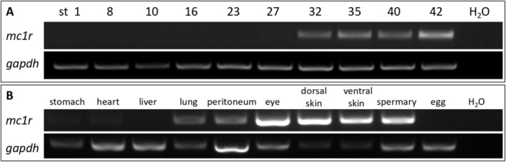
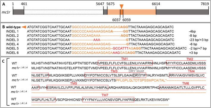
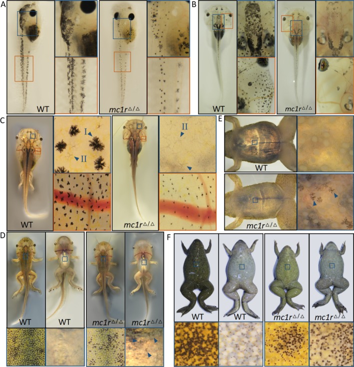
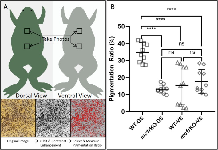
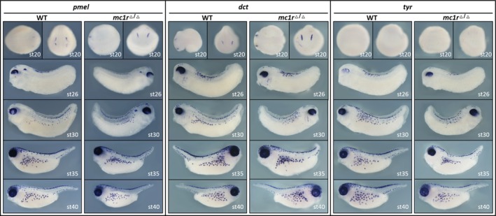
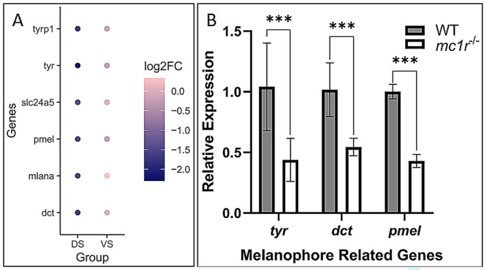
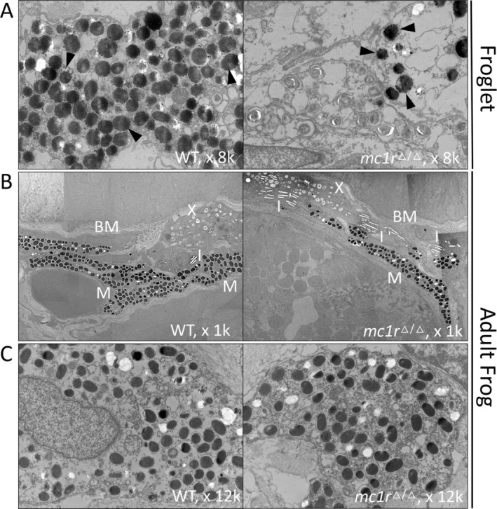
The melanocortin 1 receptor (MC1R) is well-established as a pivotal regulator of pigmentation in various species. Despite a wealth of research focused on mammals and fish, the role of Mc1r in amphibians has remained largely unexplored. This study was designed to elucidate the contribution of Mc1r in Xenopus tropicalis. Our results reveal that targeted ablation of mc1r in Xenopus tropicalis led to a significant reduction in dorsal skin pigmentation, while simultaneously accelerating the onset of melanophore pigmentation in the ventral region. This dual effect resulted in a perturbation of the canonical countershading pattern. Additionally, knockout of mc1r disrupted the expression of multiple genes primarily associated with pigmentation. Collectively, these findings underscore the critical role of MC1R in the regulation of pigmentation and the development of countershading in amphibians, contributing to the growing body of literature on the evolution and function of MC1R across vertebrate species.

Keywords: Xenopus tropicalis; Mc1r; CRISPR‐Cas9; countershading; pigmentation.


Conflict of interest statement

The authors declare no conflicts of interest.

Figures

FIGURE 1
FIGURE 1
Temporal expression pattern during tadpole development and spatial expression pattern in adult Xenopus tropicalis of mc1r through RT‐PCR. The result of RT‐PCR in (A) embryos at different stages and in (B) different organs of adult frog is shown. st, stage.
FIGURE 2
FIGURE 2
mc1r knockout through CRISPR‐Cas9. (A) The target sequence is at nucleotide 6037–6059 in the mc1r gene indicated by an orange arrow head. The orange box indicates the exon, grey boxes indicate the untranslated regions (UTR) and the dotted line indicates the intron. (B) The sequence of the wild‐type target sequence (shown in orange) and some different insert/deletion (INDEL) situations in G0. bp, base pair (C) The mutated amino acid sequence led by a 4 bp deletion. TM, transmembrane; DRY, Asp‐Arg‐Tyr.
FIGURE 3
FIGURE 3
mc1r knockout disturbs pigment pattern of post‐stage 43 tadpoles and frogs. (A) The tadpoles at stage 43, (B) the tadpoles at stage 52, (C) the tadpoles during metamorphosis at Stage 56–57, (D) the tadpoles at the end of metamorphosis at stage 59, (E) the froglets have finished metamorphosis, and (F) the adult frogs are shown in black boxes. The detailed pigment pattern marked with blue or orange boxes on tadpoles or frogs is shown in the boxes on right or bottom with corresponding colors respectively. The blue arrowheads indicate melanophores.
FIGURE 4
FIGURE 4
Numerical analysis shows the reduction of pigmentation in the dorsal skin of mc1r knockout frogs and the high inter‐individual variation in the ventral skin. (A) The method of numerical analysis. Photos are taken of equivalent dorsal and ventral regions from adult Xenopus tropicalis individuals. ImageJ is used to process the images. The ratio of pigmented areas relative to the total image area is quantified and analyzed. (B) Pigmentation ratio is compared among WT‐DS, mc1rKO‐DS, WT‐VS, and mc1rKO‐VS. DS, dorsal skin, VS, ventral skin.
FIGURE 5
FIGURE 5
Whole mount in situ hybridization result of pmel, dct, and tyr in wild‐type and mc1r null tadpoles at different stages is shown.
FIGURE 6
FIGURE 6
The differentiated expression genes related with melanophores are analyzed by RNA‐seq and verified by qPCR. (A) The essential genes related with melanophore are selected and analyzed based on the RNA‐seq result. The blue dots indicate the genes are downregulated while the pink dots mean the genes are not disturbed obviously. (B) Three essential melanophore related genes downregulated in DS analyzed by RNA‐seq (tyr, dct, and pmel) are selected and tested by qPCR, accorded with RNA‐seq result.
FIGURE 7
FIGURE 7
The GO and KEGG pathways analysis of differentiated expression genes. The analysis results of (A) GO‐BP (biological process), (B) the GO‐CC (cellular component), (C) the MF (molecular function), and (D) KEGG pathways are shown.
FIGURE 8
FIGURE 8
STRING analysis of PPI (protein–protein interaction) and visualized by Cytoscape. Darker color means higher degree.
FIGURE 9
FIGURE 9
Transmission electron microscopy result is shown. (A) The ultrastructure of melanophores in wild‐type and mc1r null froglets (×8k). (B) The overall chromatophore distribution under basal membrane (×1k) and (C) ultrastructure of melanosomes (×12k) in wild‐type and mc1r null adult frogs. BM, basal membrane; I, iridophore; M, melanophore; X, xanthophore. Black arrowheads indicate melanosomes.

Similar articles

References

    1. Aberdam, E. , Bertolotto C., Sviderskaya E. V., et al. 1998. “Involvement of Microphthalmia in the Inhibition of Melanocyte Lineage Differentiation and of Melanogenesis by Agouti Signal Protein.” Journal of Biological Chemistry 273, no. 31: 19560–19565. 10.1074/jbc.273.31.19560. - DOI - PubMed
    1. Alan, G.‐E. , Heather L. R., Jaren M. A., et al. 2024. “A Ball Python Colour Morph Implicates MC1R in Melanophore‐Xanthophore Distribution and Pattern Formation.” Pigment Cell & Melanoma Research 38, no. 1: e13215. 10.1111/pcmr.13215. - DOI - PubMed
    1. Alessandro, B. , and Elizabeth P.. 2024. “Melanocyte Lineage Dynamics in Development, Growth and Disease.” Development (Cambridge, England) 151, no. 15: dev201266. 10.1242/dev.201266. - DOI - PMC - PubMed
    1. Bagnara, J. T. , Taylor J. D., and Hadley M. E.. 1968. “Dermal Chromatophore Unit.” Journal of Cell Biology 38, no. 1: 67–79. 10.1083/jcb.38.1.67. - DOI - PMC - PubMed
    1. Baxter, L. L. , and Pavan W. J.. 2003. “Pmell17 Expression Is Mitf‐Dependent and Reveals Cranial Melanoblast Migration During Murine Development.” Gene Expression Patterns 3, no. 6: 703–707. 10.1016/j.modgep.2003.07.002. - DOI - PubMed

MeSH terms

Substances

LinkOut - more resources