

Hypersomatotropism and Hypercortisolism Caused by a Plurihormonal Pituitary Adenoma in a Dog
- PMID: 40524601
- PMCID: PMC12171238
- DOI: 10.1111/jvim.70177
Hypersomatotropism and Hypercortisolism Caused by a Plurihormonal Pituitary Adenoma in a Dog
Abstract
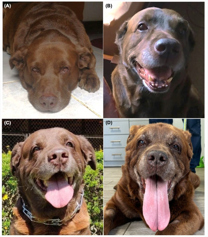
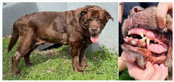
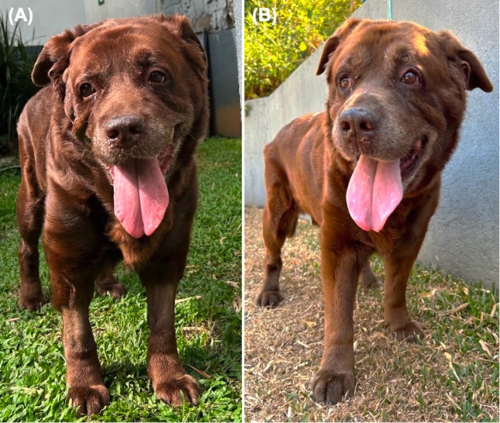
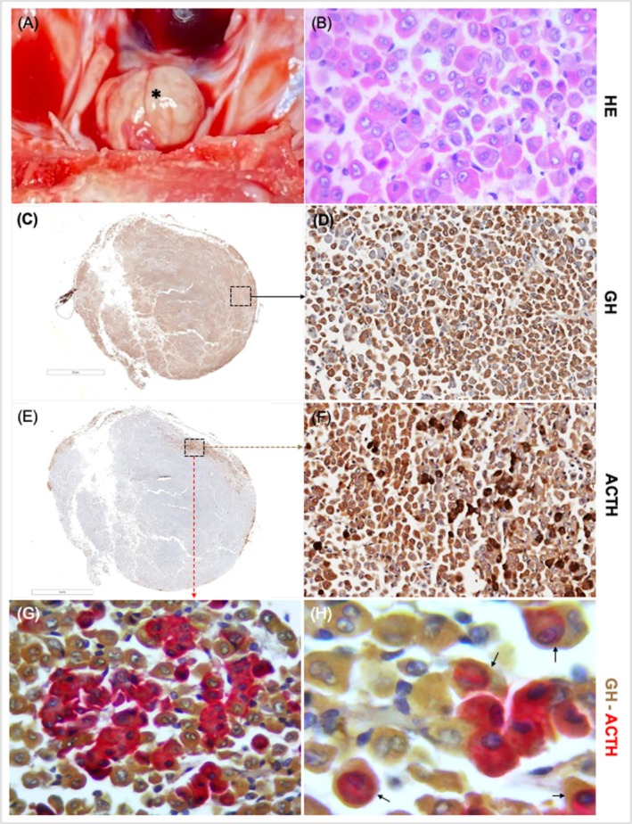
A 12-year-old, male Labrador Retriever was presented because of polyuria, polydipsia, polyphagia, joint pain, and physical features consistent with acromegaly. Circulating insulin-like growth factor-1 (IGF-1) concentration was increased (> 1000 ng/mL; reference interval [RI], 42-449), suggestive of hypersomatotropism. An abnormal low-dose dexamethasone suppression test and increased circulating adrenocorticotropic (ACTH) concentration indicated pituitary-dependent hypercortisolism. Computed tomography identified an enlarged pituitary gland. Treatment with cabergoline initially decreased circulating IGF-1 and ACTH concentrations and urinary cortisol-to-creatinine ratio (UCCR), with a notable reduction in acromegalic physical features. However, 7 months after the start of cabergoline treatment, IGF-1, ACTH, and UCCR had increased again, although pituitary gland size remained stable. Because of worsening joint pain, euthanasia was performed. On necropsy, double immunohistochemistry identified pituitary tumor cells with cytoplasmic co-expression of both growth hormone (GH) and ACTH, consistent with a monomorphic plurihormonal macroadenoma. This case shows that concurrent hypersomatotropism and hypercortisolism can occur in dogs caused by a plurihormonal pituitary adenoma.
Keywords: acromegaly; caber goline; corticotropinoma; cushing's syndrome; growth hormone; insulin‐like growth factor‐1; somatotropinoma.
© 2025 The Author(s). Journal of Veterinary Internal Medicine published by Wiley Periodicals LLC on behalf of American College of Veterinary Internal Medicine.
Conflict of interest statement
The authors declare no conflicts of interest.
Figures

References
-
- Kooistra H. S., “Chapter 40—Acromegaly in Dogs,” in Clinical Endocrinology of Companion Animals, 1st ed., ed. Rand J. (Wiley, 2013), 421–426.
-
- Selman P. J., Mol J. A., Rutteman G. R., van Garderen E., and Rijnberk A., “Progestin‐Induced Growth Hormone Excess in the Dog Originates in the Mammary Gland,” Endocrinology 134 (1994): 287–292. - PubMed
-
- Jaillardon L., Martin L., Nguyen P., and Siliart B., “Serum Insulin‐Like Growth Factor Type 1 Concentrations in Healthy Dogs and Dogs With Spontaneous Primary Hypothyroidism,” Veterinary Journal 190 (2011): e95–e99. - PubMed
-
- Daly A. F. and Beckers A., “Chapter 12—Medical Management of Pituitary Gigantism and Acromegaly,” in Gigantism and Acromegaly, 1st ed., ed. Stratakis C. A. (Academic Press, Elsevier, 2021), 245–257.
-
- van Keulen L. J., Wesdorp J. L., and Kooistra H. S., “Diabetes Mellitus in a Dog With a Growth Hormone‐Producing Acidophilic Adenoma of the Adenohypophysis,” Veterinary Pathology 33 (1996): 451–453. - PubMed
Publication types
MeSH terms
Substances
LinkOut - more resources
Full Text Sources
Medical
Miscellaneous
