

Epithelioid angiosarcoma of the cervical spine: A case report
- PMID: 40524767
- PMCID: PMC11866273
- DOI: 10.12998/wjcc.v13.i17.101593
Epithelioid angiosarcoma of the cervical spine: A case report
Abstract
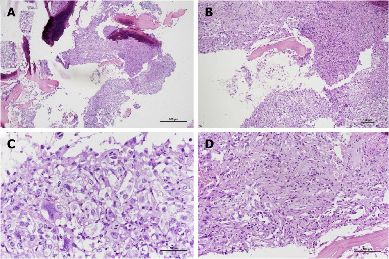
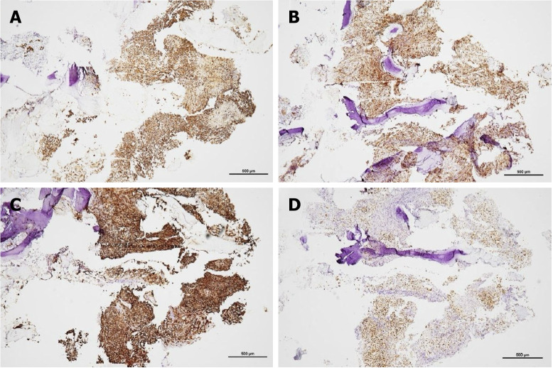
Background: Epithelioid angiosarcoma (EA) is an aggressive, malignant endothelial-cell tumor of vascular or lymphatic origin. EA often arises from deep soft tissues such as pleura, breast, bone and gastrointestinal tract. It usually affects patients aged 60-70 years and is associated with high recurrence and metastasis rates with surgical resection as the primary treatment of choice. Overall survivals are generally poor, ranging from 6 to 16 months. More than 50% of patients died of disease within 2 to 3 years of diagnosis.
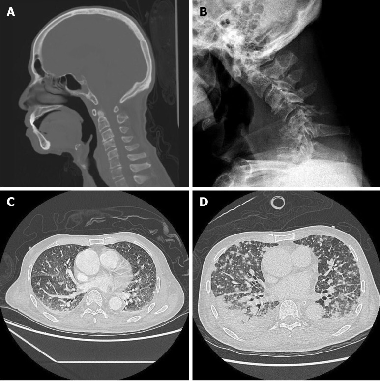
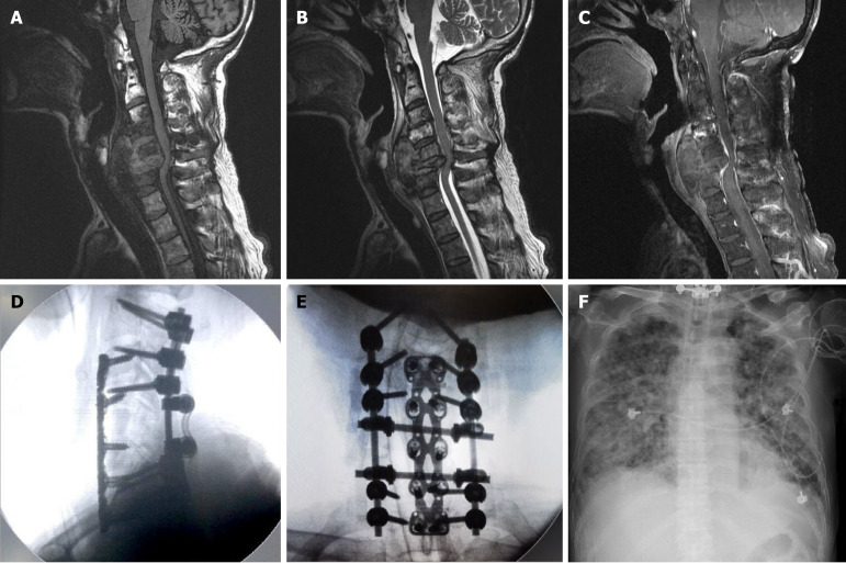
Case summary: We present a rare case of EA of the cervical spine causing a C6 pathological fracture complicated by severe kyphosis. The patient received C4-7 posterior laminectomy and C2/3/4/7/T1 transpedicular screw fixation, followed by anterior C5-6 corpectomy with allograft bone fusion and cervical plate fixation. Postoperative radiotherapy was administered without delay. However, the patient died of rapidly progressive acute respiratory distress syndrome 3 weeks after the second surgery.
Conclusion: EA with spinal involvement is extremely rare. Early detection and cord decompression may prevent neurological deterioration and preserve better quality of life.
Keywords: Case report; Cervical spine; Epithelioid angiosarcoma; Pathologic fracture; Spinal cord compression; Vertebral body.
©The Author(s) 2025. Published by Baishideng Publishing Group Inc. All rights reserved.
Conflict of interest statement
Conflict-of-interest statement: All the authors report no relevant conflicts of interest for this article.
Figures
Similar articles
-
Pediatric cervical kyphosis in the MRI era (1984-2008) with long-term follow up: literature review.Childs Nerv Syst. 2022 Feb;38(2):361-377. doi: 10.1007/s00381-021-05409-z. Epub 2021 Nov 22. Childs Nerv Syst. 2022. PMID: 34806157 Review.
-
Posterior cervicothoracic instrumentation in spine tumors.Spine (Phila Pa 1976). 2004 Jun 1;29(11):1246-53. doi: 10.1097/00007632-200406010-00015. Spine (Phila Pa 1976). 2004. PMID: 15167665
-
Multicentric epithelioid angiosarcoma of the spine: a case report of a rare bone tumor.Spine J. 2007 Nov-Dec;7(6):716-9. doi: 10.1016/j.spinee.2006.08.013. Epub 2006 Dec 22. Spine J. 2007. PMID: 17998131
-
Epithelioid angiosarcoma of the spine: A case report of a rare bone tumor.Oncol Lett. 2014 Jun;7(6):2170-2174. doi: 10.3892/ol.2014.2055. Epub 2014 Apr 9. Oncol Lett. 2014. PMID: 24932310 Free PMC article.
-
Primary extradural epithelioid leiomyosarcoma of the cervical spine: case report and literature review.Neurosurgery. 2005 Aug;57(2):E372; discussion E372. doi: 10.1227/01.neu.0000166695.89757.a4. Neurosurgery. 2005. PMID: 16094142 Review.
References
-
- Young RJ, Brown NJ, Reed MW, Hughes D, Woll PJ. Angiosarcoma. Lancet Oncol. 2010;11:983–991. - PubMed
-
- Deshpande V, Rosenberg AE, O'Connell JX, Nielsen GP. Epithelioid angiosarcoma of the bone: a series of 10 cases. Am J Surg Pathol. 2003;27:709–716. - PubMed
-
- Marthya A, Patinharayil G, Puthezeth K, Sreedharan S, Kumar A, Kumaran CM. Multicentric epithelioid angiosarcoma of the spine: a case report of a rare bone tumor. Spine J. 2007;7:716–719. - PubMed
Publication types
LinkOut - more resources
Full Text Sources
Research Materials
Miscellaneous
