

Diagnosis of Female Urethral Cancer Based on Multimodal Ultrasound: A Case Report
- PMID: 40524812
- PMCID: PMC12168908
- DOI: 10.2147/IJWH.S517310
Diagnosis of Female Urethral Cancer Based on Multimodal Ultrasound: A Case Report
Abstract
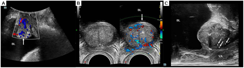
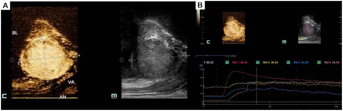
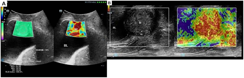
While histopathology remains the diagnostic gold standard for urothelial carcinoma, this case highlights the emerging role of advanced ultrasonographic techniques in characterizing urethral malignancies. We present a 51-year-old female with a one-month history of refractory lower urinary tract symptoms (urinary frequency, urgency, dysuria, and nocturia) unresponsive to conventional anti-inflammatory therapy. Multimodal ultrasonographic evaluation, incorporating conventional sonography, transrectal ultrasound, elastography, and contrast-enhanced ultrasonography (CEUS), revealed a complex proximal urethral mass with malignant features. Subsequent cystoscopic and histopathological examinations confirmed the diagnosis of primary urethral adenocarcinoma. This case underscores the diagnostic value of comprehensive ultrasound protocols in evaluating female urethral neoplasms.
Keywords: contrast-enhanced ultrasonography; female urethral adenocarcinoma; multimodal ultrasound; shear wave elasticity; transrectal biplane ultrasound.
© 2025 Wang et al.
Conflict of interest statement
The authors have no conflicts of interest to declare for this work.
Figures
Similar articles
-
The Value of Ultrasound and Contrast-Enhanced Ultrasound in a Young Asymptomatic Patient With Duodenal Neuroendocrine Neoplasms: A Case Report.J Clin Ultrasound. 2025 Jun;53(5):1176-1181. doi: 10.1002/jcu.23957. Epub 2025 Mar 22. J Clin Ultrasound. 2025. PMID: 40119627
-
Vesicoureteral Reflux.2024 Apr 30. In: StatPearls [Internet]. Treasure Island (FL): StatPearls Publishing; 2025 Jan–. 2024 Apr 30. In: StatPearls [Internet]. Treasure Island (FL): StatPearls Publishing; 2025 Jan–. PMID: 33085409 Free Books & Documents.
-
A novel multimodal approach to C2 vertebroplasty in the setting of osteolytic metastases: A case report.Radiol Case Rep. 2025 Jun 4;20(9):4196-4202. doi: 10.1016/j.radcr.2025.05.042. eCollection 2025 Sep. Radiol Case Rep. 2025. PMID: 40528910 Free PMC article.
-
Endorectal ultrasound with elastography for differentiating benign and malignant rectal tumors: a systematic review and meta-analysis.Am J Transl Res. 2025 May 15;17(5):3830-3841. doi: 10.62347/BJMS2565. eCollection 2025. Am J Transl Res. 2025. PMID: 40535670 Free PMC article. Review.
-
Prevalence and odds of anxiety and depression in cutaneous malignant melanoma: a proportional meta-analysis and regression.Br J Dermatol. 2024 Jun 20;191(1):24-35. doi: 10.1093/bjd/ljae011. Br J Dermatol. 2024. PMID: 38197404
References
-
- Wenzel M, Nocera L, Collà Ruvolo C, et al. Sex - related differences include stage, histology, and survival in urethral cancer patients. Clin Genitourin Cancer. 2021;19(2):135–143. - PubMed
-
- João Pedro Paulino Ruas T, Vasconcellos Andrade E, Costa Scopacasa F, et al. Urethral mucinosous adenocarcinoma in a female patient - a case report. Urogynecology. 2024;30:1005–1009. - PubMed
Publication types
LinkOut - more resources
Full Text Sources
