

Alterations on Microcirculation of Optic Nerve Head Before and After OSA Surgery
- PMID: 40524901
- PMCID: PMC12169017
- DOI: 10.2147/NSS.S493508
Alterations on Microcirculation of Optic Nerve Head Before and After OSA Surgery
Abstract
Objective: Obstructive sleep apnea/hypopnea syndrome (OSA) can compromise oxygenation of the optic nerve and cause glaucomatous optic neuropathy; however, there were no studies to investigate the changes of optic nerve microcirculation in patients with OSA before and after treatment. We conducted the first study to assess whether OSA surgery will change the optic nerve microcirculation in patients with OSA.
Study design: Prospective single-blind study.
Setting: Tertiary medical center.
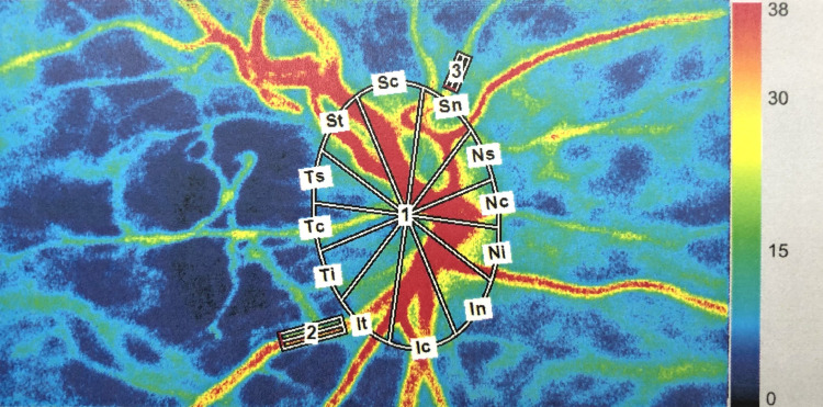
Methods: The enrolled patients were completed for overnight polysomnography (PSG) and comprehensive ophthalmologic evaluation, including laser speckle flowgraphy (LSFG) for microcirculation of optic nerve head (ONH) before and 3 months after OSA surgery. LSFG measurements were summarized as mean blur rate in all areas (MA), in big vessel area (MV) and in tissue area (MT) of ONH.
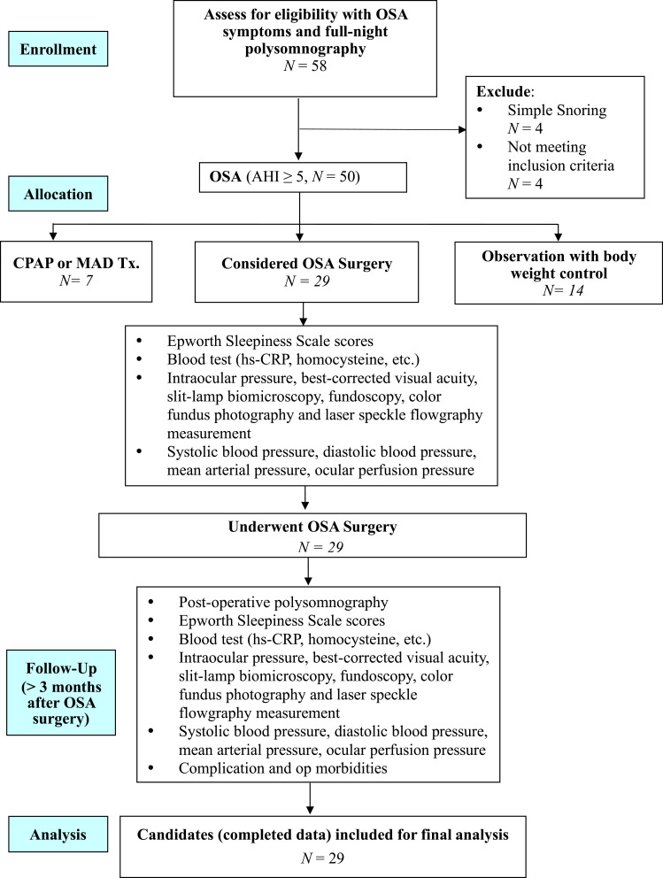
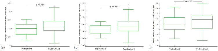
Results: Twenty-nine patients underwent upper airway surgery were included. Three months after surgery, 75.9% (22/29) patients, including 15 of 20 patients with severe OSA and 7 of 9 patients with moderate OSA had improvement in apnea/hypopnea index (AHI). The major parameters of PSG significantly improved. Regarding the LSFG parameters, MA (p = 0.023), MV (p = 0.033) and MT (p = 0.026) significantly increased 3 months after surgery. Moreover, there were significant differences in MA (p = 0.035) and MT (p = 0.045) in the AHI-improved subgroup after surgery.
Conclusion: The ONH microcirculation significantly improved in the AHI-improved patients with OSA 3 months after upper airway surgery. Upper airway surgery may ameliorate the ONH microcirculation in patients with OSA.
Keywords: laser speckle flowgraphy; obstructive sleep apnea surgery; obstructive sleep apnea syndrome; optic nerve head microcirculation; snoring.
© 2025 Lin et al.
Conflict of interest statement
The authors report no conflicts of interest in this work.
Figures
References
LinkOut - more resources
Full Text Sources
