

Sjögren's Syndrome Associated With Erythema Dyschromicum Perstans: A Rare Dermatological Manifestation
- PMID: 40524988
- PMCID: PMC12168861
- DOI: 10.7759/cureus.84234
Sjögren's Syndrome Associated With Erythema Dyschromicum Perstans: A Rare Dermatological Manifestation
Abstract
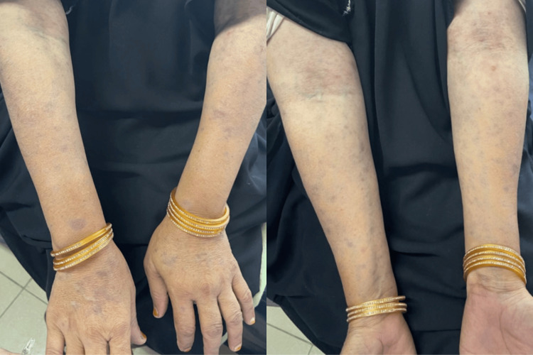
Sjögren's syndrome is an autoimmune disorder with a complex, multifactorial etiopathogenesis that predominantly affects women, typically in their middle-aged years. The condition is associated with a variety of skin manifestations beyond the characteristic skin tightening and thickening. These include erythema multiforme, lichen planus, erythema nodosum (dermo-panniculitis), chilblain-like erythema, vasculitis, livedo reticularis, and granuloma annulare. One rare dermatological manifestation of Sjögren's syndrome is erythema dyschromicum perstans (EDP), also known as ashy dermatosis or dermatosis cenicienta, which is an acquired condition characterized by symmetrical hyperpigmentation on the trunk and extremities. Although few cases of ashy dermatosis have been reported in association with Sjögren's syndrome, we present the case of a 50-year-old woman diagnosed with Sjögren's syndrome and ashy dermatosis based on biopsy, marking what appears to be the first reported case from Pakistan.
Keywords: ashy dermatosis; erythema cenicienta; erythema dyschromicum perstans; sjogren syndrome antibodies or ssa ssb; sjogren syndrome.
Copyright © 2025, Golani et al.
Conflict of interest statement
Human subjects: Consent for treatment and open access publication was obtained or waived by all participants in this study. Conflicts of interest: In compliance with the ICMJE uniform disclosure form, all authors declare the following: Payment/services info: All authors have declared that no financial support was received from any organization for the submitted work. Financial relationships: All authors have declared that they have no financial relationships at present or within the previous three years with any organizations that might have an interest in the submitted work. Other relationships: All authors have declared that there are no other relationships or activities that could appear to have influenced the submitted work.
Figures


Similar articles
-
Erythema Dyschromicum Perstans: Identical to Ashy Dermatosis or Not?Case Rep Dermatol. 2015 Jul 18;7(2):146-50. doi: 10.1159/000437414. eCollection 2015 May-Aug. Case Rep Dermatol. 2015. PMID: 26351421 Free PMC article.
-
A Delphi consensus on the nomenclature and diagnosis of lichen planus pigmentosus and related entities.Indian J Dermatol Venereol Leprol. 2023 Jan-Frebuary;89(1):41-46. doi: 10.25259/IJDVL_804_2021. Indian J Dermatol Venereol Leprol. 2023. PMID: 35593293
-
Ribociclib-Induced Erythema Dyschromicum Perstans (Ashy Dermatosis)-Like Pigmentation in a Metastatic Breast Cancer Patient.J Breast Cancer. 2021 Feb;24(1):117-121. doi: 10.4048/jbc.2021.24.e1. J Breast Cancer. 2021. PMID: 33634626 Free PMC article.
-
Literature Review of Treatment Outcomes for Lichen Planus Pigmentosus, Erythema Dyschromicum Perstans, and Ashy Dermatosis.J Cutan Med Surg. 2018 Nov/Dec;22(6):643-645. doi: 10.1177/1203475418782132. J Cutan Med Surg. 2018. PMID: 30322299 Review. No abstract available.
-
Erythema dyschromicum perstans: a case report and review.Cutis. 2001 Jul;68(1):25-8. Cutis. 2001. PMID: 11480143 Review.
References
-
- Cutaneous findings in patients with primary Sjogren's syndrome. Soy M, Piskin S. Clin Rheumatol. 2007;26:1350–1352. - PubMed
-
- Dermatological manifestations of Sjögren's syndrome (German) Ueki H, Inagaki Y, Hamasaki Y, Ono M. https://pubmed.ncbi.nlm.nih.gov/1765488/ Hautarzt. 1991;42:741–747. - PubMed
Publication types
LinkOut - more resources
Full Text Sources
Research Materials