. 2025 May 21;33(2):101494.
doi: 10.1016/j.omtm.2025.101494. eCollection 2025 Jun 12.

Common AAV gene therapy vectors show nonselective transduction of ex vivo human brain tissue

Affiliations

Common AAV gene therapy vectors show nonselective transduction of ex vivo human brain tissue

J P McGinnis et al. Mol Ther Methods Clin Dev. .

Abstract

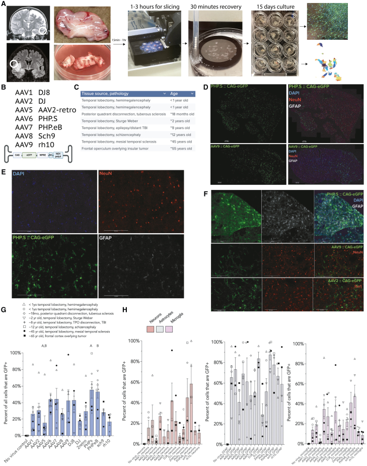
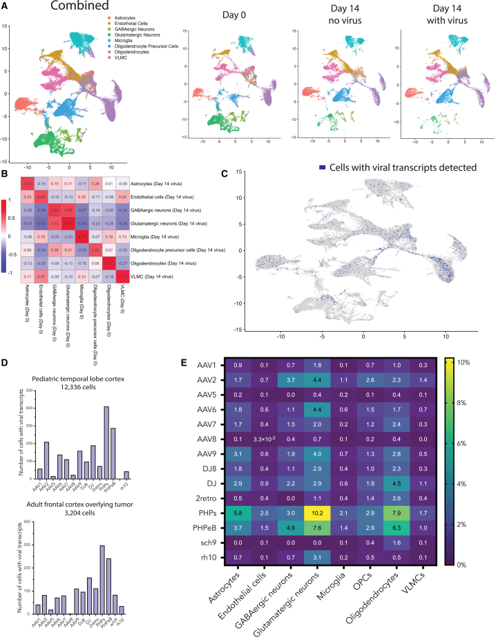
The ability to deliver a therapeutic sequence to a specific cell type in the human brain would make possible innumerable therapeutic options for some of our most challenging diseases; however, studies on adeno-associated virus (AAV) vector tropism have generally relied on animal models with limited translational utility. For this reason, establishing the tropism of common adeno-associated virus (AAV) vectors in living human brain tissue serves as an important baseline for further optimization, as well as a determination of human brain cell types transduced by clinically approved gene therapy vectors AAV2 and AAV9. We have adapted an ex vivo organotypic model to evaluate AAV transduction properties in living slices of human brain tissue. Using fluorescent reporter expression and single-nucleus RNA sequencing, we found that common AAV vectors show broad transduction of normal cell types, with protein expression most apparent in astrocytes; this work introduces a pipeline for identifying and optimizing AAV gene therapy vectors in human brain samples.

Keywords: AAV; AAV tropism; gene therapy; human brain; human brain organotypic slice culture.


Conflict of interest statement

The authors declare no conflict of interest with the study or any of the topics discussed.

Figures

None
Graphical abstract
Figure 1
Figure 1
Human brain organotypic slice model facilitates characterization of AAV tropism by immunofluorescence (A) Workflow of the human brain organotypic model: example MRIs with location of tissue resected, resected samples, vibratome slicing, NMDG-aCSF recovery, culture plates, and immunostaining or single-nucleus sequencing. (B) The AAV capsid variants studied and the CAG-eGFP construct used for immunostaining and single-nucleus sequencing. (C) The anatomical locations of patients’ samples, pathologies involved, and approximate ages. (D) Representative images of entire sections of PHP.S and AAV9 from adult frontal cortex overlying tumor. Scale bars: 200 μm. (E) Magnified images of PHP.S with DAPI, NeuN (neurons), GFAP (astrocytes), and transduced cells (GFP). (F) Representative magnified images showing PHP.S and astrocytes, AAV9 and neurons, and AAV2 and microglia in pediatric temporal lobectomy samples. Scale bars: 200 μm. (G) Percent transduction of all cells present in the tissue (measured as percentage of DAPI-based ROIs that show GFP signal higher than the brightest control ROIs). (“A” indicates significant differences from the other bar labeled “A”, as does “B”. Kruskal-Wallis test with Dunn correction for multiple comparisons, adjusted p < 0.05). (H) Percentage of NeuN-positive (neurons), GFAP-positive (astrocytes), and Iba1-positive (microglia) cells that co-express GFP, indicating successful transduction of that cell type by the respective capsid variants. Note that the neuronal transduction may be influenced by the loss of neurons over time in culture.
Figure 2
Figure 2
Single-nucleus RNA sequencing identifies human brain cell types transduced with AAV capsid variants (A) Samples from the pediatric temporal lobe and adult frontal lobe were sequenced and aligned independently and then combined for cluster analysis. Panels separated by day. Clusters assigned using SingleR and the Allen Institute’s middle temporal gyrus transcriptomic data. (B) Correlation matrix for log2 fold change by cell type, comparing day 0 tissue to day 14 transduced (vector added) tissue. Each cell contains the Pearson correlation coefficient. (C) UMAP of the day 14 with vector sample (15,540 cells), with dark blue cells indicating the presence of at least one viral transcript identified (1,432 cells). (D) Bar graphs showing total number of cells transduced by each AAV capsid variant for the pediatric and adult samples (pediatric sample missing Sch9). (E) Heatmap showing percentage of each cell type transduced by AAV capsid variant.

Update of

Similar articles

References

    1. Patel R.V., Nanda P., Richardson R.M. Neurosurgical gene therapy for central nervous system diseases. Neurotherapeutics. 2024;21 doi: 10.1016/j.neurot.2024.e00434. - DOI - PMC - PubMed
    1. Kreatsoulas D., Damante M., Cua S., Lonser R.R. Adjuvant convection-enhanced delivery for the treatment of brain tumors. J. Neuro Oncol. 2024;166:243–255. doi: 10.1007/s11060-023-04552-8. - DOI - PMC - PubMed
    1. Lonser R.R., Akhter A.S., Zabek M., Elder J.B., Bankiewicz K.S. Direct convective delivery of adeno-associated virus gene therapy for treatment of neurological disorders. J. Neurosurg. 2021;134:1751–1763. doi: 10.3171/2020.4.JNS20701. - DOI - PubMed
    1. Challis R.C., Ravindra Kumar S., Chen X., Goertsen D., Coughlin G.M., Hori A.M., Chuapoco M.R., Otis T.S., Miles T.F., Gradinaru V. Adeno-Associated Virus Toolkit to Target Diverse Brain Cells. Annu. Rev. Neurosci. 2022;45:447–469. doi: 10.1146/annurev-neuro-111020-100834. - DOI - PubMed
    1. Pekrun K., De Alencastro G., Luo Q.J., Liu J., Kim Y., Nygaard S., Galivo F., Zhang F., Song R., Tiffany M.R., et al. Using a barcoded AAV capsid library to select for clinically relevant gene therapy vectors. JCI Insight. 2019;4 doi: 10.1172/jci.insight.131610. - DOI - PMC - PubMed

LinkOut - more resources