. 2025 Jun 17;14(6):539-550.
doi: 10.1302/2046-3758.146.BJR-2024-0307.R3.

Single-cell transcriptomics analysis of the healing process of ligament rupture

Affiliations

Single-cell transcriptomics analysis of the healing process of ligament rupture

Haibo Zhao et al. Bone Joint Res. .

Abstract

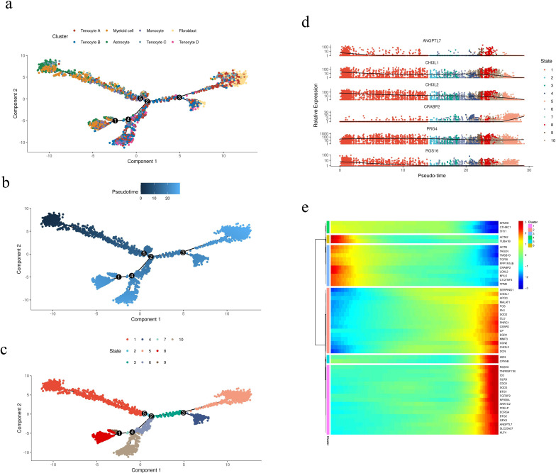
Aims: The repair process of ligament ruptures is a complex phenomenon involving three stages: early, repair, and remodelling. This study aimed to investigate the cellular and genetic aspects related to the repair and healing of ligament ruptures using single-cell RNA sequencing (scRNA-seq) on anterior cruciate ligament (ACL) tissues.

Methods: A comprehensive examination was conducted on ACL tissues from three healthy individuals and three patients with ligament ruptures at different timepoints (one week, three weeks, and six months post-injury). A deep gene expression analysis was performed on 83,195 cells obtained from the six cases, and immunohistochemistry techniques were used to identify cell types.

Results: In this study, tenocytes and fibroblasts in ligament tissues were distinctly identified for the first time. Moreover, a total of ten cell populations were discovered in ACL tissues, comprising tenocytes, fibroblasts, macrophages, stromal cells, T cells, endothelial cells, B cells, epithelial cells, chondrocytes, and monocytes. Further analysis of the tenocyte populations revealed ten distinct subtypes, highlighting the diversity of tenocytes in human ACL tissues.

Conclusion: The identification of multiple specialized tenocyte populations in human ACL tissues sheds light on potential avenues for advancing research in cell therapy for ligament injuries. These findings provide valuable insights into the cellular components involved in the repair and healing processes of ligament ruptures, paving the way for future investigations in this field.


Conflict of interest statement

The authors report that this study was supported by grants from the Qingdao Shinan District Science and Technology Bureau public domain science and technology support plan project (2023-2-011-YY) and Shandong Province medical health science and technology project (202404070961).

Figures

Fig. 1
Fig. 1
Distribution of ligament rupture cell groups (n = 6). a) A total of 83,195 ligament rupture cells were divided into 20 groups using uniform manifold approximation and projection (UMAP). b) Cell groups and the corresponding cell types. c) Marker gene identification of each cell type. d) Dot plot illustration of each marker gene expression. e) Difference in cell types between normal (left) group (n = 3) and ligament rupture (right) group (n = 3). f) Distribution of cell proportions in each cluster. ACAN, aggrecan; ACTA2, actin alpha 2; CCL14, chemokine (C-C motif) ligand 14; COL1A1, collagen type I alpha 1 chain; CXCL2, chemokine (C-X-C motif) ligand 2; HLA-DRA, major histocompatibility complex, class II, DR Alpha; IFIT3, interferon-induced protein with tetratricopeptide repeats 3; IL7R, interleukin-7 receptor subunit alpha; ITGBL1, integrin subunit beta like 1; MMP3, matrix metalloproteinase 3; MYH11, myosin heavy chain 11; RGS16, regulator of G-protein signaling 16.
Fig. 2
Fig. 2
Analysis of tenocyte subgroups and components. a) Uniform manifold approximation and projection (UMAP) grouping of tenocytes. b) Identification of cell subtypes in tenocytes cluster 10. c) Dot plots exhibit the expressions of marker genes in each cell subgroup. d) The bar chart presents a relevant component contribution of each research object to each cell cluster.
Fig. 3
Fig. 3
Gene ontology (GO) enrichment analysis of differential genes in each tenocyte subtype. a) Biological process analysis. b) Molecular function analysis. mRNA, messenger RNA.
Fig. 4
Fig. 4
Gene ontology (GO) enrichment analysis of differential genes in each tenocyte subtype. a) Cell composition analysis. b) Kyoto Encyclopedia of Genes and Genomes (KEGG) pathway analysis. c) Heat map of Metadata, Analysis, Support, and Teamwork difference analysis.
Fig. 5
Fig. 5
Difference analysis of branch points. a) Expression levels of differential genes of all cells in different branches. The influence of this gene on branches was determined in light of the position of the branch state. b) The heat map was divided into four expression modules as per the expressions of differential genes. CENPF, Centromere Protein F; CFH, Complement Factor H; CYP1B1, Cytochrome P450 Family 1 Subfamily B Member 1; EFEMP1, EGF Containing Fibulin Like Extracellular Matrix Protein 1; PLA2G2A, Phospholipase A2 Group IIA; SNED1, Sushi, Nidogen and EGF Like Domains 1.
Fig. 6
Fig. 6
Pseudotime analysis. a) Distribution of different cell clusters over time. b) Differentiation of cells at the time level. c) Different cell fate branches of cell clusters. d) Distribution of the top six differential gene expression. e) Heat map presentation of the expression levels of the top 50 differential genes. ANGPTL7, Angiopoietin Like 7; CHI3L1, Chitinase 3 Like 1; CRABP2, Cellular Retinoic Acid Binding Protein 2; PRG4, Proteoglycan 4; RGS16, Regulator Of G Protein Signaling 16.
Fig. 7
Fig. 7
Verification of tenocytes and fibroblasts via immunofluorescence. The expression of different marker genes in patients with ligament rupture at different stages to verify the subtype of cells. a) Anti-collagen type I alpha 1 chain (COL1A1) antibody, b) anti-regulator of G-protein signaling 16 (RGS16) antibody, and c) anti-matrix metalloproteinase 3 (MMP3) antibody staining tenocytes of patients at three stages. Scale bar = 50 µm. d) Statistical values were presented in the form of mean (SD) for a sample size of three. p-values were calculated using one-way analysis of variance.

Similar articles

References

    1. Ferretti A. To heal or not to heal: the ACL dilemma. J Orthop Traumatol. 2020;21(1):11. doi: 10.1186/s10195-020-00554-8. - DOI - PMC - PubMed
    1. Gumucio JP, Sugg KB, Mendias CL. TGF-β superfamily signaling in muscle and tendon adaptation to resistance exercise. Exerc Sport Sci Rev. 2015;43(2):93–99. doi: 10.1249/JES.0000000000000041. - DOI - PMC - PubMed
    1. Kjaer M, Magnusson P, Krogsgaard M, et al. Extracellular matrix adaptation of tendon and skeletal muscle to exercise. J Anat. 2006;208(4):445–450. doi: 10.1111/j.1469-7580.2006.00549.x. - DOI - PMC - PubMed
    1. Ackermann PW, Domeij-Arverud E, Leclerc P, Amoudrouz P, Nader GA. Anti-inflammatory cytokine profile in early human tendon repair. Knee Surg Sports Traumatol Arthrosc. 2013;21(8):1801–1806. doi: 10.1007/s00167-012-2197-x. - DOI - PubMed
    1. Noah AC, Li TM, Martinez LM, et al. Adaptive and innate immune cell responses in tendons and lymph nodes after tendon injury and repair. J Appl Physiol (1985) 2020;128(3):473–482. doi: 10.1152/japplphysiol.00682.2019. - DOI - PMC - PubMed