. 2025 Aug 5;152(5):313-326.
doi: 10.1161/CIRCULATIONAHA.125.074058. Epub 2025 Jun 17.

Family Screening in Relatives at Risk for Plakophilin-2-Associated Arrhythmogenic Right Ventricular Cardiomyopathy

Affiliations

Family Screening in Relatives at Risk for Plakophilin-2-Associated Arrhythmogenic Right Ventricular Cardiomyopathy

Steven A Muller et al. Circulation. .

Abstract

Background: Penetrance and risk of ventricular arrhythmias (VAs) in arrhythmogenic right ventricular cardiomyopathy (ARVC) are increasingly recognized as being genotype specific. Therefore, genotype-informed family screening protocols may lead to safer and more personalized recommendations than the current one-size-fits-all screening recommendations. We aimed to develop a safe, evidence-based plakophilin-2 (PKP2)-specific longitudinal screening algorithm.

Methods: We included 295 relatives (41% male; age 30.9 years [18.0-47.7 years]) with a pathogenic or likely pathogenic PKP2 variant from 145 families. Phenotype was ascertained with ECG, Holter monitoring, and cardiac imaging and classified by the 2010 Task Force Criteria. VA was defined as a composite of sudden cardiac arrest or death, spontaneous sustained ventricular tachycardia, ventricular fibrillation, or appropriate implantable cardioverter defibrillator intervention. We performed Cox regression to determine predictors of ARVC development and multistate modeling to assess the probability of ARVC development and occurrence of VA.

Results: At baseline, 110 relatives (37%) had definite ARVC. During 8.5 years (4.2-12.9 years) of follow-up, 62 of 185 relatives (34%) without definite ARVC at baseline progressed to definite ARVC diagnosis, and 35 of 295 of all relatives (12%) had VA. VAs occurred only in relatives who previously fulfilled definite ARVC diagnosis. Relatives with borderline ARVC (fulfillment of one minor criterion plus the major family history criterion) progressed 5 times faster in the multistate model to definite ARVC diagnosis and compared with genotype-positive/phenotype-negative (G+/P-) relatives (ie, major family history criterion alone). Relatives 20 to 40 years of age had increased risk for developing definite ARVC (hazard ratio, 2.23; P=0.012) compared with those ≥40 years of age. New Task Force Criteria fulfillment most commonly occurred first on ECGs, followed by Holter monitoring and cardiac imaging. Consequently, 3 risk profiles were identified, and appropriate screening protocols were derived: relatives with borderline ARVC (annual ECG and Holter monitoring; complete evaluation [ie, ECGs, Holter monitoring, and imaging] every 2 years), younger (<40 years of age) or symptomatic G+/P- relatives (every 2 years an ECG and Holter monitoring; complete evaluation every 4 years), and older (≥40 years of age) and asymptomatic G+/P- relatives (complete evaluation every 5 years).

Conclusions: An evidence-based longitudinal screening algorithm that integrates age, symptoms, and baseline clinical phenotype may improve patient care and improve efficiency of clinical resource allocation.

Keywords: cardiomyopathies; death, sudden, cardiac; genetics; genotype; precision medicine.


Conflict of interest statement

Dr James has received research support from Stride Bio Inc, Lexeo Therapeutics, and ARVADA Therapeutics. C. Tichnell has received salary support on these grants. Dr te Riele is a consultant for Tenaya, Rocket Pharmaceuticals, and BioMarin for unrelated work (gene therapy trials). Drs James and Gasperetti have served as compensated consultants for LEXEO Therapeutics for unrelated work. Dr Yap has received honoraria (speaker or consultancy fees) from Boston Scientific, Medtronic, Biotronik, and Acutus Medical. In addition, he has received research grants from Medtronic, Biotronik, and Boston Scientific. The other authors report no conflicts.

Figures

Figure 1.
Figure 1.
Visualization of the multistate model. A, Multistate model of progression of disease using the diagnostic 2010 Task Force Criteria (TFC) consisted of 4 states: genotype positive/phenotype negative (G+/P−), borderline arrhythmogenic right ventricular cardiomyopathy (ARVC), definite ARVC, and ventricular arrhythmia (VA). A relative could only stay in the same state or progress to a more severe state during follow-up, which is shown by the blue arrows. Numbers represent the estimated transition intensities per year. B, Multistate models of progression to new TFC by diagnostic test. Numbers represent the estimated transition intensities per diagnostic test per year.
Figure 2.
Figure 2.
Flowchart of the study population. *Complete baseline evaluation defined as at least 12-lead ECG, Holter monitoring, and imaging (cardiac magnetic resonance imaging [CMR] or echocardiography). Boxes around the study population visualize specific analyses as described in the text.
Figure 3.
Figure 3.
Progression of disease. Survival curve of definite arrhythmogenic right ventricular cardiomyopathy (ARVC) diagnosis in the overall, nondefinite population (A) and stratified by baseline clinical phenotype (B). Relatives who are genotype positive/phenotype negative (G+/P−) and who have borderline ARVC are depicted as yellow and blue lines, respectively. Shaded areas indicate 95% CI. Censoring is indicated by a vertical bar.
Figure 4.
Figure 4.
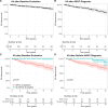
Occurrence of VA. Survival curve of ventricular arrhythmia (VA) after baseline evaluation (A), arrhythmogenic right ventricular cardiomyopathy (ARVC) diagnosis (B), baseline evaluation stratified by baseline clinical phenotype (C), and ARVC diagnosis stratified by baseline clinical phenotype (D). Relatives with and without definite ARVC diagnosis at baseline are depicted as red and turquois lines, respectively. Because only 3 relatives (1.6%) without definite ARVC had a VA during follow-up, we combined the genotype-positive/phenotype-negative (G+/P−) relatives and relatives with borderline ARVC into a “no ARVC at baseline” group for visual purposes in C and D. Shaded areas indicate 95% CI. Censoring is indicated by a vertical bar.
Figure 5.
Figure 5.
Progression to developing definite ARVC, VA, and new TFC. A, Yield of screening stratified by baseline clinical phenotype for definite arrhythmogenic right ventricular cardiomyopathy (ARVC) and ventricular arrhythmia (VA; y axis) over different screening intervals (x axis). Every screening interval consists of 3 bars, which depict the genotype-positive/phenotype-negative (G+/P−; yellow), borderline ARVC (blue), and definite ARVC (gray) groups. The bars are divided into solid and striped bars, which differentiate between the probability of definite ARVC and VA, respectively. Of note, because relatives with definite ARVC have a 100% - “probability of VA” probability of definite ARVC, we visualize only the probability of VA in that population. B, Yield of screening new Task Force Criteria (TFC) and ECG, Holter monitoring, and imaging TFC over different screening intervals. C, Yield of screening for new cardiac magnetic resonance imaging (CMR) and echocardiography TFC over different screening intervals. ECG, Holter monitoring, imaging, CMR, and echocardiography are indicated by red, green, blue, dark blue, and light blue bars, respectively. Dotted black line indicates the clinically acceptable risk for definite ARVC (ie, 6%–16%). Error bars indicate 95% CI.
Figure 6.
Figure 6.
Longitudinal screening algorithm for at-risk pathogenic or likely pathogenic PKP2 carriers. The longitudinal screening algorithm. The baseline evaluation should start between 8 and 10 years of age, and the serial evaluation depends on the baseline clinical phenotype, presence of symptoms, and age: (1) genotype-positive/phenotype-negative (G+/P−) relatives who are ≥40 years of age and are asymptomatic should be evaluated every 5 years with an ECG, Holter monitoring, and imaging test; (2) G+/P− relatives who are <40 years of age or are symptomatic should be evaluated every 2 years with an ECG and Holter monitoring and every 4 years with an ECG, Holter monitoring, and imaging test; and (3) relatives with borderline arrhythmogenic right ventricular cardiomyopathy (ARVC) should be evaluated every year with an ECG and Holter monitoring and every 2 years with an ECG, Holter monitoring, and imaging test. If a relative remains in the same clinical phenotype (G+/P− or borderline ARVC) or progresses from G+/P− to borderline ARVC, screening intervals and management should be adjusted accordingly as shown by the dotted lines. If a relative progresses to definite ARVC, management and risk stratification should follow the most current guideline recommendations. Following this screening algorithm ensures that all relatives will be screened at a similar risk for progressing to definite ARVC (G+/P−, 9%; borderline ARVC, 11%) and ventricular arrhythmia (VA; both G+/P− and borderline ARVC, <0.5%). CMR indicates cardiac magnetic resonance imaging.

References

    1. Bhonsale A, Groeneweg JA, James CA, Dooijes D, Tichnell C, Jongbloed JDH, Murray B, te Riele ASJM, van den Berg MP, Bikker H, et al. Impact of genotype on clinical course in arrhythmogenic right ventricular dysplasia/cardiomyopathy-associated mutation carriers. Eur Heart J. 2015;36:847–855. doi: 10.1093/eurheartj/ehu509 - PubMed
    1. Marcus FI, Fontaine GH, Guiraudon G, Frank R, Laurenceau JL, Malergue C, Grosgogeat Y. Right ventricular dysplasia: a report of 24 adult cases. Circulation. 1982;65:384–398. doi: 10.1161/01.cir.65.2.384 - PubMed
    1. Towbin JA, McKenna WJ, Abrams DJ, Ackerman MJ, Calkins H, Darrieux FCC, Daubert JP, de Chillou C, DePasquale EC, Desai MY, et al. 2019 HRS expert consensus statement on evaluation, risk stratification, and management of arrhythmogenic cardiomyopathy. Heart Rhythm. 2019;16:e301–e372. doi: 10.1016/j.hrthm.2019.05.007 - PubMed
    1. Corrado D, Wichter T, Link MS, Hauer RNW, Marchlinski FE, Anastasakis A, Bauce B, Basso C, Brunckhorst C, Tsatsopoulou A, et al. Treatment of arrhythmogenic right ventricular cardiomyopathy/dysplasia: an international task force consensus statement. Circulation. 2015;132:441–453. doi: 10.1161/CIRCULATIONAHA.115.017944 - PMC - PubMed
    1. Hershberger RE, Givertz MM, Ho CY, Judge DP, Kantor PF, McBride KL, Morales A, Taylor MRG, Vatta M, Ware SM. Genetic evaluation of cardiomyopathy: a Heart Failure Society of America practice guideline. J Card Fail. 2018;24:281–302. doi: 10.1016/j.cardfail.2018.03.004 - PMC - PubMed

LinkOut - more resources