Review
. 2025 Jul 24;207(7):e0007125.
doi: 10.1128/jb.00071-25. Epub 2025 Jun 17.

Milestones in the development of Myxococcus xanthus as a model multicellular bacterium

Affiliations
Review

Milestones in the development of Myxococcus xanthus as a model multicellular bacterium

Lee Kroos et al. J Bacteriol. .

Abstract

From the humblest of beginnings (i.e. a pile of dry cow dung) over 80 years ago, the Gram-negative bacterium Myxococcus xanthus has emerged as a premier model system for studying diverse fields of bacteriology, including multicellular development, sporulation, motility, cell-envelope biogenesis, spatiotemporal regulation, signaling, photoreception, kin recognition, social evolution, and predation. As the flagship representative of myxobacteria found in varied terrestrial and aquatic environments, M. xanthus research has evolved into a collaborative global effort, as reflected by the contributions to this article. In celebration of the upcoming 50th anniversary of the International Conference on the Biology of Myxobacteria, this review highlights the historical and ongoing contributions of M. xanthus as a multifaceted model bacterium.

Keywords: cell division; cell polarity; evolution; gliding motility; kin recognition; microbial ecology; multicellularity; myxobacteria; peptidoglycan; photoreception; polysaccharides; predation; secondary metabolites; signal transduction; sporulation; type 4 pilus.


Conflict of interest statement

The authors declare no conflict of interest.

Figures

Fig 1
Fig 1
Myxobacteria in various fruiting-body complexities. (A) Fruiting bodies of diverse myxobacterial species, as sketched by Roland Thaxter, 1892 (3). Used with permission from the Archives of the Farlow Herbarium of Cryptogamic Botany, Harvard University, Cambridge, MA, USA. (B) Stigmatella aurantiaca, (C) Chondromyces lanuginosus, (D) Corallococcus coralloides, (E) Cystobacter velatus, (F) Polyangium spomosum, and (G) Myxococcus stipitatus. Photo credit of B–G: Ronald Garcia/HIPS. Scale bars, 500 µm.
Fig 2
Fig 2
Relative expression profiles of significantly regulated genes at the indicated hours post-starvation induction. Genes were clustered into 10 developmental groups based on the time of peak expression and then organized according to the temporal progression of development. Developmental group number and the stages of the developmental program (with images of aggregates under a dissecting microscope and cells under a scanning electron microscope) are indicated on the left. In the images, red, blue, and yellow bars represent 2 mm, 100 µm, and 5 µm, respectively. Modified from Reference (30).
Fig 3
Fig 3
Polysaccharide-mediated M. xanthus physiology. (A) Scanning electron microscopy of WT, EPS, and BPS M. xanthus cell surfaces (scale bar: 1 µm), adopted from Reference (120) with permission. (B) T4P-dependent motility of WT, EPS, and BPS M. xanthus cells (scale bar: 2 mm). Red dashed line: relative location of slice presented in Panel C. (C) Transmission electron microscopy cross-section of M. xanthus polysaccharide mutant swarms (scale bar: 1 µm), adopted from Reference (120) with permission. (D) Fruiting-body formation of WT, EPS, and BPS M. xanthus cells (scale bar: 400 µm), adopted from Reference (55). (E) Tiling fluorescence microscopy of swarming cells expressing EPS and BPS machinery linked to green and red fluorescent reporters, respectively (scale bar: 400 µm), adopted from Reference (55). (F) Transmission electron microscopy of myxospores from WT, MASC, and Nfs-mutant cells (scale bar: 0.1 µm), adopted from Reference (128). (G) Phase-contrast microscopy (left side, scale bar: 50 µm) and 3D optical profilometry (right side) of M. xanthus trails left behind at the agar-air interface, adopted from Reference (129). (H) Wet-SEEC high-refraction microscopy (top) and fluorescence microscopy (bottom) of M. xanthus cells (treated with fluorescent ConA lectin) gliding on a chitosan-functionalized glass substratum in a PDMS microfluidic device (scale bar: 1 µm), adopted from Reference (130). (I) Transmission electron microscopy of a negatively stained “slime” trail left behind a M. xanthus cell gliding across a chitosan-functionalized copper electron microscopy grid (scale bar: 250 nm). Lipid vesicles and lipid tubes are present in the electron-dense trail material. White arrows indicate colloidal gold particles associated with biotinylated ConA lectin, adopted from Reference (131).
Fig 4
Fig 4
T4P-dependent motility and the T4PM. (A) T4P-dependent motility depends on dynamic cycles of extension, adhesion, and retraction. The T4PM is present at both cell poles, but only active at the leading pole. During reversal, the leading/lagging polarity switches, enabling the activation of the T4PM at the new leading pole. (B) The architecture of the T4PM of M. xanthus in two states. Left, unpiliated state. The core T4PM consists of four functional elements, that is, an OM pore (PilQ and TsaP), an alignment complex (PilP, PilN, and PilO) that connects the OM pore to the IM platform complex (PilC and PilM), and a priming complex (four minor pilins (orange, yellow, blue, and green) and PilY1). Right, piliated T4PM with an extended T4P capped by the tip complex. PilB and PilT associate with PilM and PilC in a mutually exclusive manner for extension and retraction, respectively. Dashed arrows indicate incorporation at, and removal from, the pilus base of PilA during extension and retraction, respectively. Proteins labeled with single letters have the Pil prefix.
Fig 5
Fig 5
Gliding motility and the bFAC. (A) Stationary bFACs (relative to the substratum) drive M. xanthus gliding motility. Motors carrying incomplete gliding complexes either diffuse or move rapidly along helical paths but do not generate propulsion. Motors stall and become nearly static relative to the substrate when they assemble into complete bFACs with other motor-associated proteins at the ventral side of the cell. Stalled motors push MreB and bFACs in opposite directions and thus exert force against OM adhesins. Overall, as motors transport bFACs toward lagging cell poles, cells move forward, but bFACs remain static relative to the substratum. Reproduced from Reference (183). (B) Structural model of the bFAC predicted by AlphaFold (184), modified from Reference (185). Note that whether and how GltG and GltI thread through PG are not investigated by experiments. IM, inner membrane; OM, outer membrane; PG, peptidoglycan.
Fig 6
Fig 6
Polarity control of M. xanthus motility systems. (A) The polarity module and the Frz chemosensory system regulate motility. All proteins in the Frz system have the Frz prefix. Homologs of individual proteins in the E. coli Che system are indicated in brackets. Phosphate flow from the FrzE kinase is indicated by arrows. Phosphorylated FrzZ and FrzX localize at the leading and lagging poles, respectively, and are indicated to act on the polarity module. The cell below schematically illustrates the polar localization of the proteins of the polarity module with T4P at the leading pole. The circle sizes indicate the relative amount of a protein at a pole. (B) MglA GTPase cycle and interaction with downstream effectors to stimulate gliding motility and T4P-dependent motility. GEF, guanine nucleotide exchange factor; GAP, GTPase-activating protein. (C) Interactions between the proteins of the polarity module establish their asymmetric polar localization at the two poles, resulting in correct leading–lagging cell polarity. Colors as in A.
Fig 7
Fig 7
Schematic of the dynamic localization of the PomXYZ complex on the nucleoid and the organization of the M. xanthus chromosome segregation machinery over the cell cycle. The schematics follow a cell over the cell cycle starting immediately after cell division (i). (ii–iv) As the cell cycle progresses, the PomXYZ complex translocates across the nucleoid to midcell; in parallel, chromosome replication and segregation proceed, resulting in a chromosome arrangement in which the ParB/parS complexes localize in the subpolar regions attached to the bactofilin/PadC complexes, and the ter regions around midcell. (v) At midcell, the PomXYZ complex recruits FtsZ and stimulates FtsZ polymerization, resulting in the formation of the Z-ring. FtsZ directly or indirectly recruits all other proteins required for cytokinesis.
Fig 8
Fig 8
Model of kin recognition and outer membrane exchange (OME). (A) 10 experimentally determined TraA recognition groups (colored triangles) govern partner binding. Colored cell envelopes represent OM phenotypes, for example, fluorescent proteins, while colored cytoplasm represents distinct genotypes, particularly in reference to the suite of sitAI loci. Following TraA homotypic binding, cells transiently fuse OMs and exchange cargo, including SitA lipoprotein toxins. Clonal cells express cognate immunity and homogenize their OMs (same color), while nonclonal cells are poisoned. (B) Merged fluorescent micrographs of M. xanthus cells expressing Lipo-GFP or Lipo-mCherry in the periplasmic leaflet of their OMs. 90 min following plating (right), cells that recognize and undergo OME contain both markers, while no OME results in a distinct phenotype. Courtesy of Sheila Walsh; scale bar, 2 µM.
Fig 9
Fig 9
M. xanthus is an active predator. (A) An M. xanthus population (top) invades an E. coli colony (bottom), kills, and lyses prey cells and consumes the released biomass. The white arrow denotes fruiting bodies, and the yellow arrow highlights rippling movement by the preying population. (B) An individual M. xanthus cell approaches E. coli, stops upon cell contact, then kills and lyses the prey cell within minutes. Adopted from Reference (265).

Similar articles

Cited by

  • Tissue-like structures formed by a bacterium.
    Ampah GML, Myers CJ, Ramírez Carbó CA, Lin IS, Nan B. Ampah GML, et al. bioRxiv [Preprint]. 2025 Jun 28:2025.06.27.661998. doi: 10.1101/2025.06.27.661998. bioRxiv. 2025. PMID: 40666919 Free PMC article. Preprint.

References

    1. Mohr KI. 2018. Diversity of myxobacteria-we only see the tip of the iceberg. Microorganisms 6:84. doi: 10.3390/microorganisms6030084 - DOI - PMC - PubMed
    1. Waite DW, Chuvochina M, Pelikan C, Parks DH, Yilmaz P, Wagner M, Loy A, Naganuma T, Nakai R, Whitman WB, Hahn MW, Kuever J, Hugenholtz P. 2020. Proposal to reclassify the proteobacterial classes Deltaproteobacteria and Oligoflexia, and the phylum Thermodesulfobacteria into four phyla reflecting major functional capabilities. Int J Syst Evol Microbiol 70:5972–6016. doi: 10.1099/ijsem.0.004213 - DOI - PubMed
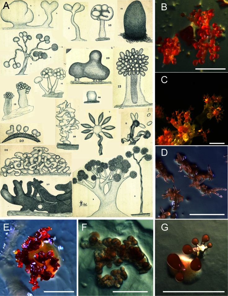
    1. Thaxter R. 1892. On the myxobacteriaceæ, a new order of schizomycetes. Bot Gaz 17:389–406. doi: 10.1086/326866 - DOI
    1. Beebe JM. 1941. The morphology and cytology of Myxococcus xanthus, N. Sp. J Bacteriol 42:193–223. doi: 10.1128/jb.42.2.193-223.1941 - DOI - PMC - PubMed
    1. Reichenbach H. 1970. Nannocystis exedens gen. nov., spec. nov., a new myxobacterium of the family sorangiaceae. Arch Mikrobiol 70:119–138. doi: 10.1007/BF00412203 - DOI - PubMed

MeSH terms

LinkOut - more resources