. 2025 Jun;29(12):e70590.
doi: 10.1111/jcmm.70590.

CuATSM Enhances Wound Repair Without Scarring via Hippo/YAP Signalling Pathway to Reduce Ferroptosis and Macrophage Polarisation

Affiliations

CuATSM Enhances Wound Repair Without Scarring via Hippo/YAP Signalling Pathway to Reduce Ferroptosis and Macrophage Polarisation

Yingdan Tang et al. J Cell Mol Med. 2025 Jun.

Abstract

Skin wound healing is a complex biological process involving haemostasis, inflammation, proliferation/repair and remodelling. However, skin scarring, as one of the important stages in the healing process, can adversely affect the structure and function of related organs. Currently, effective treatments to address such scars remain insufficient. In this study, we established a full-thickness skin excision wound model using male ICR mice, which were randomly divided into a Control group and a CuATSM group. The CuATSM group received CuATSM (30 mg/kg) via gavage, with daily treatments continuing throughout the observation period. The Control group received an equivalent volume of 0.9% sodium chloride solution. Wound healing progression was evaluated through macroscopic photography, histological analyses, Western blotting and quantification of relevant biochemical markers at different healing stages. Our study reveals that CuATSM not only promotes rapid skin wound healing but also reduces scar formation in the late healing phase. Furthermore, our findings suggest that this effect is mediated through the ferroptosis-induced Hippo/YAP signalling pathway and macrophage polarisation. These findings highlight CuATSM as a promising therapeutic candidate for achieving scarless wound repair in clinical applications.

Keywords: CuATSM; ferroptosis; hippo/YAP pathway; scarless; wound healing.


Conflict of interest statement

The authors declare no conflicts of interest.

Figures

FIGURE 1
FIGURE 1
CuATSM improves wound healing in mice. (A) The healing condition of skin wounds in Control and CuATSM groups of mice at days 0, 3, 6, 9, 12 and 15; (B) Simulating the range of wounds based on wound images to visually display the contours of the wounds; (C) Experimental flowchart; (D) Statistical analysis of wound area at different time points for Control and CuATSM groups, p < 0.0001, n = 10.
FIGURE 2
FIGURE 2
CuATSM accelerates granulation tissue formation and enhances collagen deposition in mouse skin wounds. (A, B) Haematoxylin and Eosin (H&E) staining and Masson's trichrome staining in days 14 and 28. scale bar = 200 μm, n = 4.
FIGURE 3
FIGURE 3
CuATSM enhances angiogenesis, cellular proliferation and collagen deposition during cutaneous wound healing in mice. (A) Representative fluorescence images of skin stained with CD31 (green) and DAPI (blue) in mice from the two groups at day 7 after full‐thickness skin excision, digitalized pictures were selected in the central regions, scale bars = 150 μm, enlarge scale bars = 50 μm; (B) Relative fluorescence intensity of CD31, CD31 presence significantly differed between the two groups, p = 0.0093, n = 4; (C) Representative fluorescence images of skin stained with Col I (green) and DAPI (blue) in mice from the two groups at day 7 after full‐thickness skin excision, digitalized pictures were selected in the central regions, scale bars = 150 μm, enlarge scale bars = 50 μm; (D) Relative fluorescence intensity of Col I, Col I presence significantly differed between the two groups, p = 0.0012, n = 4; (E) Representative fluorescence images of skin stained with Ki67 (green) and DAPI (blue) in mice from the two groups at day 7 after full‐thickness skin excision, digitalized pictures were selected in the central regions, scale bars = 150 μm, enlarge scale bars = 50 μm; (F) Relative fluorescence intensity of Ki67, Ki67 presence significantly differed between the two groups, p = 0.0024, n = 4.
FIGURE 4
FIGURE 4
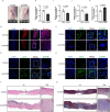
CuATSM enhance scarless wound healing in vivo. (A) On day 35, the healing and hair growth of skin wounds in mouse from the Control group and the CuATSM group, n = 3; (B–E) Representative fluorescence images of skin stained with En‐1 (red) and DAPI (blue) in mice from the two groups at day 35 after full‐thickness skin excision, scale bars = 300 μm, enlarge scale bars = 100 μm; Representative fluorescence images of skin stained with CD26 (green), Sox9 (green) and CK19 (green) and DAPI (blue) in mice from the two groups at day 35 after full‐thickness skin excision, digitalized pictures were selected in the central regions, scale bars = 150 μm, enlarge scale bars = 50 μm; (H–K) Relative fluorescence intensity of En‐1, CD26, Sox9 and CK19, En‐1, CD26, Sox9 and CK19 presence significantly differed between the two groups p = 0.0002, 0.0122, 0.0009 and 0.0016, respectively; n = 4; (F, G) Haematoxylin and Eosin (H&E) staining and Masson's trichrome staining in the 35th day, scale bar = 200 μm, n = 4.
FIGURE 5
FIGURE 5
CuATSM inhibits ferroptosis during wound healing. (A) Perl's iron staining in days 14 and 28, scale bar = 200 μm; (B) Iron‐stained positive area presence significantly differed between the two groups, p < 0.0001, n = 4; (C) Western blotting was used to detect the expression of ferroptosis‐related proteins; (D–H) Quantitative analysis of the expression levels of related proteins in the CuATSM group compared to the Control group, p = 0.0171, 0.0067, 0.0234, 0.0125, 0.0109 and 0.0106; n = 4. (I) The MDA content in skin wound tissues of mice in the Control and CuATSM groups on day 7, p < 0.0001, n = 3; (J) The tissue iron content in skin wound tissues of mice in the Control and CuATSM groups on day 7, p = 0.0043, n = 5; (K) The GSH content in skin wound tissues of mice in the Control and CuATSM groups on day 7, p = 0.0009, n = 4; (L) Representative fluorescence images of skin stained with GPX4 (red) and DAPI (blue) in mice from the two groups at day 7 after full‐thickness skin excision, digitalized pictures were selected in the central regions, scale bars = 150 μm, enlarge scale bars = 50 μm; (M) Relative fluorescence intensity of GPX4, GPX4 presence significantly differed between the two groups, p = 0.0402, n = 4.
FIGURE 6
FIGURE 6
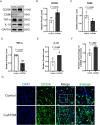
CuATSM promotes macrophage polarisation, attenuating inflammatory responses and enhancing cutaneous wound healing. (A) Western blotting was used to detect the expression of macrophage‐related proteins and inflammatory factors; (B–E) Quantitative analysis of the expression levels of related proteins in the CuATSM group compared with the Control group. p = 0.0045, 0.0164, 0.0012 and 0.0260, n = 4 ; (F) Relative fluorescence intensity of CD206, CD206 presence significantly differed between the two groups, p = 0.0062, n = 4; (G) Representative fluorescence images of skin stained with CD206 (green) and DAPI (blue) in mice from the two groups at day 7 after full‐thickness skin excision, digitalized pictures were selected in the central regions, scale bars = 150 μm, enlarge scale bars = 50 μm.
FIGURE 7
FIGURE 7
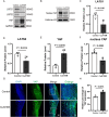
CuATSM modulates Hippo/YAP pathway during wound healing. (A) Western blotting was used to detect the expression of Hippo‐related proteins outside the nucleus; (B) Western blotting was used to detect the expression of YAP within the nucleus; (C–F) Quantitative analysis of the expression levels of related proteins in the CuATSM group compared to the Control group, p = 0.0136, 0.0314, 0.0016, and 0.0003; n = 4; (G) Representative fluorescence images of skin stained with YAP (green) and DAPI (blue) in mice from the two groups at day 35 after full‐thickness skin excision, scale bars = 300 μm, enlarge scale bars = 100 μm; (H) Relative fluorescence intensity of YAP, YAP presence significantly differed between the two groups, p = 0.0016, n = 4.

Similar articles

References

    1. Gauglitz G. G., “Management of Keloids and Hypertrophic Scars: Current and Emerging Options,” Clinical, Cosmetic and Investigational Dermatology 6 (2013): 103–114, 10.2147/CCID.S35252. - DOI - PMC - PubMed
    1. Macintyre L. and Baird M., “Pressure Garments for Use in the Treatment of Hypertrophic Scars—A Review of the Problems Associated With Their Use,” Burns 32, no. 1 (2006): 10–15, 10.1016/j.burns.2004.06.018. - DOI - PubMed
    1. Wang S., Xu C., Yu Y., et al., “Immunoregulative Coating for Scarless Healing in Anterior Cruciate Ligament Reconstruction,” Bioactive Materials 45 (2025): 71–87, 10.1016/j.bioactmat.2024.11.007. - DOI - PMC - PubMed
    1. Xing D., Xia G., Tang X., et al., “A Multifunctional Nanocomposite Hydrogel Delivery System Based on Dual‐Loaded Liposomes for Scarless Wound Healing,” Advanced Healthcare Materials 13, no. 28 (2024): e2401619, 10.1002/adhm.202401619. - DOI - PubMed
    1. Talbott H. E., Mascharak S., Griffin M., Wan D. C., and Longaker M. T., “Wound Healing, Fibroblast Heterogeneity, and Fibrosis,” Cell Stem Cell 29, no. 8 (2022): 1161–1180, 10.1016/j.stem.2022.07.006. - DOI - PMC - PubMed

MeSH terms