

Sex differences in the chronic autoimmune response to myocardial infarction
- PMID: 40526105
- PMCID: PMC12238818
- DOI: 10.1042/CS20243091
Sex differences in the chronic autoimmune response to myocardial infarction
Abstract
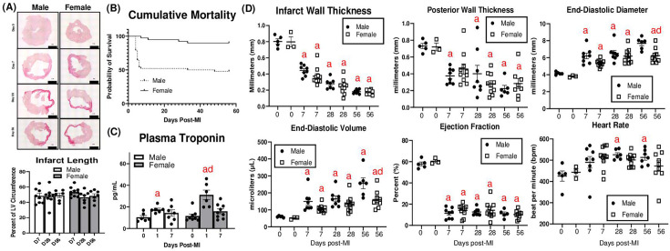
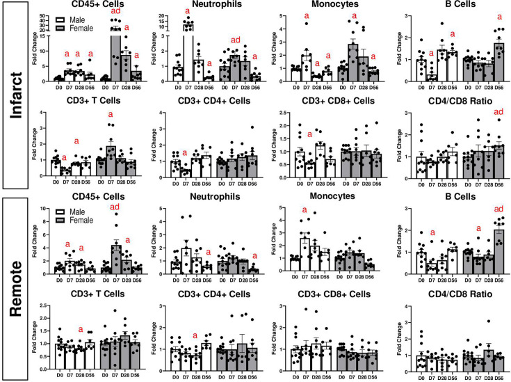
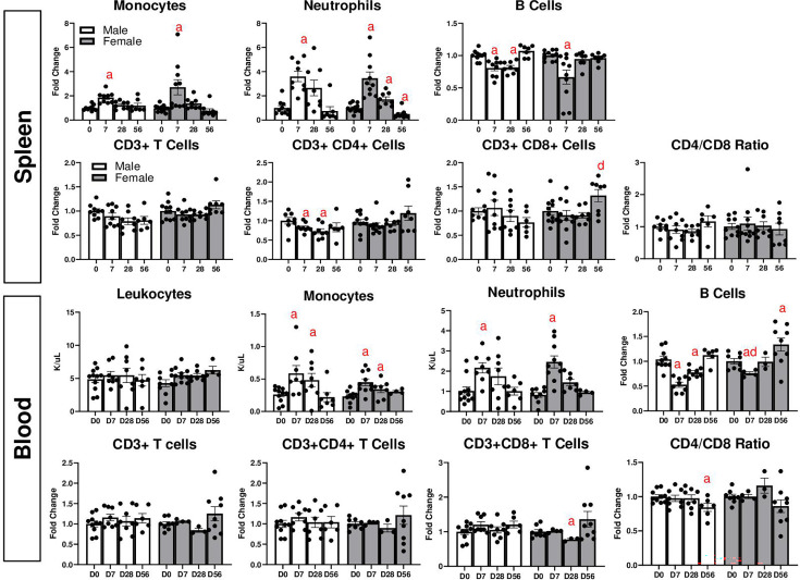
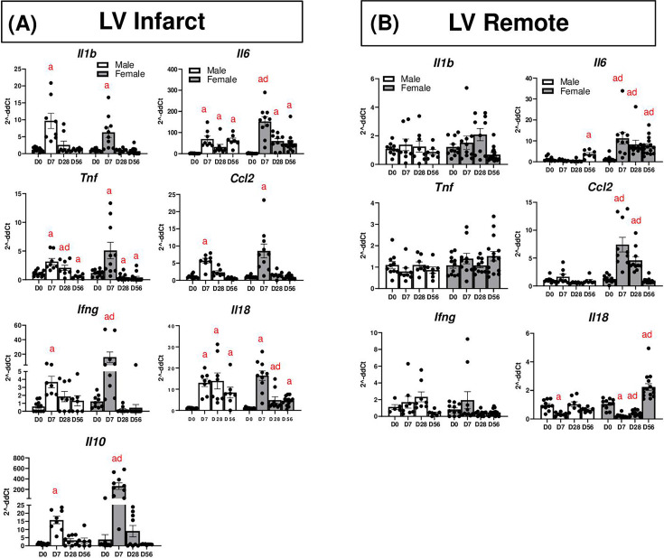
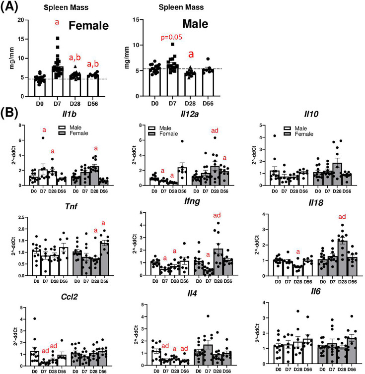
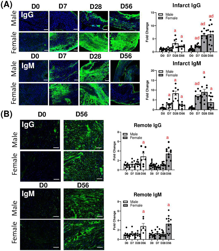
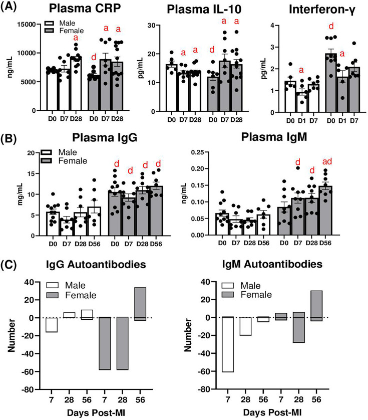
Myocardial infarction (MI) causes a robust inflammatory response, which is necessary for remodeling and scar formation of the infarcted left ventricle (LV). However, this can lead to chronic systemic inflammation and persistent autoimmune responses. In this study, we analyzed sex differences in the inflammatory autoimmune response to chronic MI. MI was induced by permanent left coronary artery ligation in adult male and female C57BL/6J mice for one, four, and eight weeks. Both sexes exhibited similar declines in LV function. Females had higher levels of total immune cells and T cells in the infarct and remote area at D7 post-MI, and B cells at D56. MI increased levels of pro-inflammatory cytokines (Il1b, Il6, Tnf, Ccl2, Ifng, Il18) in the LV infarct that peaked at one week, which was exaggerated in females for Il6, Ifng, and Il10. In the remote LV, females had higher levels of Il6, Tnf, Ccl2, and Il18. MI increased spleen mass in females only, and splenic cytokines were higher in females at several time points, including Il1b, Il12a, Il10, Ifng, Il18, Ccl2, and Il4. IgG and IgM deposition in the LV infarct increased over time in both sexes, but more so in females. In the remote area, both sexes had increased IgG and IgM at eight weeks. Plasma IgM was higher in females at one, four, and eight weeks post-MI compared with males. Plasma IgG and IgM autoantibodies were detected in males and females after MI, but the number of autoantibodies displaying reactivity to autoantigens was much higher in females, particularly at week 8. In summary, MI leads to the development of systemic and myocardial autoimmune activation, which is more pronounced in females.
Keywords: autoimmune disease; heart failure; inflammation; myocardial infarction; sex differences.
© 2025 The Author(s).
Conflict of interest statement
The authors declare that there are no competing interests associated with the manuscript.
Figures
Similar articles
-
Macro- and microinjury define the heart failure progression after permanent coronary ligation or ischemia-reperfusion in young healthy mice.Am J Physiol Heart Circ Physiol. 2025 Aug 1;329(2):H521-H533. doi: 10.1152/ajpheart.00267.2025. Epub 2025 Jul 16. Am J Physiol Heart Circ Physiol. 2025. PMID: 40668645
-
Glucocorticoids impair T lymphopoiesis after myocardial infarction.Am J Physiol Heart Circ Physiol. 2024 Aug 1;327(2):H533-H544. doi: 10.1152/ajpheart.00195.2024. Epub 2024 Jul 12. Am J Physiol Heart Circ Physiol. 2024. PMID: 38995212 Free PMC article.
-
Systemic pharmacological treatments for chronic plaque psoriasis: a network meta-analysis.Cochrane Database Syst Rev. 2021 Apr 19;4(4):CD011535. doi: 10.1002/14651858.CD011535.pub4. Cochrane Database Syst Rev. 2021. Update in: Cochrane Database Syst Rev. 2022 May 23;5:CD011535. doi: 10.1002/14651858.CD011535.pub5. PMID: 33871055 Free PMC article. Updated.
-
Exercise-based cardiac rehabilitation for coronary heart disease.Cochrane Database Syst Rev. 2021 Nov 6;11(11):CD001800. doi: 10.1002/14651858.CD001800.pub4. Cochrane Database Syst Rev. 2021. PMID: 34741536 Free PMC article.
-
Sex differences in myocardial infarction care and outcomes: a longitudinal Scottish National Data-Linkage Study.Eur J Prev Cardiol. 2025 Jun 3;32(8):696-707. doi: 10.1093/eurjpc/zwae333. Eur J Prev Cardiol. 2025. PMID: 39592008
References
MeSH terms
Substances
Grants and funding
LinkOut - more resources
Full Text Sources
Medical
Miscellaneous