Review
. 2025 Jun 17;45(1):58.
doi: 10.1007/s10571-025-01563-z.

Role of Nuclear Receptors on the Progression of Multiple Sclerosis: A Review

Affiliations
Review

Role of Nuclear Receptors on the Progression of Multiple Sclerosis: A Review

Behina Babaalizadeh et al. Cell Mol Neurobiol. .

Abstract

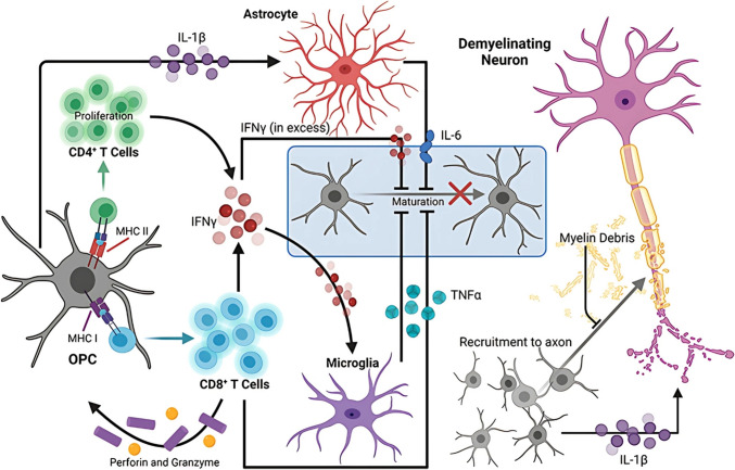
Multiple sclerosis (MS) is the consequence of early-onset inflammatory demyelination and progressive neurodegenerative lesions in the central nervous system (CNS). Despite the unclear pathogenesis of MS, two main processes occur, oligodendrocyte repair and lipid metabolism. Nuclear receptors have been observed to have a role as ligand sensors, regulators of transcriptional factors, and modulators of gene expression. One member of this family, the retinoid X receptor (RXR), which forms a heterodimeric complex with liver X receptor (LXR), have been implicated in the pathophysiology of MS. Moreover, it has been expressed that LXR receptors play pivotal role in the regulation of inflammation, oxidative stress responses, and cholesterol metabolism in phagocytes in active MS lesions. This review aimed to study role of RXR and LXR in MS pathogenesis.

Keywords: Inflammation; LXR; Multiple sclerosis; Neurodegeneration; RXR.


Conflict of interest statement

Declarations. Competing Interests: The authors declare no competing interests. Ethical Approval: No ethic approval required.

Figures

Fig. 1
Fig. 1
OPCs play a role in the maintenance of inflammation, which in turn accelerates neurodegeneration in multiple sclerosis. Impaired antigen presentation to CD4 + and CD8 + T lymphocytes can be enhanced during neuroinflammation by upregulating the MHC I and II antigen presentation molecules on OPCs, respectively, through IFNγ. The antigen-MHC I complex has the ability to trigger the production of TNFα and IFNγ by CD8 + T cells, along with the secretion of perforin and granzyme, all of which can lead to the death of specific OPC cells in the area. Through the presentation of MHC II antigens, OPCs also stimulate the proliferation and cytokine production of CD4 + memory and effector T lymphocytes. Microglia also have the ability to hinder OPC development through the production of TNFα in response to IFNγ signaling by T cells. Inhibition of OPC maturation and oligodendrocyte cell death are linked to excess extracellular IFNγ. OPC differentiation capacity can be directly diminished since IL-6 synthesis by astrocytes is stimulated by IFNγ signaling, which in turn increases OPC IL-1β expression. It is possible that IL-1β, which is produced by OPCs, directly causes neuronal cytotoxicity by improving the transmission of toxic glutamatergic synapses. In order to prevent OPC recruitment and maturation, myelin fragments from deteriorating myelin sheaths act as a repellent. All things considered, OPCs themselves maintain a neuroinflammatory state, which limits the ability of OL lineage cells to provide axonal support, repair, and remyelination (Psenicka, et al. 2021)
Fig. 2
Fig. 2
A schematic showing the main genes that are active when RA is present. RA activates a number of genes, including those involved in cell differentiation and cancer cell proliferation, when it binds to its receptors, RAR, and RXR (Lavudi 2023)
Fig. 3
Fig. 3
Expression patterns and cellular location of endogenous RXR and SIRT6 in the brain following SAH are examined. an Analyses of quantitative western blot bands and the temporal course of RXR in the left hemisphere following SAH. b Quantitative analysis of the left hemisphere’s SIRT6 time course following SAH and representative western blot bands. **Compared to the control group (sham), *P < 0.05. The mean ± SD was used to illustrate the error bars. for each group, n = 6. c At 24 h post-SAH, red-labeled neuronal (NeuN), astrocyte (GFAP), and microglia (Iba-1) cells in the left basal cortex were stained with double immunofluorescence for RXR (green). d Twenty-four hours following SAH, double immunofluorescence labeling for SIRT6 (red) in cells of the left basal cortex, specifically in neurons (NeuN, green), astrocytes (GFAP, green), and microglia (Iba-1, green). Each group has n = 2. A 50 μm scale bar is used. Iba-1 is an ionized calcium binding adaptor molecule, NeuN is a neuronal nuclear protein, and DAPI is 4′,6-diamidino2-phenylindole. GFAP is a glial fibrillary acidic protein (Zuo et al. 2019)
Fig. 4
Fig. 4
When they bind to LXR response elements (LXREs) in the regulatory areas of the genes they target, liver X receptors (LXRs) and retinoid X receptors (RXRs) create heterodimers. Separated by four nucleotides (N), the target regions include variants of the repeating sequence AGGTCA. When ligands are not present, the LXR-RXR complex binds to co-repressors and shuts down gene expression. Oxysterols and synthetic ligands activate LXRs, and ligands for RXRs, like 9-cis-retinoic acid, activate RXRs, which in turn activate target genes involved in lipid metabolism by replacing co-repressors with co-activators. ASC2, histone acetyltransferase p300, NCoR, SMRT, and thyroid hormone receptor are all acronyms for activating signal co-integrator 2 and nuclear receptor co-repressor, respectively (Wang and Tontonoz 2018)
Fig. 5
Fig. 5
Bile acid, lipid, and glucose metabolism-related genes are targeted by FXR and LXR (Ding et al. 2014)

Similar articles

References

    1. AGonzalez N et al (2013) The nuclear receptor LXRα controls the functional specialization of splenic macrophages. Nat Immunol 14(8):831–839 - PMC - PubMed
    1. Alnaaim SA et al (2023) Role of brain liver X receptor in Parkinson’s disease: hidden treasure and emerging opportunities. Mol Neurobiol. 10.1007/s12035-023-03561-y - PMC - PubMed
    1. Al-Nusaif M et al (2022) The role of NURR1 in metabolic abnormalities of Parkinson’s disease. Mol Neurodegener 17(1):46 - PMC - PubMed
    1. Altucci L et al (2007) RAR and RXR modulation in cancer and metabolic disease. Nat Rev Drug Discov 6(10):793–810 - PubMed
    1. Andavar A et al (2025) Current research in drug-free cancer therapies. Bioengineering 12(4):341 - PMC - PubMed

MeSH terms

Substances

LinkOut - more resources