. 2025 Jul 10;68(13):13295-13320.
doi: 10.1021/acs.jmedchem.4c02953. Epub 2025 Jun 17.

Structure-Guided Design of a KMT9 Inhibitor Prodrug with Cellular Activity

Affiliations

Structure-Guided Design of a KMT9 Inhibitor Prodrug with Cellular Activity

Sheng Wang et al. J Med Chem. .

Abstract

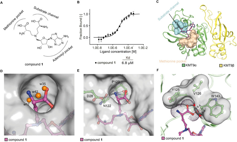
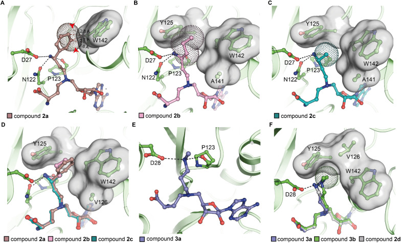
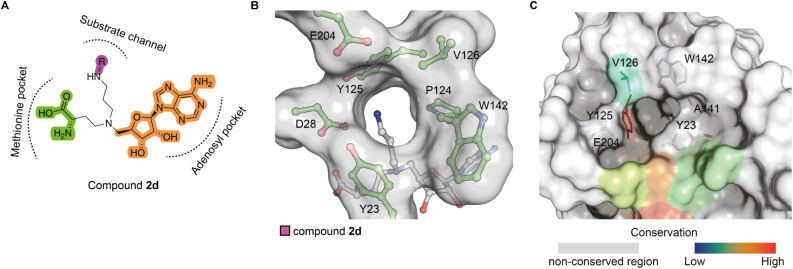
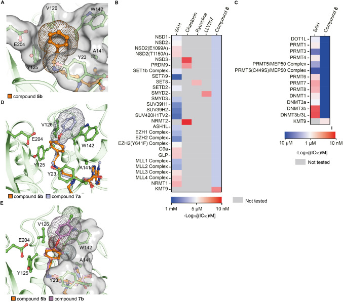
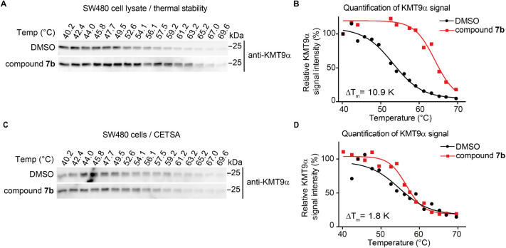
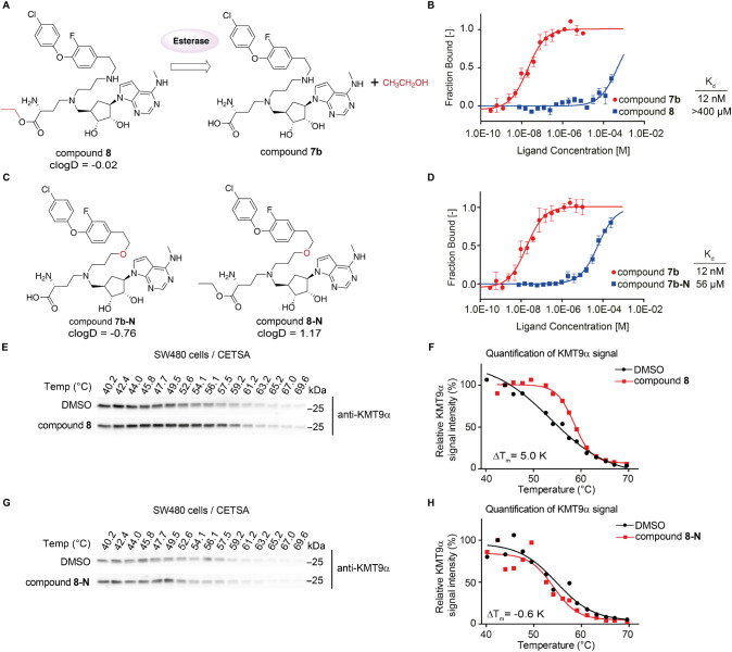
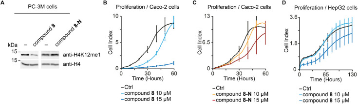
Lysine methyltransferase 9 (KMT9), an obligate heterodimer (KMT9α/KMT9β), belongs to the few described Rossmann-fold histone lysine methyltransferases and monomethylates histone H4 at lysine 12 (H4K12me1). KMT9 depletion or inhibition impairs the proliferation of tumors, including prostate, lung, colon, and bladder cancer cells, underscoring its therapeutic potential. Here, we show the development of branched cofactor analogues with a methionine side chain as highly potent KMT9 inhibitors. Through structure-guided design, a basic nitrogen and 4-chlorophenoxy-2-fluorobenzene in the substrate branch contribute most to the high potency and selectivity. Due to the zwitterionic methionine side chain, the inhibitors did not show cellular activity. Importantly, an ethyl ester prodrug 8 exhibits cellular target engagement and effectively blocks the proliferation of colon cancer cell lines, further validating pharmacological inhibition of KMT9 as a promising strategy for cancer therapy.


Figures

1
1
Identification of a hit compound of KMT9. (A) Chemical structure of compound 1 with predicted binding mode. (B) MST assay to determine the dissociation constant (Kd) of compound 1 binding to KMT9. Data represent means ± s.d (n = 3). (C) Overall structure of the KMT9/compound 1 complex (PDB code: 9FIM). KMT9α (green) and KMT9β (yellow) proteins are represented as ribbons. Arrows indicate the substrate channel (cyan) and the methionine pocket (brown) illustrated by surface view. Compound 1 is shown as sticks (magenta). (D) Superimposition of compound 1/KMT9 and SAH/KMT9 complex structures. Compound 1 (magenta) and SAH (blue) are shown by sticks. Water molecules are shown by spheres (orange). (E) Hydrogen bonds between the amino group of compound 1 (magenta) and KMT9α (green) in the substrate branch. (F) Unfavored contacts between the carbonyl group of compound 1 (magenta) and W142 of KMT9α. Key residues and ligands are depicted as sticks. Water molecules are shown as orange spheres. Contacts are represented by black dashed lines. Van-der-Waals radius of compound 1 are shown as dots. Substate channel is shown as surface. KMT9α is shown as ribbon.
2
2
Ligand bound KMT9 structures. (A) Unfavored contacts between compound 2a and W142 of KMT9α­(PDB code: 9FKG) (B, C) Hydrophobic contacts between compound 2b (B)/2c (C) and KMT9α in the substrate channel (PDB code: 9FKM and 9FKV). (D) Superimposition of compound 2a-/2b-/2c- bound KMT9 structures.(E) Hydrogen bonds between the methylamine of compound 3a and KMT9α (PDB code: 9FKW).(F) Superimposition of compounds 2d-, 3a-, and 3b- bound KMT9 structures. Compound 2a (brown), 2b (pink), 2c (cyan), 2d (white), 3a (blue), 3b (green) and protein residues (green) are represented as sticks. KMT9α is shown as ribbon (green). Part of the substrate channel of KMT9α is shown as surface (gray). Van-der-Waals radius of compounds are shown as dots. Hydrogen bonds between ligands and KMT9 residues are indicated as gray dash lines. Unfavored contacts between compound 2a and W142 of KMT9α are represented as red dash lines.
3
3
Lead optimization of compound 2d. (A) Strategies of lead optimization for compound 2g. (B) Unoccupied substrate pocket in compound 2g-bound KMT9 complex structure. (C) Structural conservation of KMT9a substrate pocket across Rossmann-fold MTs. Residue conservation is shown with a color scheme ranging from blue (low conservation) to red (high conservation); The residues which are not found in structural conserved region are colored in white.
4
4
Lead optimization of compound 5b. (A) Hydrophobic contact between the phenyl moiety of compound 5b and substrate channel of KMT9 (PDB code: 9FL4). (B, C) Selectivity of compound 6 against SET-domain containing and Rossmann-fold MTs. (D, E) Predicted binding mode of compound 7a-/7b- bound KMT9 structures based on compound 5b/KMT9 crystal structure. Ligands and key residues are represented by sticks. Substrate channel of KMT9a is shown as surface. Van-der-Waals radius of corresponding moiety are shown as dots.
5
5
Cellular target engagement of compound 7b. (A, B) CETSA for KMT9a in SW480 cell lysate treated with vehicle (DMSO), 10 μM compound 7b. Representative Western blots (A) and quantification (B) showing increased melting temperatures (ΔT m) of endogenous KMT9a upon treatment with compound 7b compared to DMSO. (C, D) CETSA for KMT9a in SW480 cells treated with vehicle (DMSO), 10 μM compound 7b. Representative Western blots (C) and quantification (D) showing almost no increased melting temperatures (ΔT m) of endogenous KMT9a upon treatment with compound 7b compared to DMSO.
6
6
Cellular target engagement of compound 8. (A) Scheme of the cleavage of ethyl ester prodrug, compound 8, by esterase. (B) MST assay to determine the K d of compound 8 and compound 7b binding to KMT9. Data represent means ± s.d (n = 3). (C) Chemical structures of compound 7b-N and compound 8-N. (D) MST assay to determine the K d of compound 7b and compound 7b-N binding to KMT9. Data represent means ± s.d (n = 3). (E, F) CETSA for KMT9a in SW480 cells treated with vehicle (DMSO), 10 μM compound 8. Representative Western blots (E) and quantification (F) showing increased melting temperatures (ΔT m) of endogenous KMT9a upon treatment with compound 8 compared to DMSO. (G, H) CETSA for KMT9a in SW480 cells treated with vehicle (DMSO), 10 μM compound 8-N. Representative Western blots (G) and quantification (H) showing no increased melting temperatures (ΔT m) of endogenous KMT9a upon treatment with compound 8-N compared to DMSO.
7
7
Cellular activity of prodrug compound 8. (A) Levels of H4K12me1 in PC-3 M prostate tumor cells cultured in the presence of DMSO (−), 10 μM compound 8 or compound 8-N for 3 days were analyzed by Western blot using the indicated antibodies. Histone H4 was used as control. (B, C) Proliferation of Caco-2 cells treated with DMSO (Ctrl), compound 8, or compound 8-N were measured by Xcelligence. Data represent means ± s.d (n = 3). (D) Proliferation of HepG2 cells upon treatment of DMSO (Ctrl) and compound 8.
1
1. Synthesis of the Key Intermediate si4
2
2. Synthesis of the “Methionine-Like” Amino Acid Side Chain
3
3. Synthesis of the Final Compounds 2/3/5 Derivatives
4
4. Synthesis of the Carbasugar Derivative si47
5
5. Synthesis of the Final Compounds 6, 7a Aand 7b

Similar articles

References

    1. Kaniskan H. U., Martini M. L., Jin J.. Inhibitors of Protein Methyltransferases and Demethylases. Chem. Rev. 2018;118(3):989–1068. doi: 10.1021/acs.chemrev.6b00801. - DOI - PMC - PubMed
    1. Luo M.. Chemical and Biochemical Perspectives of Protein Lysine Methylation. Chem. Rev. 2018;118(14):6656–6705. doi: 10.1021/acs.chemrev.8b00008. - DOI - PMC - PubMed
    1. Shen H., Laird P. W.. Interplay between the cancer genome and epigenome. Cell. 2013;153(1):38–55. doi: 10.1016/j.cell.2013.03.008. - DOI - PMC - PubMed
    1. Polak P., Karlic R., Koren A., Thurman R., Sandstrom R., Lawrence M., Reynolds A., Rynes E., Vlahovicek K., Stamatoyannopoulos J. A., Sunyaev S. R.. Cell-of-origin chromatin organization shapes the mutational landscape of cancer. Nature. 2015;518(7539):360–364. doi: 10.1038/nature14221. - DOI - PMC - PubMed
    1. Chi P., Allis C. D., Wang G. G.. Covalent histone modifications--miswritten, misinterpreted and mis-erased in human cancers. Nat. Rev. Cancer. 2010;10(7):457–469. doi: 10.1038/nrc2876. - DOI - PMC - PubMed

MeSH terms

LinkOut - more resources