

Not Enough Cells: How Insufficient Cytological Specimens Are Mirrored by Reporting Systems - Journey from the Bethesda to the WHO Reporting Systems
- PMID: 40527306
- PMCID: PMC12283066
- DOI: 10.1159/000546947
Not Enough Cells: How Insufficient Cytological Specimens Are Mirrored by Reporting Systems - Journey from the Bethesda to the WHO Reporting Systems
Abstract
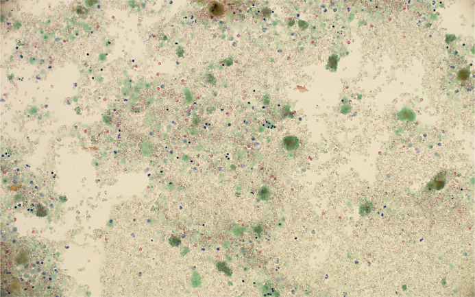
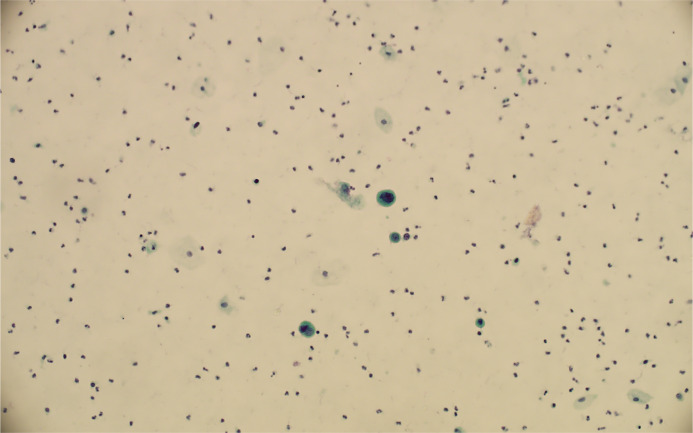
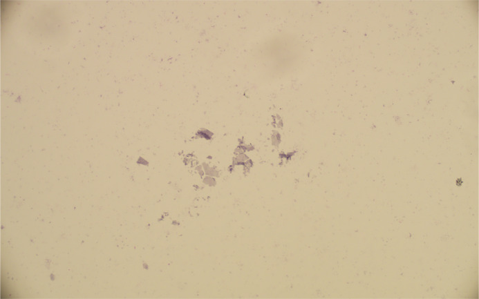
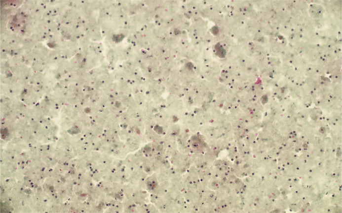
Background: Pap classes have been replaced by organ-specific reporting systems in recent decades; however, part of the cytological specimens is insufficient. The present review summarizes how different organ-specific systems define the insufficient category: both quantitative and qualitative criteria are used. In addition, the sample volume may be evaluated in certain specimens.
Summary: The reasons for an insufficient sample may vary and depend either on the lesion itself or the sampling procedure.
Key messages: The management recommendations for insufficient specimens improve communication between cytopathologists and treating physicians.
Keywords: Cytology; Fine-needle aspirate biopsies; Insufficient specimens; WHO reporting systems for cytopathology.
© 2025 The Author(s). Published by S. Karger AG, Basel.
Conflict of interest statement
The author was a member of the journal’s editorial board at the time of submission. The author has no other conflicts of interest to declare.
Figures

Similar articles
-
The Black Book of Psychotropic Dosing and Monitoring.Psychopharmacol Bull. 2024 Jul 8;54(3):8-59. Psychopharmacol Bull. 2024. PMID: 38993656 Free PMC article. Review.
-
Morphological, functional and neurological outcomes of craniectomy versus cranial vault remodeling for isolated nonsyndromic synostosis of the sagittal suture: a systematic review.JBI Database System Rev Implement Rep. 2015 Sep;13(9):309-68. doi: 10.11124/jbisrir-2015-2470. JBI Database System Rev Implement Rep. 2015. PMID: 26470674
-
The effect of sample site and collection procedure on identification of SARS-CoV-2 infection.Cochrane Database Syst Rev. 2024 Dec 16;12(12):CD014780. doi: 10.1002/14651858.CD014780. Cochrane Database Syst Rev. 2024. PMID: 39679851 Free PMC article.
-
Short-Term Memory Impairment.2024 Jun 8. In: StatPearls [Internet]. Treasure Island (FL): StatPearls Publishing; 2025 Jan–. 2024 Jun 8. In: StatPearls [Internet]. Treasure Island (FL): StatPearls Publishing; 2025 Jan–. PMID: 31424720 Free Books & Documents.
-
[Volume and health outcomes: evidence from systematic reviews and from evaluation of Italian hospital data].Epidemiol Prev. 2013 Mar-Jun;37(2-3 Suppl 2):1-100. Epidemiol Prev. 2013. PMID: 23851286 Italian.
References
-
- Kurman RJ, Solomon D, editors. The Bethesda System for Reporting Cervical/Vaginal Cytologic Diagnoses. Definitions, criteria, and explanatory notes for terminology and specimen adequacy. New York: Springer; 1994.
-
- Solomon D, Nayar R, editors. The Bethesda System for Reporting Cervical Cytology. Definitions, criteria, and explanatory notes. New York: Springer; 2004.
-
- Nayar R, Wilbur D, editors. The Bethesda System for reporting cervical cytology. 3rd ed. New York: Springer; 2015. p. 1–313.
-
- Ali SZ, Cibas E, editors. The Bethesda System for reporting thyroid cytopathology. 1st ed. New York: Springer; 2010.
Publication types
LinkOut - more resources
Full Text Sources
Research Materials
