

Giant mature teratoma of the retroperitoneal pelvic floor in an adult with combined developmental malformations of the reproductive system: A case report
- PMID: 40527775
- PMCID: PMC12173249
- DOI: 10.1097/MD.0000000000042889
Giant mature teratoma of the retroperitoneal pelvic floor in an adult with combined developmental malformations of the reproductive system: A case report
Abstract
Rationale: Teratoma is a germ cell tumor with multidirectional differentiation potential, but retroperitoneal and sacrococcygeal teratomas are relatively rare.
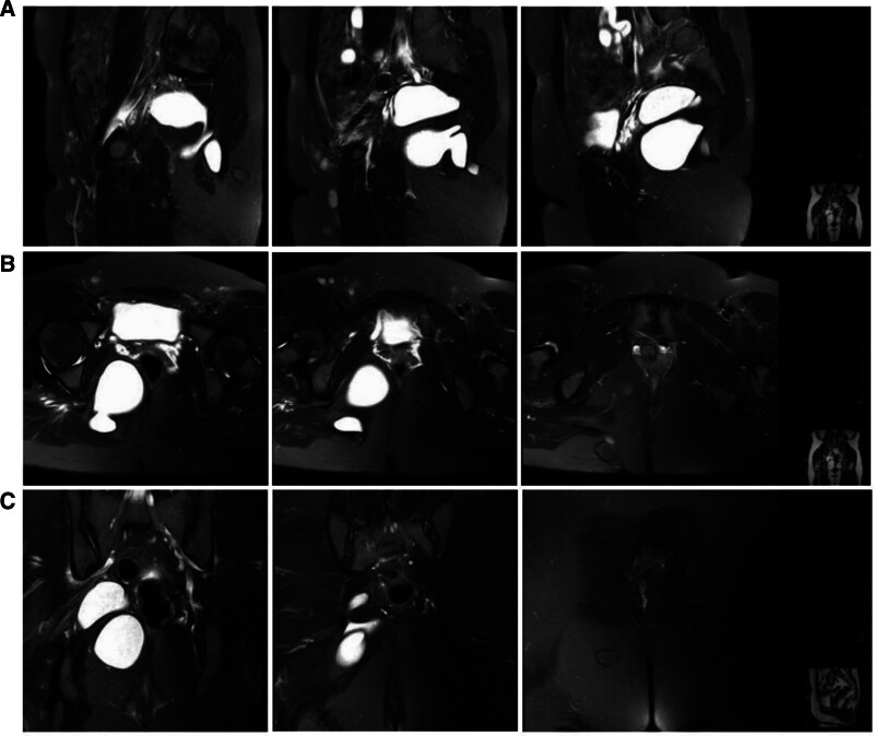
Patient concerns: A 32-year-old adult woman with a right duplicated ureter and left unicornuate uterus was found to have had a large pelvic mass for more than 8 years, which was increasing in size year by year.
Diagnoses: After 2 surgeries that failed to remove the pelvic mass completely, and the biopsy of the mass only suggested that it was a mature teratoma.
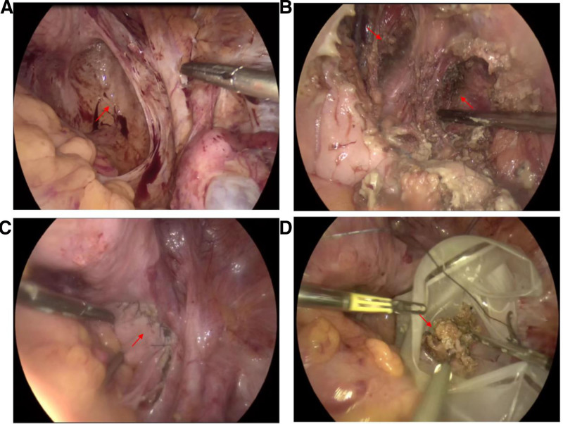
Interventions: The third time, after the transabdominal and transsacral combined pathway surgery, the mass was finally removed completely and found to have grown from the pelvic floor to the presacral area and finally to the right pelvic wall.
Outcomes: At 1-year follow-up, the patient had no complications, and serial pelvic ultrasounds showed no recurrence or metastasis.
Lessons: Thus, we demonstrate the feasibility of a combined transabdominal and transsacral approach for resecting this large mature retroperitoneal teratoma of the pelvic floor, providing a reference for surgeons managing similarly complex cases.
Keywords: pelvic floor tumor; retroperitoneal tumor; teratoma.
Copyright © 2025 the Author(s). Published by Wolters Kluwer Health, Inc.
Conflict of interest statement
The authors have no funding and conflicts of interest to disclose.
Figures
Similar articles
-
Molecular feature-based classification of retroperitoneal liposarcoma: a prospective cohort study.Elife. 2025 May 23;14:RP100887. doi: 10.7554/eLife.100887. Elife. 2025. PMID: 40407808 Free PMC article.
-
Mature hyperdense teratomas in the posterior fossa.Childs Nerv Syst. 2025 May 28;41(1):191. doi: 10.1007/s00381-025-06854-w. Childs Nerv Syst. 2025. PMID: 40437292
-
Mature Cystic Teratoma of Anterior Mediastinum in a Child: A Case Report and Literature Review.J Investig Med High Impact Case Rep. 2024 Jan-Dec;12:23247096241274510. doi: 10.1177/23247096241274510. J Investig Med High Impact Case Rep. 2024. PMID: 39230157 Free PMC article. Review.
-
Prenatal administration of progestogens for preventing spontaneous preterm birth in women with a multiple pregnancy.Cochrane Database Syst Rev. 2019 Nov 20;2019(11):CD012024. doi: 10.1002/14651858.CD012024.pub3. Cochrane Database Syst Rev. 2019. PMID: 31745984 Free PMC article.
-
Surveillance for Violent Deaths - National Violent Death Reporting System, 50 States, the District of Columbia, and Puerto Rico, 2022.MMWR Surveill Summ. 2025 Jun 12;74(5):1-42. doi: 10.15585/mmwr.ss7405a1. MMWR Surveill Summ. 2025. PMID: 40493548 Free PMC article.
References
-
- Luo CC, Huang CS, Chu SM, Chao HC, Yang CP, Hsueh C. Retroperitoneal teratomas in infancy and childhood. Pediatr Surg Int. 2005;21:536–40. - PubMed
-
- Panageas E. General diagnosis case of the day. Primary retroperitoneal teratoma. AJR Am J Roentgenol. 1991;156:1292–4. - PubMed
-
- Gatcombe HG, Assikis V, Kooby D, Johnstone PA. Primary retroperitoneal teratomas: a review of the literature. J Surg Oncol. 2004;86:107–13. - PubMed
-
- Ratan SK, Ratan J, Kalra R. Large benign cystic teratoma of the mesosigmoid causing intestinal obstruction: report of a case. Surg Today. 2002;32:922–4. - PubMed
Publication types
MeSH terms
LinkOut - more resources
Full Text Sources
