. 2025 Jun 17;3(1):56.
doi: 10.1038/s44259-025-00121-3.

Impact of stereochemical replacement on activity and selectivity of membrane-active antibacterial and antifungal cyclic peptides

Affiliations

Impact of stereochemical replacement on activity and selectivity of membrane-active antibacterial and antifungal cyclic peptides

Sandeep Lohan et al. NPJ Antimicrob Resist. .

Abstract

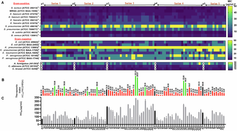
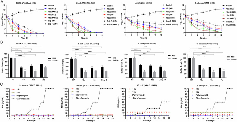
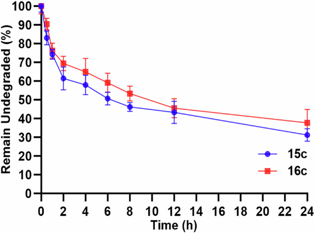
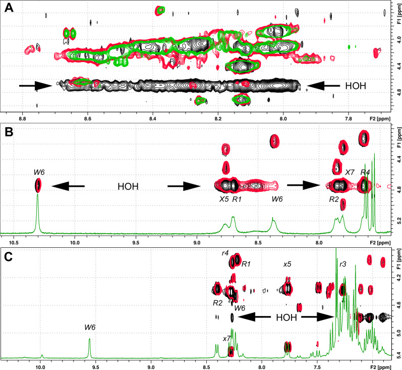
Herein, we report a library of 7-mer macrocyclic peptides designed by systematically replacing one, multiple, or all L-amino acids with their D-isomers in our previously identified hit compounds. Lead peptides, 15c and 16c, showed broad-spectrum activity against bacteria (Gram-positive minimum inhibitory activity (MIC 1.5-6.2 µg/mL and Gram-negative MIC 6.2-25 µg/mL) and fungi (MIC = 3.1-25 µg/mL). Additionally, peptides 15c and 16c showed rapid kill kinetics and biofilm degradation potential against both bacteria and fungi, while resistance development was not observed. The antimicrobial effect of these macrocyclic peptides was attributed to their membranolytic action, which was confirmed by calcein dye leakage assay and scanning electron microscopy analysis. Both peptides, 15c (HC50 = 335 µg/mL) and 16c (HC50 = 310 µg/mL), exhibited significantly lower hemolytic activity compared to their parent peptide p3 (HC50 = 230 µg/mL). At 100 µg/mL, both peptides showed >90% cell viability after 24 h incubation across four normal mammalian cell lines. Both peptides showed plasma stability (t1/2 ≥ 6 h), further supporting their therapeutic potential. Finally, the molecular mechanisms determining the pharmacological properties of a number of typical representatives of each series of synthesized peptides were investigated by NMR spectroscopy and computer simulations. The study revealed specific combinations of structural, dynamic, and hydrophobic parameters of these amphiphilic peptides that allow a reasonable prediction of their hemolytic activity. This Structure-Activity Relationship provides a basis for the rational design of peptides or peptidomimetics with predefined pharmacological profiles.


Conflict of interest statement

Competing interests: The authors declare no competing interests.

Figures

Scheme 1
Scheme 1
Overview of the steps involved in the solid phase synthesis of cyclic peptides. Amino acid residues are represented in three-letter notation: Arg, L-arginine; Trp, L-tryptophan; Dip, 3,3-diphenyl-L-alanine; Nal, 3-(2-naphthyl)-L-alanine. D-amino acids, arg, trp, dip, and nal are indicated in bold font with an underline. Cationic and hydrophobic residues are depicted in blue and red color, respectively.
Fig. 1
Fig. 1. Antibacterial, antifungal, and hemolytic activity data of Series 1 (1a-16a), Series 2 (1b-16b), series 3 (1c-24c), Series 4 (1d-24d), and Series 5 (1e-11e), cyclic peptides and parent cyclic peptides (p1, p2, p3, p4, and p5) of each series.
A aThe minimum inhibitory concentration (MIC) heatmap of cyclic peptides against Gram-positive and Gram-negative bacterial strains and fungal strains. MIC is the lowest concentration of the peptides that inhibited bacterial growth. bNon-resistant (wild type) bacterial strain. cMethicillin-resistant Staphylococcus aureus. dVancomycin-resistant enterococci strain. eMulti-drug resistant (Penicillin, Tetracycline, and Erythromycin) bacterial strain. fNDM-1, gCarbapenem, hCiprofloxacin, iImipenem resistant bacterial strains. jMulti-drug resistant clinical isolate of A. fumigatus. kNon-resistant (wild type) Candida strains. The data represents the results of the experiments performed in triplicate. B The therapeutic index of each cyclic peptide is calculated by dividing HC50 with the MIC (observed against a representative Gram-positive strain S. aureus (ATCC 29213)) of each peptide. C The peptides ‘ hemolytic activity was determined using human red blood cells (hRBC) as HC50 values. The black color bar in each graph represents the HC50 and therapeutic index of parent peptides (p1, p2, p3, p4, and p5). Green color bars represent the peptides with high therapeutic index within the respective series. The data represents the average of experiments performed in triplicate.
Fig. 2
Fig. 2. Cytotoxicity profiles of cyclic peptides across human cell lines.
Percentage cell viability data for selected cyclic peptides from A Series 1 (7a and 12a), B Series 2 (7b and 12b), C Series 3 (15c, 16c, and 20c), and D Series 4 (15d and 16d), each compared to their respective parent peptide (p1, p2, p3, and p4). A1–D4 show viability after 24-h incubation against: human lung fibroblasts (MRC-5; A1, B1, C1, D1), embryonic kidney cells (HEK-293; A2, B2, C2, D2), liver cells (HPRGC10; A3, B3, C3, D3), and skin fibroblasts (HeKa; A4, B4, C4, D4). All results are presented as mean ± SD from experiments performed in triplicate.
Fig. 3
Fig. 3. Antimicrobial activity and resistance profile of lead cyclic peptides.
A Time-kill kinetics of lead peptides (15c and 16c) compared to standard antibiotics (daptomycin and polymyxin B) against MRSA (ATCC BAA-1556), E. coli (ATCC BAA-2452), A. fumigatus (Af-293), and C. albicans (ATCC 60193), measured at MIC and 4×MIC over time. B Antibiofilm efficacy of 15c and 16c against the same pathogens at MIC and 2×MIC. NT represents the negative control (untreated cells in PBS), while positive controls include standard antibiotics: daptomycin (Dap), polymyxin B (Poly B), amphotericin B (Amp B), and fluconazole (Flz). Data were analyzed using a two-tailed unpaired Student’s t-test ((***p < 0.001; ****p < 0.0001). C Resistance development upon repeated exposure (18 passages) to lead peptides (15c and 16c) and standard antibiotics (daptomycin, polymyxin B, and ciprofloxacin) in S. aureus (ATCC 29213), MRSA (ATCC BAA-1556), and E. coli strains (ATCC 25922 and BAA-2452). All data represent triplicate experiments.
Fig. 4
Fig. 4. Membrane disruption and morphological effects of lead peptides.
A Concentration-dependent calcein dye leakage from liposomes mimicking bacterial (A1A3) and mammalian (A4A6) membranes following treatment with 15c (A1, A4), 16c (A2, A5), and daptomycin (A3, A6). B Field-emission scanning electron microscopy (FE-SEM) images of MRSA (ATCC BAA-1565; B1B3), E. coli (ATCC BAA-2452; B4B6), A. fumigatus (Af-293; B7B9), and C. albicans (ATCC 60193; B10B12). Untreated controls are shown in B1, B4, B7, and B10; cells treated with 15c appear in B2, B5, B8, and B11; and cells treated with 16c appear in B3, B6, B9, and B12. All treatments were conducted at 4×MIC for 1 h, and images were captured with a 2 μm scale bar.
Fig. 5
Fig. 5. In vitro plasma stability assay of the lead cyclic peptides 15c and 16c.
The data represents the percentage of undegraded peptides measured using Q-TOF LC/MS as the area under the curve in the extracted ion chromatogram in three independent experiments.
Fig. 6
Fig. 6. Oligomeric state of peptides in water.
1H-NMR spectra of peptides p1 (A), p3 (B), and 15c (C) in water at different concentrations (shown with the spectrum color). The spectra are scaled to match signal intensity.
Fig. 7
Fig. 7. Water accessibility of backbone HN groups of the peptides.
2D 1H-NMR TOCSY spectra of peptides p1 (A), p3 (B), and 15c (C) in water.
Fig. 8
Fig. 8. Stabilization of backbone structure of the peptides in presence of liposomes.
2D 1H-NMR NOESY spectra of peptides p3 (Left column, A, C, E) and 15c (right column, B, D, F) in water (top row, A, B) and in the presence of mammalian membrane mimics (DOPC/cholesterol, middle row, C, D) and bacterial membrane mimics (DOPC/DOPG, bottom row, E, F).
Fig. 9
Fig. 9. Principal conformational states of the peptides and distribution of MHP on their surfaces.
(Left) Spatial structures in stick mode represent the most populated conformations of the peptides in water. The most common hydrogen bonds (dashed lines) and residues involved (residue numbers) are shown. (Right) MD-averaged MHP spherical projection maps are displayed for the most populated MD-states in both fold types. Structures and MHP maps for the peptides in fold2-6 and fold1-4 conformations are shown on the top and bottom, respectively. Peptide-induced MHP values on the peptide molecular surface are color-coded according to the scale bar. Peptide names and corresponding 3D structures are in the same color.
Fig. 10
Fig. 10. Organization of the hydrophobic pattern for peptides p1, 7a, and 15c in an aqueous environment.
Preferred backbone conformations of peptides p1 (A, parent) and 7a (B, having D-isomer of Arg1, stereoisomeric substitution is indicated by the location of R1_HA atom), and the lead cyclic peptide 15c (C). Peptides p1 (A) and 7a (B) have very similar fold2-6 conformations with an elongated hydrophobic patch, compared with the alternative fold1-4 conformation of peptide 15c (C). Molecular surfaces of the peptides in bend (A, C) and flat (B) conformations, with surfaces color-coded based on molecular hydrophobicity potential (MHP) values: blue for hydrophilic regions and brown for hydrophobic regions. D Normalized histograms of distributions of dihedral angle φ of Dip6. The association of two major peaks with the different geometry of the apolar pattern (flat or bend) is demonstrated by dashed lines indicating the distribution of φ of Dip6 in flat and bend populations of the peptide.
Fig. 11
Fig. 11. Hemolytic activity (HC50) of the studied peptides and their attribution to one of four conformational/hydrophobicity types based on the population ratios of MD-derived backbone configurations.
Peptides are color-coded to indicate their levels of hemolytic activity: red for high, brown for medium, and green for low activity. Ration values on the X and Y axes are given in a logarithmic scale.

References

    1. de Kraker, M. E., Stewardson, A. J. & Harbarth, S. Will 10 million people die a year due to antimicrobial resistance by 2050? PLoS Med.13, e1002184 (2016). - PMC - PubMed
    1. Ventola, C. L. The antibiotic resistance crisis: part 1: causes and threats. Pharm. Ther.40, 277 (2015). - PMC - PubMed
    1. Zhen, X., Lundborg, C. S., Sun, X., Hu, X. & Dong, H. Economic burden of antibiotic resistance in ESKAPE organisms: a systematic review. Antimicrob. Resist. Infect. Control8, 1–23 (2019). - PMC - PubMed
    1. Roberts, R. R. et al. Hospital and societal costs of antimicrobial-resistant infections in a Chicago teaching hospital: implications for antibiotic stewardship. Clin. Infect. Dis.49, 1175–1184 (2009). - PubMed
    1. Hancock, R. & Patrzykat, A. Clinical development of cationic antimicrobial peptides: from natural to novel antibiotics. Curr. Drug Targets Infect. Disord.2, 79–83 (2002). - PubMed