. 2025 Jun 17;16(1):1137.
doi: 10.1007/s12672-025-02988-0.

Identification of intracranial solitary fibrous tumor and atypical meningioma by multi-parameter MRI-based radiomics model

Affiliations

Identification of intracranial solitary fibrous tumor and atypical meningioma by multi-parameter MRI-based radiomics model

Yanghua Fan et al. Discov Oncol. .

Abstract

Purposes: The preoperative distinction between atypical meningioma (AM) and intracranial solitary fibrous tumor (SFT) holds significant importance in guiding surgical approach decisions and prognostic assessments.

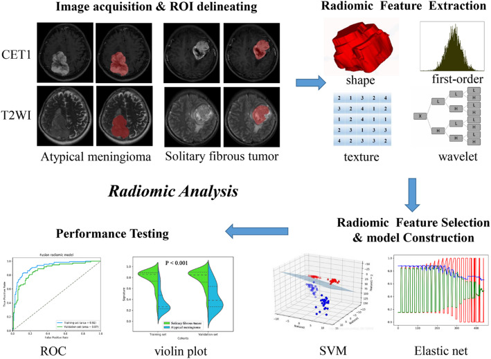
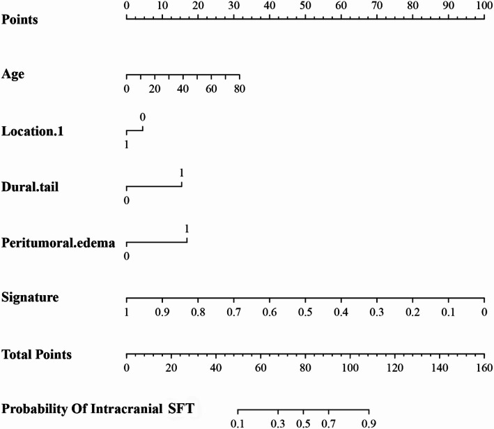
Methods: A total of 310 SFT patients and 203 AM patients were retrospectively included and stratified into training and validation cohorts. Employing the elastic net algorithm, relevant features were identified to form the fusion radiomic model. Subsequently, a clinical-radiomic combined model was developed by integrating the fusion radiomic model with significant clinical variables through multivariate logistic regression analysis. The models' calibration, discriminative capacity, and clinical utility were thoroughly assessed.

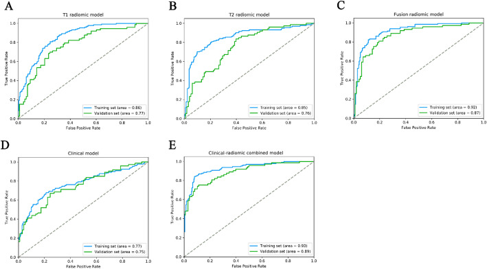
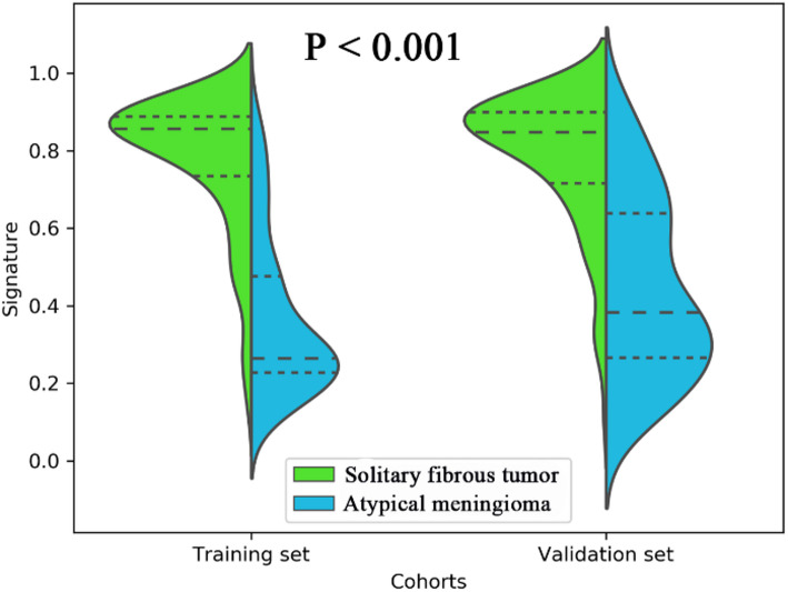
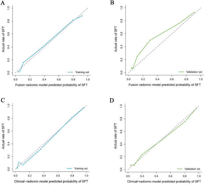
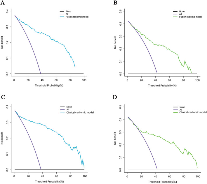
Results: The fusion radiomic model was crafted from 17 radiomic features, achieving AUC values of 0.920 in the training set and 0.870 in the validation set. Subsequently, the clinical-radiomic combined model exhibited AUC values of 0.930 and 0.890 in the training and validation sets, indicating commendable discrimination and calibration. Assessment through decision curve analysis underscored the clinical utility of both the fusion radiomic model and the clinical-radiomic combined model for individuals with intracranial SFT and AM.

Conclusions: The clinical-radiomic combined model exhibited notable sensitivity and exceptional efficacy in the distinctive diagnosis of intracranial SFT and AM, holding promise for the non-invasive advancement of personalized diagnostic and therapeutic strategies.

Keywords: Algorithm; Atypical meningioma; Diagnosis; Intracranial solitary fibrous tumor; Radiomics.


Conflict of interest statement

Declarations. Ethics approval and consent to participate: The need for patients’ informed consent was waved. All investigations conformed to the principles outlined in the Declaration of Helsinki and were performed with permission by the responsible Ethics Committee of the Institutional Review Board of Beijing Tiantan Hospital. Consent for publication: Not applicable. Competing interests: The authors declare no competing interests.

Figures

Fig. 1
Fig. 1
The flow chart of the present study. (I) a. Brain MR images acquisition (axial T2WI and sagittal CE-T1WI). b. ROI segmentation by ITK-SNAP software. (II) Four categories radiomics features extracted by PyRadiomics algorithm. (III) Radiomic Feature selection by elastic net and support vector machine (SVM) algorithm. (IV) And model training and testing
Fig. 2
Fig. 2
The performance of ROC curves for the three predictive models the training and validation sets. (A) T1 radiomic model; (B) T2 radiomic model; (C) fusion radiomic model; (D) Clinical model; (E) Clinical-radiomic combined model
Fig. 3
Fig. 3
A violin plot comparing the signature distribution of the fusion radiomic model between intracranial solitary fibrous tumor (SFT) and atypical meningioma (AM) patients
Fig. 4
Fig. 4
Calibration curve analysis for the fusion radiomic model (A: training set, B: validation set) and clinical-radiomic combined model (C: training set, D: validation set)
Fig. 5
Fig. 5
Decision curve analysis for for the fusion radiomic model (A: training set, B: validation set) and clinical-radiomic combined model (C: training set, D: validation set). The Y-axis measures the net benefit. The blue (A, C: training set) and green (B, D: validation set) line represents the radiomics model. The purple line represents the assumption that all patients were diagnosed as intracranial solitary fibrous tumor (SFT). The black line represents the assumption that all patients diagnosed as atypical meningioma
Fig. 6
Fig. 6
Bar plots for the clinical-radiomic combined model in the training (A) and validation sets (B). The blue histogram above the horizontal axis and the green histogram below the horizontal axis indicate the patients with correct diagnosis of the clinical-radiomic combined model
Fig. 7
Fig. 7
A nomogram derived from the clinical-radiomic combined model. This nomogram is used based on the value of signature of radiomic model and four clinical characteristics, including age, location 1 (supratentorial or infratentorial), dural tail, and peritumoral edema. Draw a vertical line from the corresponding axis of each factor until it reaches the first “Points” line. Next, summarize the points of all risk factors, then draw a vertical line that falls vertically from the “Total Points” axis until it reaches the last axis to the diagnostic probability of intracranial solitary fibrous tumor (SFT)

Similar articles

References

    1. Cohen-Inbar O. Nervous system Hemangiopericytoma. Can J Neurol Sci 2019: 1–12. - PubMed
    1. Louis DN, Perry A, Wesseling P, Brat DJ, Cree IA, Figarella-Branger D, et al. The 2021 WHO classification of tumors of the central nervous system: a summary. Neuro Oncol. 2021;23:1231–51. - PMC - PubMed
    1. Gubian A, Ganau M, Cebula H, Todeschi J, Scibilia A, Noel G, et al. Intracranial solitary fibrous tumors: A heterogeneous entity with an uncertain clinical behavior. World Neurosurg. 2019;126:e48–56. - PubMed
    1. Gritsch S, Batchelor TT, Gonzalez Castro LN. Diagnostic, therapeutic, and prognostic implications of the 2021 world health organization classification of tumors of the central nervous system. Cancer. 2022;128:47–58. - PubMed
    1. Yu Y, Gu F, Luo YL, Li SG, Jia XF, Gu LX, et al. The role of tumor parenchyma and brain cortex signal intensity ratio in differentiating solitary fibrous tumors and meningiomas. Discov Oncol. 2024;15:32. - PMC - PubMed

LinkOut - more resources