. 2025 Jun 17;23(1):660.
doi: 10.1186/s12967-025-06730-w.

Interleukin-1β mediates a tumor-supporting environment prompted by IGF1 in triple-negative breast cancer (TNBC)

Affiliations

Interleukin-1β mediates a tumor-supporting environment prompted by IGF1 in triple-negative breast cancer (TNBC)

Domenica Scordamaglia et al. J Transl Med. .

Abstract

Background: The intricate mechanisms that associate obesity with triple-negative breast cancer (TNBC) remain to be disclosed. Considering that obesity is linked with an increased bioavailability of the insulin-like growth factor 1 (IGF1), we evaluated whether IGF1 triggers aggressive features in TNBC cells and the molecular paths involved.

Methods: Gene expression and Chromatin Immunoprecipitation experiments, ELISA, immunoblotting, immunoprecipitation and immunofluorescence assays, combined with two-dimensional and three-dimensional in vitro model-based studies, were used to investigate the molecular mechanisms through which IGF1 may promote proliferative and motile responses in TNBC cells and reprogram normal fibroblasts into cancer-associated fibroblasts (CAFs)-like cells. The translational relevance of the results obtained was supported by bioinformatics analyses leveraging data from extensive TNBC patient databases.

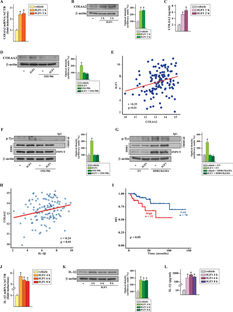
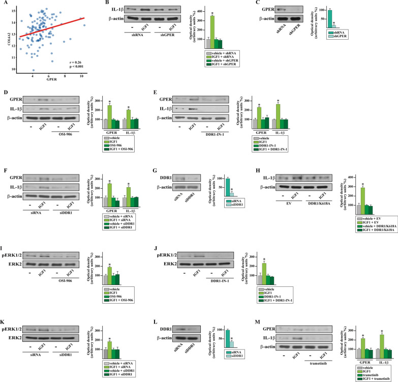
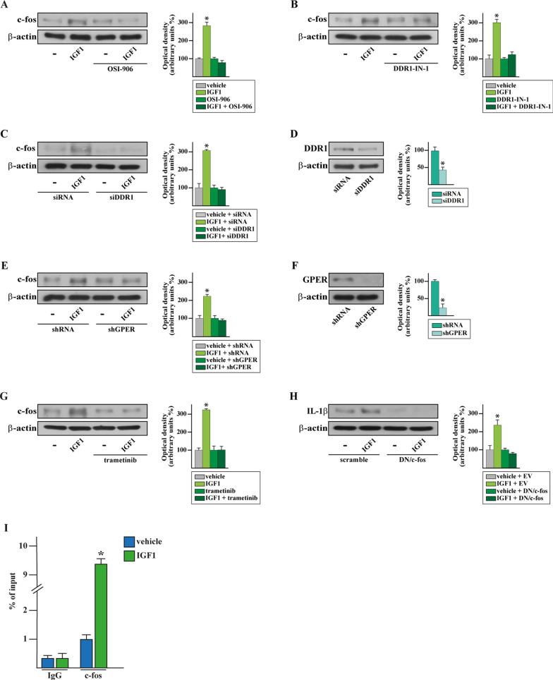
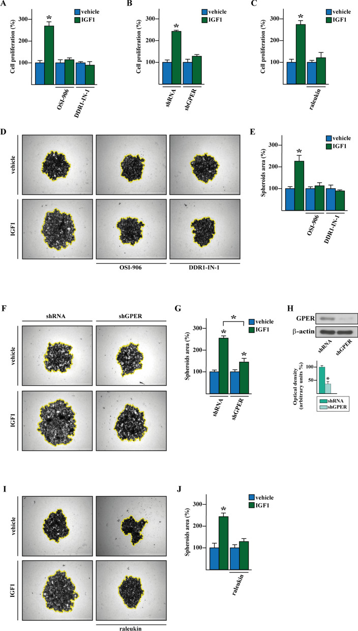
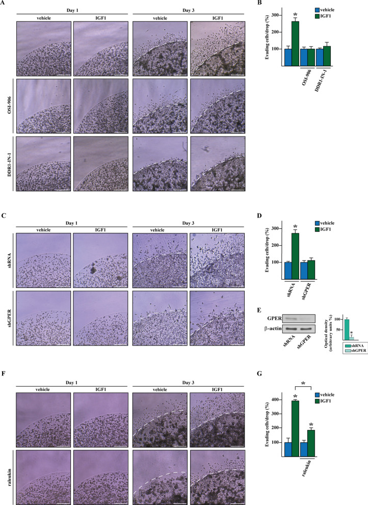
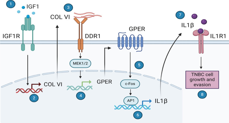
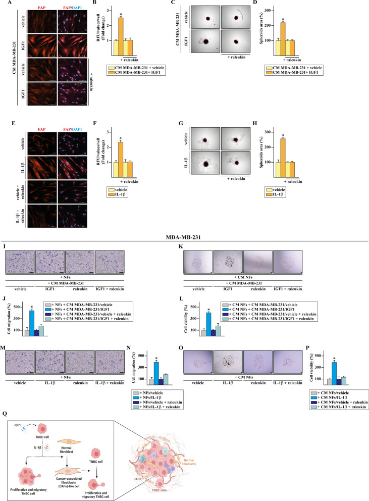
Results: We found that the cytokine interleukin-1β (IL-1β) mediates certain IGF1 actions within the tumor microenvironment, hence facilitating the TNBC landscape. Mechanistically, we assessed that the IGF1/IGF1 receptor (IGFIR) axis induces the collagen VI-dependent activation of discoidin domain receptor 1 (DDR1) and the subsequent increase of the G protein estrogen receptor (GPER), toward IL-1β regulation and secretion. Consequently, IL-1β promoted both the autocrine stimulation of TNBC cells and the differentiation of normal fibroblasts into cancer-associated fibroblasts (CAFs)-like cells, which in turn achieved a proliferative profile and enhanced the motility of TNBC cells.

Conclusions: IL-1β may be considered as a therapeutic target in more comprehensive approaches in obese TNBC patients exhibiting high IGF-1 bioavailability.

Keywords: Cancer associated fibroblasts (CAFs); Discoidin domain receptor 1 (DDR1); G protein estrogen receptor (GPER); Insulin-like growth factor 1 (IGF1); Interleukin 1β (IL1β); Triple-negative breast cancer (TNBC).


Conflict of interest statement

Declarations. Ethics approval and consent to participate: Not applicable. Consent for publication: Not applicable. Competing interests: The authors declare that they have no competing interests.

Figures

Fig. 1
Fig. 1
IGF1 induces the COL6A2-dependent activation of DDR1 along with the up-regulation of IL1-β in TNBC cells. mRNA (A) and protein (B) levels of COL6A2 evaluated respectively by real-time PCR and immunoblotting in MDA-MB-231 cells exposed to vehicle ( −) or 50 ng/ml IGF1, as indicated. C COL6A2 levels evaluated by ELISA in the supernatants collected from MDA-MB-231 exposed to vehicle ( −) or 50 ng/ml IGF1. D Protein levels of COL6A2 in MDA-MB-231 cells exposed for 1 h to vehicle ( −) or 50 ng/ml IGF1 alone or in combination with 1 µM IGF1R inhibitor OSI-906. E Correlation analysis of IGF1 and COL6A2 in TNBC patients of the TCGA cohort. The Pearson correlation coefficient (r) and the relative p-value are shown in the panel. F Immunoprecipitation assays performed in MDA-MB-231 cells exposed to vehicle ( −) or 50 ng/ml IGF1 alone or in combination with 1 µM IGF1R inhibitor OSI-906. Cell lysates were immunoprecipitated with an anti-DDR1 antibody and DDR1 phosphorylation was assessed using a phosphotyrosine antibody (p-Tyr). In control samples, nonspecific IgG was used instead of the primary antibody. An equal amount of the total lysates (input) was blotted for DDR1 as loading control. G MDA-MB-231 cells were transiently transfected with a constitutive empty vector (EV) or the mutant K618A DDR1 (DDR1/K618A) expressing plasmid. After 48 h, cells were exposed to vehicle ( −) or 50 ng/ml IGF1 for 1 h, lysed and subjected to immunoprecipitation and immunoblotting assays to detect DDR1 phosphorylation levels, as described above. H Correlation between the mRNA expression levels of COL6A2 and IL1-β in TNBC patients of the TCGA dataset, as ascertained by calculating the Pearson correlation coefficient (r). p-value is indicated in the panel. (I) Kaplan–Meier curve showing the correlation of high cumulative expression of COL6A2 and IL1-β with worse disease-free interval (DFI) in TNBC patients of TCGA. p-value is indicated in the panel. mRNA (J) and protein (K) levels of IL1-β evaluated respectively by real-time PCR and immunoblotting in MDA-MB-231 cells exposed to vehicle ( −) or 50 ng/ml IGF1, as indicated. L IL1-β levels evaluated by ELISA in the supernatants deriving from MDA-MB-231 cells exposed to vehicle ( −) or 50 ng/ml IGF1. In RNA experiments, values are normalized to the actin beta (ACTB) expression and presented as fold changes of mRNA expression upon treatments relative to the vehicle. Side panels show densitometric analysis of the blots normalized to the loading control. Results shown are representative of at least three independent experiments. (*) indicates p < 0.05
Fig. 2
Fig. 2
The DDR1/ERK/GPER signaling is involved in the induction of IL1-β by IGF1 in TNBC cells. A Scatter plot depicting the correlation between COL6A2 and GPER gene expression in TNBC patients of the TCGA dataset, the Pearson correlation coefficient (r) and the relative p-value are indicated within the panel. B Immunoblot of IL1-β from GPER-silenced MDA-MB-231 cells exposed to vehicle ( −) or 50 ng/ml IGF1. C Efficacy of GPER silencing in MDA-MB-231 cells. Protein levels of GPER and IL1-β in MDA-MB-231 cells exposed for 4 h to vehicle or 50 ng/ml IGF1 alone or in combination with 1 µM IGF1R inhibitor OSI-906 (D) or DDR1 inhibitor DDR1-IN-1 (E). F Immunoblots of GPER and IL1-β from DDR1-silenced MDA-MB-231 cells exposed to vehicle ( −) or 50 ng/ml IGF1. G Efficacy of DDR1 silencing in MDA-MB-231 cells. H Protein levels of IL1-β in MDA-MB-231 cells transiently transfected with a constitutive empty vector (EV) or the mutant K618A DDR1 (DDR1/K618A) expressing plasmid, and thereafter exposed to vehicle ( −) or 50 ng/ml IGF1 for 4 h. ERK1/2 phosphorylation in MDA-MB-231 cells treated for 1 h with vehicle ( −) or 50 ng/ml IGF1 in the presence of 1 µM IGF1R inhibitor OSI-906 (I) or 1 µM DDR1 inhibitor DDR1-IN-1 (J) or in MDA-MB-231 cells transiently transfected with scramble siRNA or siRNA targeting DDR1 (K). L Efficacy of DDR1 silencing in MDA-MB-231 cells. M Immunoblots of GPER and IL1-β in MDA-MB-231 cells exposed for 4 h to vehicle or 50 ng/ml IGF1 in the presence or absence of 100 nM trametinib. Side panels show densitometric analyses of the blots normalized to β-actin or ERK2 that served as loading controls, as indicated. Values represent the mean ± SD of three independent experiments performed in triplicate. (*) indicates p < 0.05
Fig. 3
Fig. 3
The IGF1R/DDR1/GPER/ERK-dependent increase of c-fos mediates the up-regulation of IL1-β in TNBC cells. Immunoblots of c-fos in MDA-MB-231 cells exposed for 4 h to vehicle (–) or 50 ng/ml IGF1 in the presence of 1 µM IGF1R inhibitor OSI-906 (A) or DDR1 inhibitor DDR1-IN-1 (B). c-Fos protein levels in DDR1 (C) and GPER-silenced (E) MDA-MB-231 cells. Efficacy of DDR1 (D) and GPER (F) silencing in MDA-MB-231 cells. G Immunoblot of c-fos in MDA-MB-231 cells exposed for 4 h to vehicle or 50 ng/ml IGF1 in the presence or absence of 100 nM trametinib. H IL-1β protein levels in MDA-MB-231 cells transfected with a scramble or a dominant-negative c-fos construct (DN/c-fos) and thereafter exposed for 4 h to vehicle (–) or 50 ng/ml IGF1. Side panels show densitometric analyses of the blots normalized to β-actin that served as loading control. I Recruitment of c-fos to the AP-1 site located within the IL1-β promoter, as ascertained by ChIP assay in MDA-MB-231 cells exposed for 4 h to vehicle or 50 ng/ml IGF1. In control samples, nonspecific IgG were used instead of the primary antibody. The amplified sequences were evaluated by real-time PCR. Values represent the mean ± SD of three independent experiments performed in triplicate. (*) indicates p < 0.05
Fig. 4
Fig. 4
The IL1-β/IL1R1 signaling is implicated in the IGF1-induced growth of TNBC cells. A Proliferation of MDA-MB-231 cells after 5 days treatment with vehicle or 50 ng/ml IGF1 alone or in combination with 1 µM IGF1R inhibitor OSI-906 or DDR1 inhibitor DDR1-IN-1, as indicated. (B) Growth assays in MDA-MB-231 cells transfected every 2 days with shRNA or shGPER, treated every day with vehicle or 50 ng/ml IGF1 and then counted after 5 days. C Proliferation of MDA-MB-231 cells after 5 days treatment with vehicle or 50 ng/ml IGF1 alone or in combination with 5 µg/ml IL-1R1 antagonist raleukin. Values of vehicle-treated cells were set as 100% upon which cell viability was determined. D Representative pictures of spheroids (a single spheroid/well) from the MDA-MB-231 spheroid cultures grown on agar-coated plates and exposed for 8 days to vehicle or 50 ng/ml IGF1 alone or in combination with 1 µM IGF1R inhibitor OSI-906 or 1 µM DDR1 inhibitor DDR1-IN-1. (F) Representative pictures of spheroids (a single spheroid/well) from GPER-silenced MDA-MB-231 cells grown for 8 days on agar-coated plates. Spheroids were transfected every 2 days with shRNA or shGPER, treated every 2 days and then counted on day 6. Scale bar: 1000 μm. (H) Efficacy of GPER silencing in MDA-MB-231 cells. The bottom panel shows densitometric analyses of the blots normalized to β-actin that served as loading control. I Representative pictures of spheroids (a single spheroid/well) from the MDA-MB-231 spheroid cultures grown on agar-coated plates and exposed for 8 days to vehicle or 50 ng/ml IGF1 alone or in combination with 5 µg/ml IL-1R1 antagonist raleukin. E, G, J Quantification of spheroid growth. Values of vehicle-treated MDA-MB-231 cells were set as 100% upon which spheroid growth was determined. Values represent the mean ± SD of three independent experiments performed in triplicate. (*) indicates p < 0.05
Fig. 5
Fig. 5
IGF1 promotes the motile behavior of TNBC cells by engaging the IL1-β/IL1R1 axis. A Representative pictures from the matrigel drops evasion assays in MDA-MB-231 cells treated with vehicle or 50 ng/ml IGF1 alone or in combination with 1 µM IR inhibitor OSI-906 or DDR1 inhibitor DDR1-IN-1, as indicated. C Representative pictures from the matrigel drops evasion assays in GPER-silenced MDA-MB-231 cells treated vehicle or 50 ng/ml IGF1. E Efficacy of GPER silencing in MDA-MB-231 cells. Side panel shows densitometric analyses of the blots normalized to β-actin that served as loading control. F Representative pictures from the matrigel drops evasion assay in MDA-MB-231 cells treated with vehicle or 50 ng/ml IGF1 alone or in combination with or 5 µg/ml IL-1R1 antagonist raleukin. B, D, G Percentage of cells around the drop upon 3 days treatment from three independent experiments performed in triplicate. Values of vehicle-treated MDA-MB-231 cells were set as 100% upon which evading cell percentage was determined. Dotted line indicates the matrigel drop border. Scale bar: 200 μm. (*) indicates p < 0.05
Fig. 6
Fig. 6
Schematic showing model whereby IGF1/IGF1R signaling induces collagen VI-dependent DDR1 activation, ERK-mediated GPER and c-fos increase toward the up-regulation of IL-1β. IL-1β/IL1R1 signaling, in turn, contributes to proliferative and motile characteristics in TNBC cells through an autocrine mechanism. Created in https://BioRender.com
Fig. 7
Fig. 7
IL-1β stimulates the reprogramming of normal fibroblasts into CAFs-like cells that in turn stimulate aggressive features in TNBC cells. A FAP expression evaluated by immunofluorescence assays in WI38 cells cultured for 72 h with conditioned medium (CM) collected from MDA-MB-231 cells previously exposed for 4 h to vehicle or 50 ng/ml IGF1 alone or in combination with 5 µg/ml IL-1R1 antagonist raleukin. Nuclei were stained by DAPI. Scale bar: 100 μm. C Representative pictures of spheroids (a single spheroid/well) grown for 6 days on agar-coated plates from WI38 cells cultured for 72 h with conditioned medium (CM) collected from MDA-MB-231 cells previously exposed for 4 h to vehicle or 50 ng/ml IGF1 alone or in combination with 5 µg/ml IL-1R1 antagonist raleukin. Scale bar: 1000 μm. E FAP expression evaluated by immunofluorescence assays in WI38 cells exposed to vehicle or IL-1β (10 ng/ml) alone or in combination with 5 µg/ml IL-1R1 antagonist raleukin. Nuclei were stained by DAPI. Scale bar: 100 μm. B, F Fluorescence intensities were quantified in 20 random fields for each condition and results are expressed as fold change of relative fluorescence units (RFU) over vehicle-treated cells (set as one-fold induction). G Representative pictures of spheroids (a single spheroid/well) grown for 6 days on agar-coated plates from WI38 cells exposed for 72 h to vehicle or IL-1β (10 ng/ml) alone or in combination with 5 µg/ml IL-1R1 antagonist raleukin. Scale bar: 1000 μm. D, H Quantification of spheroid growth. Values of vehicle-treated WI38 cells were set as 100% upon which spheroid growth was determined. I Transwell migration assays in MDA-MB-231 cells co-cultured for 8 h with WI38 cells (NFs) cultured for 72 h with conditioned medium (CM) collected from MDA-MB-231 cells, which were previously exposed for 4 h to vehicle or 50 ng/ml IGF1 alone or in combination with 5 µg/ml IL-1R1 antagonist raleukin. K Representative pictures of sodium alginate beads encapsulating MDA-MB-231 cells cultured for 10 days in bioreactors in the presence of the conditioned medium (CM) of WI38 cells (NFs) treated for 72 h with the conditioned medium (CM) collected from MDA-MB-231 cells, which were previously exposed for 4 h to vehicle or 50 ng/ml IGF1, alone or in combination with 5 µg/ml IL-1R1 antagonist raleukin. M Transwell migration assay in MDA-MB-231 cells co-cultured for 8 h with WI38 cells (NFs) previously exposed for 72 h to vehicle ( −) or IL-1β (10 ng/ml) alone or in combination with 5 µg/ml IL-1R1 antagonist raleukin. J, N Cells were counted in at least 5 random fields in three independent experiments performed in triplicate. Scale bar: 100 μm. O Representative pictures of sodium alginate beads encapsulating MDA-MB-231 cells cultured for 10 days in bioreactors with the conditioned medium (CM) of WI38 cells (NFs) exposed for 72 h to vehicle ( −) or IL-1β (10 ng/ml) alone or in combination with 5 µg/ml IL-1R1 antagonist raleukin. (L, P) Cell viability of MDA-MB-231 in the sodium alginate beads. Scale bar: 1000 μm. Values of vehicle-treated MDA-MB-231 cells were set as 100% upon which cell viability was determined. Data shown are the mean ± SD of three independent experiments performed in triplicate. (*) indicates p < 0.05. Q Cartoon depicting the molecular events and the biological responses triggered by IL-1β within the TNBC microenvironment. Created with BioRender.com

Similar articles

References

    1. Nguyen H-L, Geukens T, Maetens M, Aparicio S, Bassez A, Borg A, et al. Obesity-associated changes in molecular biology of primary breast cancer. Nat Commun. 2023;14:4418. - PMC - PubMed
    1. Picon-Ruiz M, Morata-Tarifa C, Valle-Goffin JJ, Friedman ER, Slingerland JM. Obesity and adverse breast cancer risk and outcome: Mechanistic insights and strategies for intervention. CA Cancer J Clin. 2017;67:378–97. - PMC - PubMed
    1. Schleh MW, Caslin HL, Garcia JN, Mashayekhi M, Srivastava G, Bradley AB, et al. Metaflammation in obesity and its therapeutic targeting. Sci Transl Med. 2023;15:9382. - PMC - PubMed
    1. Jin X, Qiu T, Li L, Yu R, Chen X, Li C, et al. Pathophysiology of obesity and its associated diseases. Acta Pharm Sin B. 2023;13:2403–24. - PMC - PubMed
    1. Brown KA. Metabolic pathways in obesity-related breast cancer. Nat Rev Endocrinol. 2021;17:350–63. - PMC - PubMed

MeSH terms

LinkOut - more resources