. 2025 May 29:33:101917.
doi: 10.1016/j.mtbio.2025.101917. eCollection 2025 Aug.

Thermosensitive hydrogel composite with si-Cx43 nanoparticles and anti-VEGF agent for synergistic treatment of diabetic retinopathy

Affiliations

Thermosensitive hydrogel composite with si-Cx43 nanoparticles and anti-VEGF agent for synergistic treatment of diabetic retinopathy

Lihui Wen et al. Mater Today Bio. .

Abstract

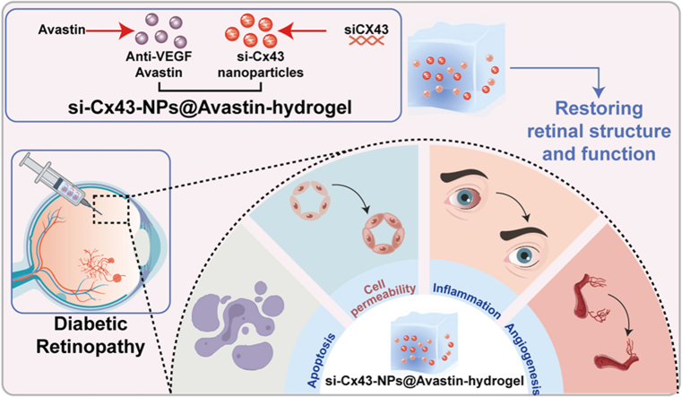
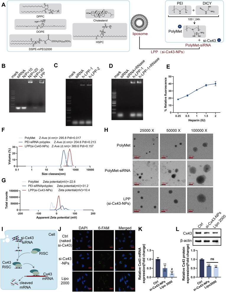
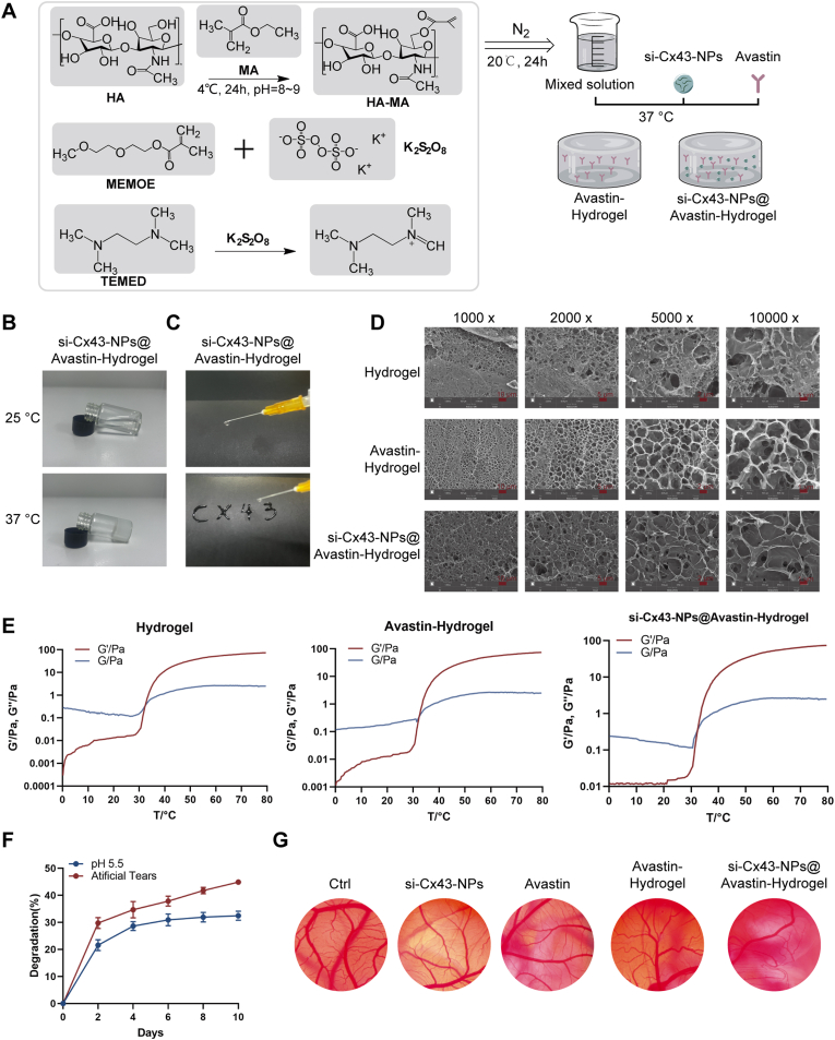
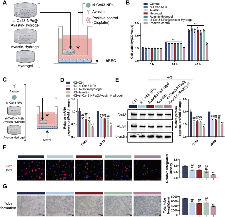
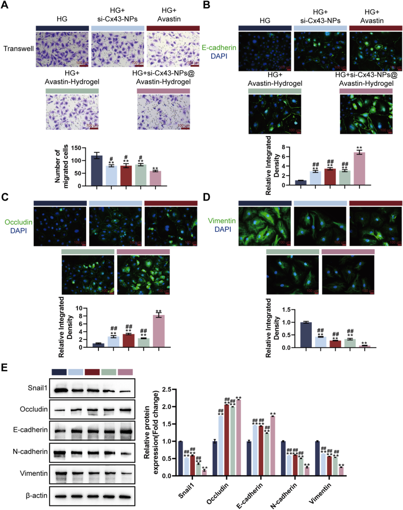
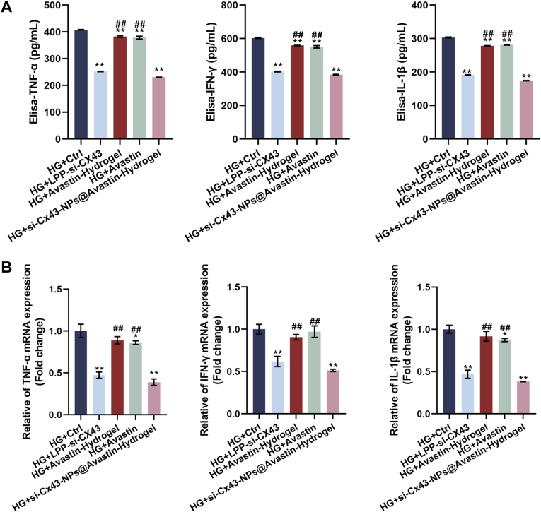
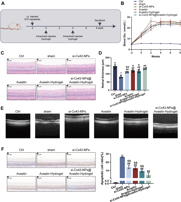
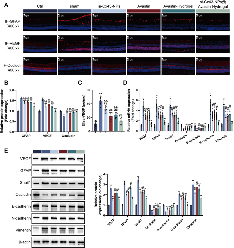
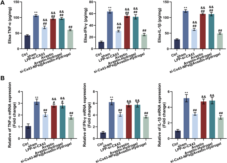
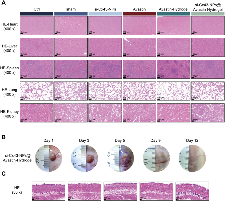
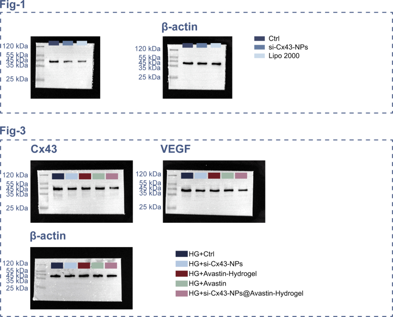
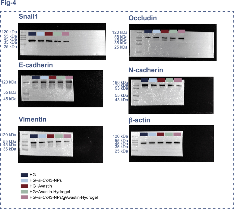
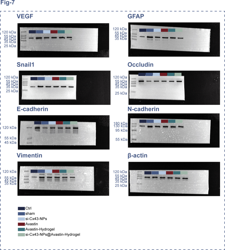
Diabetic retinopathy (DR) is characterized by pathological angiogenesis, inflammation, and retinal neurodegeneration, leading to vision loss. Current therapies, such as anti-VEGF agents, face challenges of low bioavailability and frequent invasive injections. Connexin43 (Cx43), a gap junction protein, plays a key role in DR progression through its modulation of inflammation and vascular dysfunction. A thermosensitive hydrogel composite was developed to encapsulate siRNA targeting Cx43 (si-Cx43) nanoparticles (NPs) and anti-VEGF (Avastin). The hydrogel was characterized for gelation, injectability, and degradation. In vitro studies evaluated the cytotoxicity, anti-angiogenic effects, and permeability regulation in hyperglycemic retinal cells under hyperglycemic conditions. In vivo therapeutic efficacy was assessed in a diabetic retinopathy rat model. si-Cx43-NPs demonstrated high siRNA encapsulation efficiency and stability, effectively silencing Cx43 expression in retinal endothelial cells. The hydrogel exhibited excellent injectability, temperature-sensitive gelation, and controlled degradation. In vitro, si-Cx43-NPs@Avastin-hydrogel significantly suppressed VEGF expression, reduced angiogenesis, and restored cell permeability under hyperglycemic conditions. In vivo, the hydrogel composite reduced neovascularization, inflammation, and apoptosis, restoring retinal structure and function more effectively than either single-agent treatment alone. Biocompatibility studies confirmed minimal toxicity and favorable degradation. The si-Cx43-NPs@Avastin-hydrogel provides a synergistic and minimally invasive therapeutic strategy for DR by targeting angiogenesis, inflammation, and neuroprotection with sustained drug delivery.

Keywords: Angiogenesis; Anti-VEGF; Diabetic retinopathy; Inflammation; Thermosensitive hydrogel; si-Cx43 nanoparticles.


Conflict of interest statement

The authors declare that they have no known competing financial interests or personal relationships that could have appeared to influence the work reported in this paper.

Figures

Image 1
Graphical abstract
Fig. 1
Fig. 1
Preparation and characterization of si-Cx43 nanoparticles (LPP-siRNA) (A) Schematic illustration of the synthesis of LPP-siRNA using cationic polymer (PEI) and lipid nanoparticles. (B) Gel retardation assay showing the optimal N/P ratio for the complexation of siRNA with PEI and subsequent formation of LPP-siRNA. (C) Serum stability assay evaluating the protection of siRNA by LPP in the presence or absence of fetal bovine serum. (D) RNase A protection assay assessing the integrity of siRNA in LPP after RNase A treatment. (E) siRNA release assay under competitive ligand conditions (heparin). (F) Dynamic light scattering (DLS) analysis for size distribution, polydispersity index (PDI), and zeta potential of LPP-siRNA. (G) Zeta potential measurements of various formulations. (H) TEM images of LPP-siRNA nanoparticles. (I) Schematic representation of the cellular transfection of si-Cx43 and its subsequent inhibition of Cx43 expression. (J) Confocal microscopy images showing the internalization and endosomal escape of si-Cx43-NPs in REC cells. (K) qRT-PCR analysis of Cx43 mRNA levels after treatment with si-Cx43-NPs, naked si-Cx43 or si-Cx43 with Lipo2000 transfection. (L) Immunoblotting for Cx43 expression levels in REC cells treated with si-Cx43-NPs, naked si-Cx43 or si-Cx43 with Lipo2000 transfection. ∗∗p < 0.01, vs control; #p < 0.05 vs si-Cx43-NPs.
Fig. 2
Fig. 2
Preparation and characterization of si-Cx43-NPs@Avastin-hydrogel (A) Schematic illustration of hydrogel synthesis (Avastin-hydrogel and si-Cx43-NPs@Avastin-hydrogel). (B) Images showing hydrogel gelation at room temperature and 37 °C. (C) Evaluation of hydrogel injectability. (D) SEM images of hydrogel surface morphology. (E) Rheological analysis measuring storage modulus (G′) and loss modulus (G″) of hydrogels across a temperature gradient. (F) Degradation properties of hydrogels in artificial tear fluid and citrate buffer (pH 5.5). (G) CAM assay evaluating anti-angiogenic activity of si-Cx43-NPs, single Avastin, and hydrogels.
Fig. 3
Fig. 3
Evaluation of si-Cx43-NPs@Avastin-hydrogel in hREC cells and its anti-angiogenic effects in vitro (A) Schematic illustration of cell experiments. (B) CCK-8 assay assessing cell viability after treatment with nanoparticles and hydrogels. (C) Schematic illustration of cell experiments under HG conditions. (D) qRT-PCR analysis of Cx43 and VEGF mRNA levels in hREC cells treated with nanoparticles or hydrogels under HG conditions. (E) Immunoblotting for Cx43 and VEGF protein levels in treated hREC cells. (F) Immunofluorescent staining (IF staining) detectsing Ki67 for cell proliferation. (G) Tube formation assay evaluating the anti-angiogenic effects of hydrogels in vitro. ∗∗p < 0.01, vs control; ##p < 0.01 vs si-Cx43-NPs@Avastin-hydrogel.
Fig. 4
Fig. 4
Effect of hydrogels on HG-stimulated cell permeability and migration in vitro (A) Transwell assay for cell migration. (B–D) IF staining for E-cadherin, occludin, and vimentin levels in treated cells. (E) Immunoblotting for Snail1, occludin, E-cadherin, N-cadherin, and vimentin protein levels in treated cells. ∗∗p < 0.01, vs HG; ##p < 0.01 vs si-Cx43-NPs@Avastin-hydrogel.
Fig. 5
Fig. 5
Anti-inflammatory effects of si-Cx43-NPs@Avastin-hydrogel on REC cells (A) ELISA for TNF-α, IFN-γ, and IL-1β protein levels in REC cell homogenates. (B) qRT-PCR analysis of TNF-α, IFN-γ, and IL-1β mRNA levels in retinal homogenates. ∗∗p < 0.01, vs HG + control; ##p < 0.01 vs si-Cx43-NPs@Avastin-hydrogel.
Fig. 6
Fig. 6
Evaluation of si-Cx43-NPs@Avastin-hydrogel in a diabetic retinopathy (DR) rat model (A) Schematic illustration of the DR rat modeling and corresponding treatments. (B) Blood glucose levels of rats in different groups at weeks −2, 0, 2, 4, 6, 8 of the first intravitreal injection of hydrogels. (C) H&E staining of retinal tissue sections showing structural changes at the end of week 8. (D) Morphometric analysis of retinal and basement membrane thickness according to H&E staining. (E) Optical coherence tomography (OCT) images of retinal structure and neovascularization. (F) TUNEL staining for apoptosis in retinal tissues and quantitative analysis based on staining results. ∗∗p < 0.01, vs control; ##p < 0.01, vs sham, && p < 0.01 vs si-Cx43-NPs@Avastin-hydrogel.
Fig. 7
Fig. 7
In vivo inhibition of angiogenesis and neuroprotection in the retina by si-Cx43-NPs@Avastin-hydrogel (A–B) IF staining for GFAP, VEGF, and occludin levels in rats' retinal sections. (C) ELISA for VEGF levels in rats' retinal tissues. (D) qRT-PCR analysis of VEGF, GFAP, occludin, Snail1, E-cadherin, N-cadherin, and vimentin mRNA levels in rats' retinal tissues. (E) Immunoblotting for VEGF, GFAP, occludin, Snail1, E-cadherin, N-cadherin, and vimentin protein levels in rats' retinal tissues. ∗∗p < 0.01, vs control; ##p < 0.01, vs sham, && p < 0.01 vs si-Cx43-NPs@Avastin-hydrogel.
Fig. 8
Fig. 8
Reduction of retinal inflammation by si-Cx43-NPs@Avastin-hydrogel in the DR rat model (A) ELISA for TNF-α, IFN-γ, and IL-1β levels in rats' retinal lysates. (B) qRT-PCR analysis of TNF-α, IFN-γ, and IL-1β mRNA levels in rats' retinal tissues. ∗∗p < 0.01, vs control; ##p < 0.01, vs sham, && p < 0.01 vs si-Cx43-NPs@Avastin-hydrogel.
Fig. 9
Fig. 9
In vivo degradation and biocompatibility of hydrogels (A) H&E staining of rats' major organs: heart, liver, spleen, lungs, kidneys, for toxicity assessment at the end of the in vivo experiments. (B) Time-dependent degradation of hydrogels subcutaneously implanted in rats, with histopathological evaluation of dermal and muscle tissues. (C) Assessment of local inflammation and tissue compatibility in the surrounding muscle tissue by H&E staining.
figs1
figs1
figs2
figs2
figs3
figs3

Similar articles

References

    1. Avogaro A., Fadini G.P. Microvascular complications in diabetes: a growing concern for cardiologists. Int. J. Cardiol. 2019;291:29–35. - PubMed
    1. Bourne R.R.A., Jonas J.B., Bron A.M., Cicinelli M.V., Das A., Flaxman S.R., Friedman D.S., Keeffe J.E., Kempen J.H., Leasher J., Limburg H., Naidoo K., Pesudovs K., Peto T., Saadine J., Silvester A.J., Tahhan N., Taylor H.R., Varma R., Wong T.Y., Resnikoff S. Vision loss expert group of the global burden of disease S. Prevalence and causes of vision loss in high-income countries and in Eastern and central Europe in 2015: magnitude, temporal trends and projections. Br. J. Ophthalmol. 2018;102:575–585. - PMC - PubMed
    1. Miller D.J., Cascio M.A., Rosca M.G. Diabetic retinopathy: the role of mitochondria in the neural retina and microvascular disease. Antioxidants. 2020;9 - PMC - PubMed
    1. Gomulka K., Ruta M. The role of inflammation and therapeutic concepts in diabetic retinopathy-A short review. Int. J. Mol. Sci. 2023;24 - PMC - PubMed
    1. Li Y., Liu Y., Liu S., Gao M., Wang W., Chen K., Huang L., Liu Y. Diabetic vascular diseases: molecular mechanisms and therapeutic strategies. Signal Transduct. Targeted Ther. 2023;8:152. - PMC - PubMed

LinkOut - more resources