

Spontaneous hepatic hemorrhage in fascioliasis: Imaging and diagnostic challenges in a rare case series from East Africa
- PMID: 40528911
- PMCID: PMC12171559
- DOI: 10.1016/j.radcr.2025.05.038
Spontaneous hepatic hemorrhage in fascioliasis: Imaging and diagnostic challenges in a rare case series from East Africa
Abstract
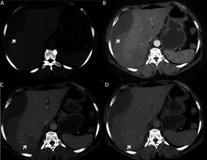
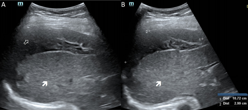
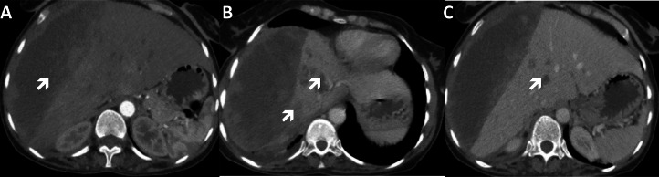
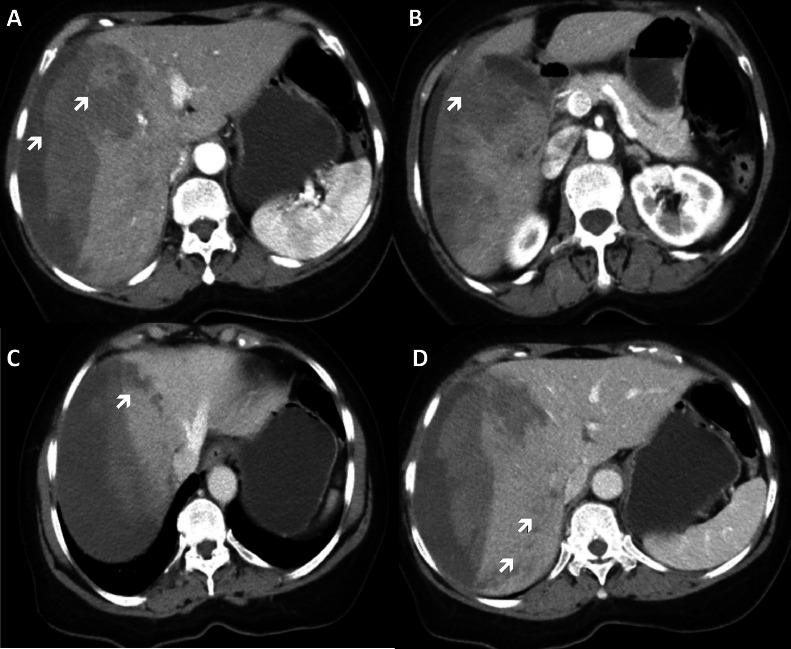
Fascioliasis is a zoonotic infection caused by the hepatic trematodes Fasciola hepatica and Fasciola gigantica. It presents with nonspecific symptoms, which can occasionally be complicated by rare but severe manifestations such as spontaneous hepatic hemorrhage. This study describes 3 cases of fascioliasis in female patients aged 35-56 years, who presented with right upper quadrant pain and nonspecific symptoms, including anorexia, nausea, and weakness. Laboratory investigations revealed hypereosinophilia and elevated liver enzymes. Imaging showed massive subcapsular and intrahepatic hematomas, along with multiple linear hypodense liver lesions. Given the lack of availability of immunological testing locally, empirical treatment with triclabendazole was initiated. The diagnosis in all cases was later confirmed through serological tests sent abroad. All patients showed significant clinical improvement with resolution of the eosinophilia and reduction of hepatic hemorrhage upon follow-up. These cases emphasize the importance of considering fascioliasis in patients presenting with hepatic hemorrhage and eosinophilia, and highlight the role of imaging and fine-needle aspiration cytology (FNAC) in guiding early empirical treatment when definitive serological testing is unavailable.
Keywords: Eosinophilia; Fasciola hepatica; Fascioliasis; Hepatic hemorrhage; Subcapsular Hematoma.
© 2025 The Authors. Published by Elsevier Inc. on behalf of University of Washington.
Figures
Similar articles
-
Spontaneous rupture of a subcapsular hepatic hematoma due to fascioliasis: A rare parasitic complication managed conservatively - Case report.IDCases. 2025 Jun 17;41:e02287. doi: 10.1016/j.idcr.2025.e02287. eCollection 2025. IDCases. 2025. PMID: 40607083 Free PMC article.
-
Fasciola hepatica Diagnosis: Clinical and Laboratory Clues from A University Hospital Experience.Turkiye Parazitol Derg. 2025 Jul 18;49(2):58-62. doi: 10.4274/tpd.galenos.2025.42650. Turkiye Parazitol Derg. 2025. PMID: 40679076
-
Hepatic Fascioliasis Complicated by Multisite Venous Thromboembolism: A Rare Association and Implications for Parasite-Associated Coagulopathy.Clin Case Rep. 2025 Jul 21;13(7):e70647. doi: 10.1002/ccr3.70647. eCollection 2025 Jul. Clin Case Rep. 2025. PMID: 40692659 Free PMC article.
-
Imported fascioliasis in non-endemic countries: a case series and review of the literature.Travel Med Infect Dis. 2025 Jul 26;67:102883. doi: 10.1016/j.tmaid.2025.102883. Online ahead of print. Travel Med Infect Dis. 2025. PMID: 40721066 Review.
-
Imaging findings of human hepatic fascioliasis: a case report and review of the literature.J Med Case Rep. 2021 Jun 24;15(1):324. doi: 10.1186/s13256-021-02945-9. J Med Case Rep. 2021. PMID: 34162437 Free PMC article. Review.
References
-
- Bayu B., Asnake S., Woretaw A., Ali J., Gedefaw M., Fente T., et al. Cases of human fascioliasis in North-West Ethiopia. Ethiop J Health Develop. 2005;19(3):237–240. doi: 10.4314/ejhd.v19i3.10004. - DOI
Publication types
LinkOut - more resources
Full Text Sources
