. 2023 Jun 2;5(3):1047-1062.
doi: 10.1016/j.fmre.2023.02.029. eCollection 2025 May.

Engineered in-situ-forming biomimetic hydrogel with self-regulated immunostimulatory capacity promotes postoperative tumor treatment

Affiliations

Engineered in-situ-forming biomimetic hydrogel with self-regulated immunostimulatory capacity promotes postoperative tumor treatment

Zhuo Cheng et al. Fundam Res. .

Abstract

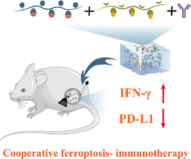
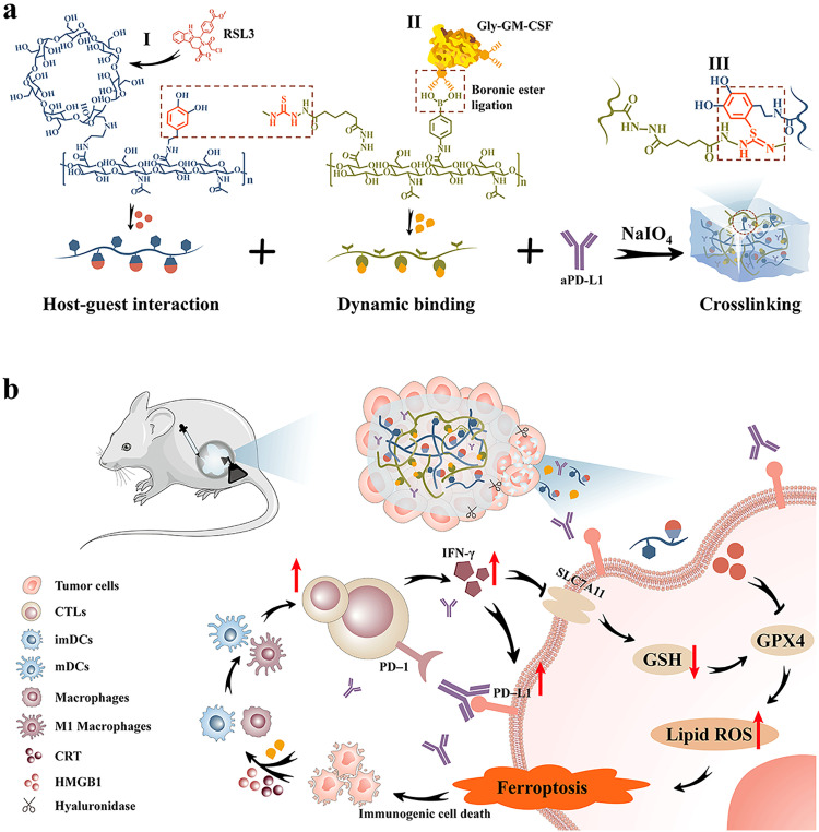
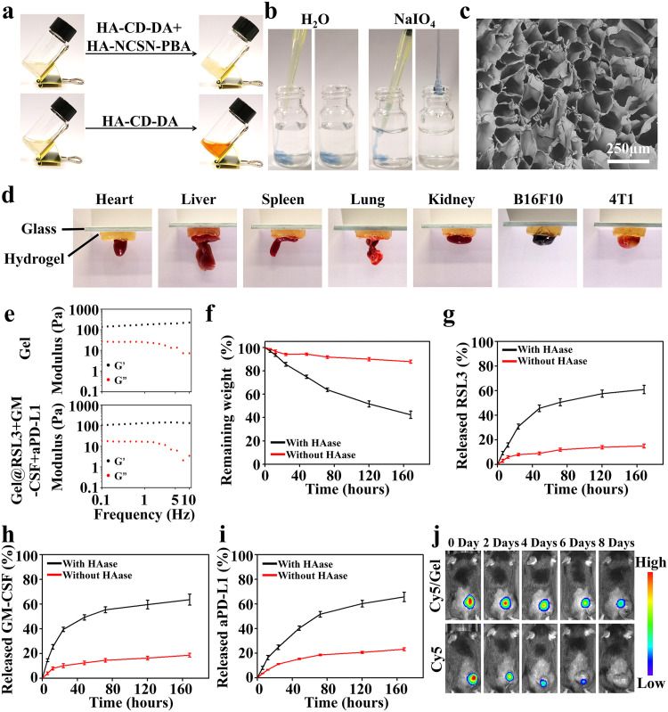
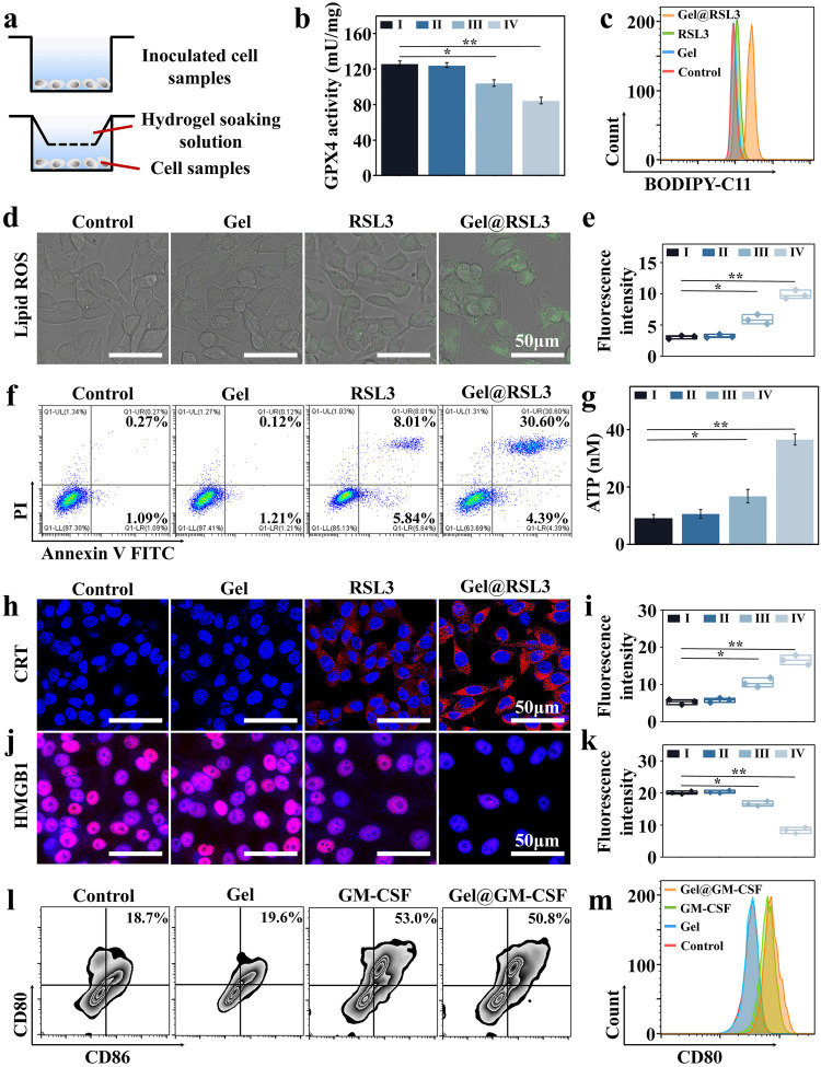
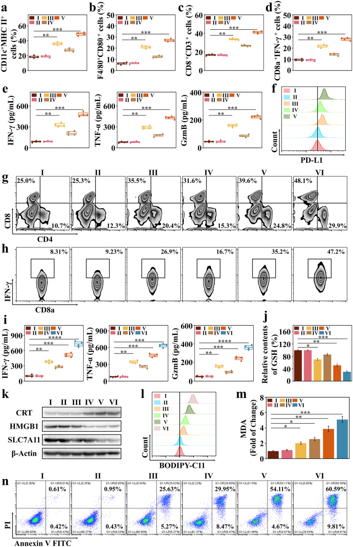
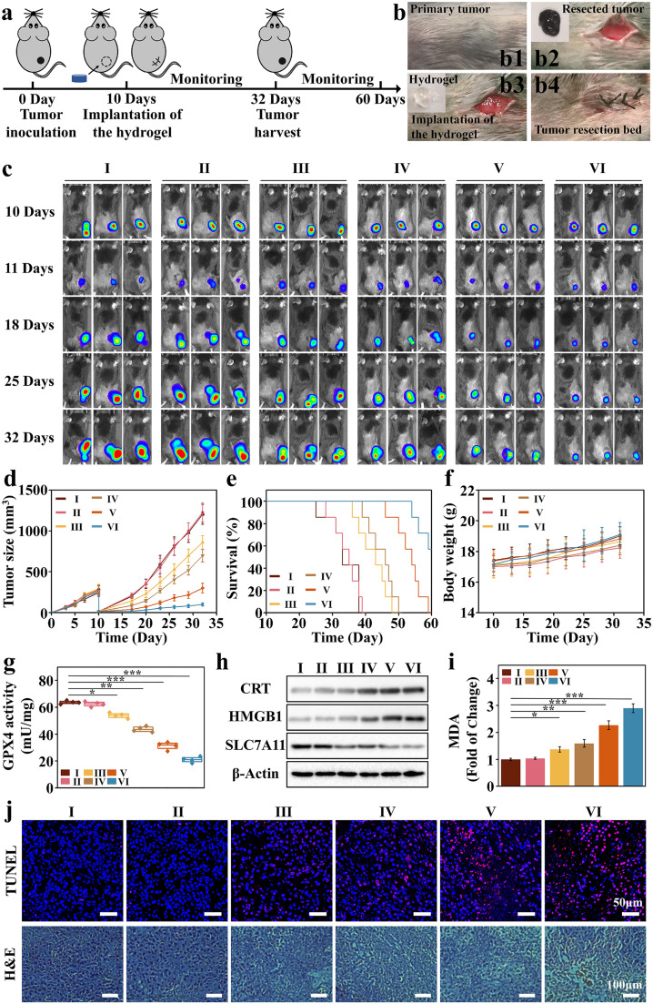
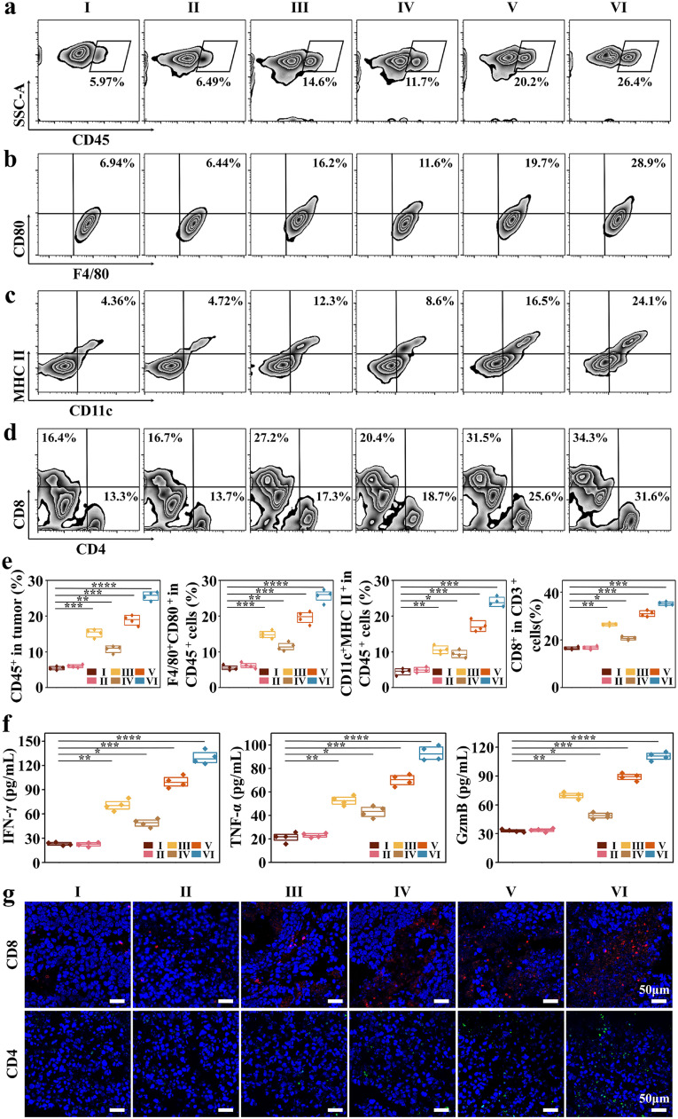
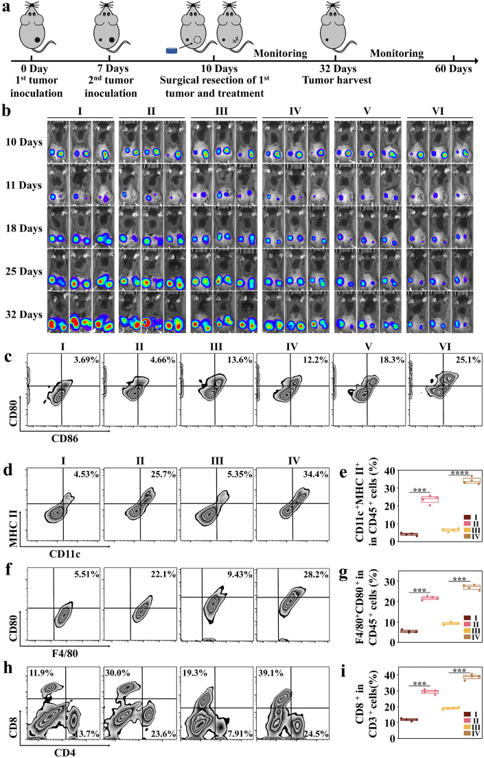
Post-resection tumors with microscopic foci and immunosuppressive microenvironments have high risk of recurrence and metastasis but respond poorly to various therapies. Herein, we propose a biomimetic hydrogel as a biocompatible, biodegradable and bioadhesive postoperative dressing that could be formed in situ by NaIO4-initiated thiourea-catechol crosslinking after syringe-injection into the resection cavity. The thiourea or catechol-bearing hyaluronic acid precursors are also separately engineered with phenylboronic acid and β-cyclodextrin (β-CD) groups, potentiating the reversible immobilization of (1S, 3R) RAS-selective lethal 3 (RSL3) and glycosylated granulocyte macrophage-colony stimulating factor (GM-CSF) without invasive chemical reactions. Meanwhile, the interconnected porous superstructure of the hydrogels allows the incorporation and self-regulated delivery of PD-L1 antibody (aPD-L1). RSL3-induced immunogenic ferroptosis and GM-CSF could cooperatively trigger robust adaptive tumor-specific immune responses, while aPD-L1 further alleviates the accumulated immunoresistance of tumor cells due to interferon γ-mediated PD-L1 upregulation, thus stimulating potent local and whole-body antitumor immunity to prevent postoperative tumor recurrence and metastasis. The biomimetic hydrogel may serve as a promising solution for the postoperative treatment of solid tumors.

Keywords: Cooperative ferroptosis-immunotherapy; Injectable in-situ forming hydrogel; Microenvironment remodeling; Postoperative tumor therapy; Supramolecular bioresponsive prodrug.


Conflict of interest statement

The authors declare that they have no conflicts of interest in this work.

Figures

Image, graphical abstract
Graphical abstract
Fig 1
Fig. 1
Injectable in-situ forming biomimetic hydrogel mediates cooperative ferroptosis-immunotherapy to elicit durable protection against postoperative tumor recurrence and metastasis. (a) Molecular structures of the HA-based precursors and the gelation mechanics. (b) Schematic illustration of hydrogel-mediated cooperative ferroptosis-immunotherapy after in-situ implantation.
Fig 2
Fig. 2
The HA-based biomimetic hydrogel demonstrates fast gelation rate, biomimetic mechanical properties, controlled drug release kinetics and biodegradability. (a) Gelation behaviors of HA-CD-DA and HA-NCSN-PBA precursors. (b) Photographical illustration of the NaIO4-initiated rapid gelation in aqueous environment. The mixture solution of the HA precursors was directly injected into NaIO4 solution (w/v 0.05%). (c) SEM image of the hydrogels after freeze-drying. (d) Adhesion behavior of hydrogel on various organs and tissues. (e) Comparative analysis on the mechanical properties of the hydrogel with or without drug loading. (f) Degradation profiles of hydrogel with or without hyaluronidase treatment. (g-i) Release kinetics of RSL3, GM-CSF and aPD-L1 from the hydrogel with or without HAase stimulation. (j) In vivo fluorescence images showing the degradation and drug release properties of the hydrogel after implantation.
Fig 3
Fig. 3
Ferroptosis-inducing and immunostimulatory capabilities of the biomimetic hydrogel. (a) Schematic diagram for the tumor cell/immune cell co-incubation system in transwell plates. The tumor cells (B16F10, 4T1) or immune cells were inoculated in the bottom chamber of the 24-well transwell culture plate, while the hydrogel soaking solution was placed in the upper chamber. (b) Changes of GPX4 activity in B16F10 cells after different treatments. (I) Control, (II) Gel, (III) RSL3, (IV) Gel@RSL3 (n = 4). (c) Flow cytometric analysis on the lipid ROS levels in B16F10 cells after different treatments. (d) CLSM imaging of lipid ROS generation in B16F10 cells after different treatments. Higher green fluorescence intensity indicates greater lipid ROS production. (e) Quantitative fluorescence analysis of lipid ROS levels in panel D (n = 4). (f) Flow cytometric analysis on the hydrogel-mediated ferroptosis levels of B16F10 cells after different treatments. (g) ATP levels in the supernatants of cell culture after different treatments. (I) Control, (II) Gel, (III) RSL3, (IV) Gel@RSL3 (n = 4). (h) CLSM imaging of CRT expression in B16F10 cells after different treatments. Stronger red fluorescence indicates higher expression levels. (i) Quantitative fluorescence analysis of CRT expression levels in panel H (n = 4). (j) CLSM imaging of cellular HMGB1 abundance after different treatments. Lower red fluorescence indicates greater HMGB1 release into the extracellular compartment. (k) Quantitative fluorescence analysis of HMGB1 release in panel J (n = 4). (l) Flow cytometric analysis on the treatment-induced maturation of BMDCs. (m) Flow cytometric analysis on the activation status of macrophages by monitoring the CD80 expression levels. * indicates significance at P < 0.05, ** indicates significance at P < 0.01.
Fig 4
Fig. 4
Gel@RSL3+GM-CSF+aPD-L1 activates immune response in vitro. (a-d) Flow cytometric analysis of the activation status of DCs (CD11c+/MHC II+), M1 macrophages (F4/80+/CD80+) and T cells (CD8+/CD3+ and CD8a+/IFN-γ+) in the co-incubation system of splenic immune cells and B16F10 cells after treatment with (I) Control, (II) Gel, (III) Gel@GM-CSF, (IV) Gel@RSL3 and (V) Gel@RSL3+GM-CSF (n = 4). (e) Secretion levels of immunostimulatory cytokines including IFN-γ, TNF-α and antitumor effector molecule GzmB in the supernatant from the co-culture system after different treatments (n = 4). (f) PD-L1 expression in tumor cells with after the hydrogel-mediated ferroptosis-immunotherapy. Group set-up for panel e-f: (I) Control, (II) Gel, (III) Gel@GM-CSF, (IV) Gel@RSL3 and (V) Gel@RSL3+GM-CSF). (g-h) Flow cytometric analysis of the expression levels of effector T cell marker CD4+/CD8+ and CD8a+/IFN-γ+ in T cells co-incubated with B16F10 cells after treatment with (I) Control, (II) Gel, (III) Gel@GM-CSF, (IV) Gel@RSL3, (V) Gel@RSL3+GM-CSF and (VI) Gel@RSL3+GM-CSF+aPD-L1. (i) Secretion levels of immunostimulatory cytokines including IFN-γ, TNF-α and antitumor effector molecule GzmB in the supernatant from the co-culture system after different treatments (n = 4). (j) Evaluation on the GSH levels in B16F10 cells after different treatments (n = 4). (k) Western blot analysis of the expression level of CRT, HMGB1 and SLC7A11 in different groups. (l) Flow cytometric analysis on the lipid ROS levels in B16F10 cells after different treatments. (m) MDA levels in B16F10 cells after different treatments (n = 4). Group set-up for panel I-M: (I) Control, (II) Gel, (III) Gel@GM-CSF, (IV) Gel@RSL3, (V) Gel@RSL3+GM-CSF and (VI) Gel@RSL3+GM-CSF+aPD-L1). (n) Flow cytometric analysis on the death rate of B16F10 cells after different treatments, including (I) Control, (II) Gel, (III) Gel@GM-CSF, (IV) Gel@RSL3, (V) Gel@RSL3+GM-CSF and (VI) Gel@RSL3+GM-CSF+aPD-L1. * indicates significance at P < 0.05, ** indicates significance at P < 0.01, *** indicates significance at P < 0.001, **** indicates significance at P < 0.0001.
Fig 5
Fig. 5
Antitumor effect of biomimetic hydrogel in vivo. (a) Schematic illustration of the treatment scheme of the B16F10-luc tumor-bearing mice (n = 7). (b) Treatment procedures on the tumor-bearing mice. (c) In vivo bioluminescence images of B16F10-luc tumor-bearing mice throughout the treatment period with (I) Control, (II) Gel, (III) Gel@GM-CSF, (IV) Gel@RSL3, (V) Gel@RSL3+GM-CSF and (VI) Gel@RSL3+GM-CSF-aPD-L1. (d) Tumor size changes during the incubation period after different treatments. (e) Survival analysis of mice after different treatments. (f) Body weight changes after treatment with different samples. (g) Evaluation on the GPX4 activity in tumor tissues after different treatments (n = 4). (h) Western blot analysis on the expression of CRT, HMGB1 and SLC7A11 in B16F10-luc tumors after treatment with (I) Control, (II) Gel, (III) Gel@GM-CSF, (IV) Gel@RSL3, (V) Gel@RSL3+GM-CSF and (VI) Gel@RSL3+GM-CSF-aPD-L1. (i) MDA levels in tumor tissue after different treatments (n = 4). (j) H&E and TUNEL staining of tumor tissue samples after treatment (I) Control, (II) Gel, (III) Gel@GM-CSF, (IV) Gel@RSL3, (V) Gel@RSL3+GM-CSF and (VI) Gel@RSL3+GM-CSF-aPD-L1. * indicates significance at P < 0.05, ** indicates significance at P < 0.01, *** indicates significance at P < 0.001.
Fig 6
Fig. 6
Hydrogel-mediated antitumor immune response in vivo. (a-e) Flow cytometric analysis on the tumor infiltration of (a) total immune cells (CD45+), (b) M1 macrophages (F4/80+/CD80+), (c) DCs (CD11c+/MHC II+) and (d) effector T cells (CD4+/CD8+) after treatment with (I) Control, (II) Gel, (III) Gel@GM-CSF, (IV) Gel@RSL3, (V) Gel@RSL3+GM-CSF and (VI) Gel@RSL3+GM-CSF-aPD-L1 in vivo (n = 4). (f) Serum levels of IFN-γ, TNF-α and GzmB in mice after treatment with (I) Control, (II) Gel, (III) Gel@GM-CSF, (IV) Gel@RSL3, (V) Gel@RSL3+GM-CSF and (VI) Gel@RSL3+GM-CSF-aPD-L1 (n = 4). (g) Immunofluorescence images of extracted tumors showing CD4+/CD8+ T cell infiltration after treatment with (I) Control, (II) Gel, (III) Gel@GM-CSF, (IV) Gel@RSL3, (V) Gel@RSL3+GM-CSF and (VI) Gel@RSL3+GM-CSF-aPD-L1. * indicates significance at P < 0.05, ** indicates significance at P < 0.01, *** indicates significance at P < 0.001, **** indicates significance at P < 0.001.
Fig 7
Fig. 7
The biomimetic hydrogel stimulates systemic antitumor immune response to suppress distal tumors. (a) Schematic illustration for establishing the bilateral tumor model and treatment schedule (n = 7). (b) In vivo bioluminescence images of B16F10-luc tumor-bearing mice throughout the treatment period with (I) Control, (II) Gel, (III) Gel@GM-CSF, (IV) Gel@RSL3, (V) Gel@RSL3+GM-CSF and (VI) Gel@RSL3+GM-CSF-aPD-L1. (c) Flow cytometric analysis on the DC maturation status in tumor-draining lymph nodes after treatment with(I) Control, (II) Gel, (III) Gel@GM-CSF, (IV) Gel@RSL3, (V) Gel@RSL3+GM-CSF and (VI) Gel@RSL3+GM-CSF-aPD-L1. (d-e) Flow cytometric analysis images (d) and quantification results (e) regarding the activation status of DCs (CD11c+/MHC II+) in primary and secondary tumors. I: Distal tumors in the untreated group. II: Distal tumors in the hydrogel-treated group. III: Primary tumors in the untreated group. IV: Primary tumors in the hydrogel-treated group (n = 4). (f-g) Flow cytometric analysis images (f) and quantification results (g) regarding the activation status of M1 macrophages (F4/80+/CD80+) in primary and secondary tumors (n = 4). I: Distal tumors in the untreated group. II: Distal tumors in the hydrogel-treated group. III: Primary tumors in the untreated group. IV: Primary tumors in the hydrogel-treated group. (h-i) Flow cytometric analysis (h) and quantification results (i) regarding the activation status of T cells (CD4+/CD8+) in different groups in primary and secondary tumors (n = 4). I: Distal tumors in the untreated group. II: Distal tumors in the hydrogel-treated group. III: Primary tumors in the untreated group. IV: Primary tumors in the hydrogel-treated group. *** indicates significance at P < 0.001, **** indicates significance at P < 0.001.

Similar articles

References

    1. Waks A.G., Winer E.P. Breast cancer treatment a review. JAMA. 2019;321(3):288–300. - PubMed
    1. Banks M., Graham D., Jansen M., et al. British Society of Gastroenterology guidelines on the diagnosis and management of patients at risk of gastric adenocarcinoma. Gut. 2019;68(9):1545–1575. - PMC - PubMed
    1. Tang F., Tie Y., Tu C., et al. Surgical trauma-induced immunosuppression in cancer: Recent advances and the potential therapies. Clin. Transl. Med. 2020;10(1):199–223. - PMC - PubMed
    1. Hiller J.G., Perry N.J., Poulogiannis G., et al. Perioperative events influence cancer recurrence risk after surgery. Nat. Rev. Clin. Oncol. 2018;15(4):205–218. - PubMed
    1. Hosein A.N., Dougan S.K., Aguirre A.J., et al. Translational advances in pancreatic ductal adenocarcinoma therapy. Nat. Cancer. 2022;3(3):272–286. - PubMed

LinkOut - more resources