. 2025 May 1;16(6):998-1007.
doi: 10.1021/acsmedchemlett.5c00038. eCollection 2025 Jun 12.

Enhanced Gram-Negative Membrane Disruption and In Vivo Efficacy via Lysine-Arginine Enrichment of Opis16a

Affiliations

Enhanced Gram-Negative Membrane Disruption and In Vivo Efficacy via Lysine-Arginine Enrichment of Opis16a

Mandelie van der Walt et al. ACS Med Chem Lett. .

Abstract

Infections complicate burn wound care, especially with the rise of antimicrobial resistance. Antimicrobial peptides (AMPs) offer the potential for advancing wound care by combating persistent infections. Opis16a, a scorpion venom-derived AMP, exhibits potent antibacterial activity by targeting Gram-negative membranes, causing rapid membrane disruption and bacterial cell death. Here, four novel Opis16a analogues were developed with improved membrane targeting and antibacterial efficacy. One analogue shows particular promise for topical application in Gram-negative burn wound infections. Enhanced peptide-lipid hydrogen bonding increases conformational stability, membrane insertion, and permeabilization rates. Substituting lysine residues in the C-terminal with arginine leads to the most consistent improvement in activity, selectivity for pathogen over HaCat cells, and stability in serum. In an in vivo Galleria mellonella burn wound model, a 5 mg/kg topical dose provides better protection than Opis16a against Enterobacter cloacae NICD 16103. These findings highlight the potential of optimized bactericidal AMPs to improve burn wound care.

Keywords: E. cloacae; Gram-negative bacteria; Opis16a; Opis16aCterKR; antimicrobial peptides; antimicrobial resistance; arginine substitution; membrane permeabilization; molecular dynamics; wound infections.


Figures

1
1
Hydrogen bonding between peptide and POPE/POPG model lipids differ among Opis16a analogues and is driven by cationic residues. Total (F) and residue-specific peptide–membrane hydrogen bonds as a function of time for Opis16a (A, red), Opis16aD5K (B, orange), Opis16aNterKR (C, lime green), Opis16aCterKR (D, dark green) and Opis16aKR (F, blue) in representative 200 ns simulations of Gram-negative model lipid bilayers.
2
2
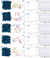
Secondary structure analysis of Opis16a and analogues in representative Gram-negative lipid bilayers. Top view snapshots showing the conformation of four peptide monomers (A, D, G, J, and M) binding to POPE/POPG bilayers in silico. Ramachandran contour plots showing the average dihedral angle concentration during the last 20 ns of 200 ns simulations (B, E, H, K, N). Psi and phi angles for each residue averaged over 200 ns of simulation and four peptides (C, F, I, L,and O). Representative data are shown for Opis16a (A–C), Opis16aD5K (D–F), Opis16aNterKR (G–I), Opis16aCterKR (J–L), and Opis16aKR (M–O).
3
3
Conformational flexibility of Opis16a and analogues in representative Gram-negative bilayers. Circular variance of psi dihedral angles for each residue, averaged over 200 ns of simulation and four peptides (A, C, E, G, I), as a measure of conformational flexibility (low = rigid; high = flexible). Changes in circular variance of psi and phi angles over time, averaged for four peptides (B, D, F, H, J). Data are for Opis16a (A, B), Opis16aD5K (C, D), Opis16aNterKR (E, F), Opis16aCterKR (G, H), and Opis16aKR (I, J).
4
4
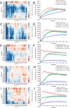
Membrane activity of Opis16a and analogues against Gram-negative bacterial membranes in silico and in vitro. (Left) Depth of Opis16a (A), Opis16aD5K (C), Opis16aNterKR (E), Opis16aCterKR (G), or Opis16aKR (I) insertion into model Gram-negative bilayers in a 200 ns simulation as the average z-position of each residue relative to the phosphate plane in the upper bilayer leaflet. Positive or negative values indicate the residues are above or below the phosphate group. (Right) E. cloacae NICD 16103 cytoplasmic membrane permeabilization by Opis16a (B), Opis16aD5K (D), Opis16aNterKR (F), Opis16aCterKR (H), and Opis16aKR (J) at 0.5×, 1×, and 2× MIC. Melittin (11 μg/mL) is used as positive control. Data show one representative experiment with mean ± SEM of a total of three biological repeats performed in triplicate.
5
5
Center of mass analysis for Opis16a and analogues relative to the midplane of model Gram-negative lipid bilayers. COM distances of the peptides from the midplane of the bilayer as an average over time for two replicate 200 ns simulations (A), with the standard deviation indicated by shaded areas. Statistical significance for mean COM averaged over the last 100 ns per peptide (B) or between Opis16a and the four new analogues, determined from one-way ANOVA with Dunnett’s test.
6
6
Bacterial time-kill kinetics of Opis16a compared with Opis16aCterKR. E. cloacae NICD 15283 treated with Opis16a (A) or Opis16aCterKR (B) at 0.5×, 1×, and 2× MIC. Data represent the mean ± SEM, for three independent experiments performed in triplicate.
7
7
Opis16a and Opis16aCterKR remain effective against E. cloacae NICD 16103 in 20% serum, with Opis16aCterKR offering improved protection in a G. mellonella burn wound infection model. MIC testing for Opis16a and Opis16aCterKR in trypsin (0.15 U/mL) or FCS (20%) (A). Data represent the mean ± SEM, for three independent experiments performed in triplicate. Survival curves over 120 h are plotted for larvae with burn wounds only (PBS), burn and E. cloacae NICD 16103 infected wounds, or infected wounds treated with Opis16a (B) or Opis16aCterKR (C). Data represent three independent experiments (30 larvae per condition).

Similar articles

References

    1. Murray C. J. L., Ikuta K. S., Rao P.. et al. Global burden of bacterial antimicrobial resistance in 2019: a systematic analysis. Lancet. 2022;399(10325):629–655. doi: 10.1016/S0140-6736(21)02724-0. - DOI - PMC - PubMed
    1. Vinaik R., Barayan D., Shahrokhi S., Jeschke M. G.. Management and prevention of drug-resistant infections in burn patients. Expert Review of Anti-infective Therapy. 2019;17(8):607–619. doi: 10.1080/14787210.2019.1648208. - DOI - PMC - PubMed
    1. Maitz J., Merlino J., Rizzo S.. et al. Burn wound infections microbiome and novel approaches using therapeutic microorganisms in burn wound infection control. Adv. Drug Delivery Rev. 2023;196:114769. doi: 10.1016/j.addr.2023.114769. - DOI - PubMed
    2. article

    1. Bennett W., Mende K., Campbell W. R.. et al. Enterobacter cloacae infection characteristics and outcomes in battlefield trauma patients. PLoS One. 2023;18(8):e0290735. doi: 10.1371/journal.pone.0290735. - DOI - PMC - PubMed
    2. article

    1. Harris P. N., Wei J. Y., Shen A. W.. et al. Carbapenems versus alternative antibiotics for the treatment of bloodstream infections caused by Enterobacter, Citrobacter or Serratia species: a systematic review with meta-analysis. J. Antimicrob. Chemother. 2016;71(2):296–306. doi: 10.1093/jac/dkv346. - DOI - PubMed

LinkOut - more resources