

Single- vs double-layer uterine closure of the cesarean scar in niche development: the Nicest Study
- PMID: 40529188
- PMCID: PMC12173043
- DOI: 10.1016/j.xagr.2025.100507
Single- vs double-layer uterine closure of the cesarean scar in niche development: the Nicest Study
Abstract
Background: There is an ongoing controversy regarding the optimal uterine closure technique for preventing niche development. Single- and double-layer closures have been considered comparable in terms of niche incidence after primary cesarean delivery. However, rather than simply the presence of a niche, its volume and residual myometrial thickness are the most potent factors in predicting gynecologic symptoms and subsequent pregnancy complications in women with cesarean scar defects. In addition, there is limited evidence on how uterine scars and niche sizes evolve over time.
Objective: This study aimed to compare the residual myometrial thickness and niche characteristics between the single-layer and double-layer uterine closure techniques and to evaluate the change in uterine scar characteristics from 6 to 12 months after cesarean delivery.
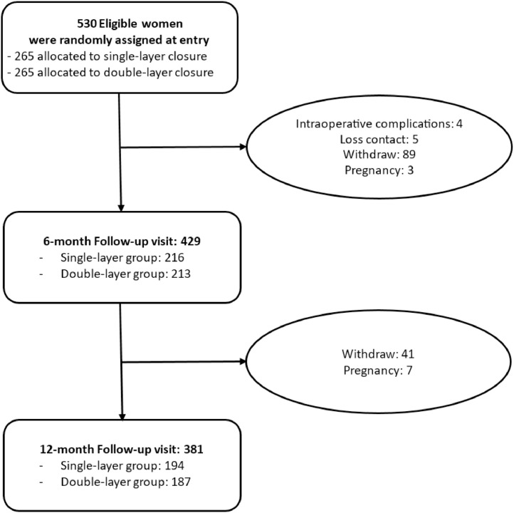
Study design: This prospective randomized study was conducted from May 2022 to December 2024 at Hanoi Obstetrics and Gynecology Hospital. A total of 530 full-term pregnant women who underwent primary cesarean delivery were randomized into single- or double-layer, unlocked, continuous suture. The exclusion criteria included previous major uterine surgery and abnormal placenta (placenta previa or placenta accreta spectrum) in the current pregnancy. Women were invited for 2 consecutive follow-up visits within 10 days of menstruation at 6 months (n=429) and 12 months (n=381) after cesarean delivery. Transvaginal ultrasound was used to evaluate uterine ultrasound characteristics. If the niche was detected, 3-dimensional transvaginal ultrasound was applied to measure the niche volume via the Virtual Organ Computer-aided AnaLysis method. Complete-case analysis was performed to evaluate the change in uterine ultrasound characteristics between the 2 visits.
Results: Of 429 women who participated in the 6-month visit, 216 had single-layer closure, and 213 had double-layer closure. The niche incidence at the first assessment was similar for both uterine closure techniques, at 35.6% in the single-layer group and 31.9% in the double-layer group, respectively (P>.05). At 6 months after delivery, the double-layer technique resulted in greater residual myometrial thickness (4.3 vs 4.0 mm; P=.007), better healing ratio (69% vs 60%; P=.048), and a lower proportion of large niches with residual myometrial thickness of <3 mm (9.9% vs 19.4%; P=.033). The median niche volume in the single-layer group (62 mm3) at 6 months after delivery was significantly higher than that in the double-layer group (39 mm3) (P=.003). Of 381 women who completed both assessments, 194 had single-layer closure, and 187 had double-layer closure. The results between the single-layer and double-layer groups of the second visit at 12 months after delivery mirrored those at the first visit. In longitudinal follow-up evaluation, uterine scar characteristics were stable, and the overall proportion of niches remained consistent from 6 months to 12 months after delivery, at 34.4% and 36.0%, respectively (P>.05). There was an increase in niche length (5.0 vs 5.5 mm; P=.000) and niche volume (47 vs 55 mm3; P=.000) among the assessments.
Conclusion: Although the niche incidence was similar between the 2 uterine closure techniques, the double-layer technique showed superior benefits, with greater residual myometrial thickness and healing ratio, lower large niche proportion, and smaller niche volume. The uterine scar characteristics were stable at 6 months after cesarean delivery, but the niche volume significantly increased over time. Future long-term follow-up research is needed to elucidate the relationship between niche size and clinical symptoms and to investigate the factors contributing to the temporal evolution of niche volume.
Keywords: double-layer uterine closure; large niche; niche volume; residual myometrial thickness; single-layer uterine closure.
© 2025 The Authors.
Similar articles
-
Risk of Cesarean scar defect following single- vs double-layer uterine closure: systematic review and meta-analysis of randomized controlled trials.Ultrasound Obstet Gynecol. 2017 Nov;50(5):578-583. doi: 10.1002/uog.17401. Epub 2017 Oct 9. Ultrasound Obstet Gynecol. 2017. PMID: 28070914
-
The association of endometrial closure during cesarean section to the risk of developing uterine scar defect: a randomized control trial.Arch Gynecol Obstet. 2024 May;309(5):2063-2070. doi: 10.1007/s00404-024-07417-1. Epub 2024 Mar 18. Arch Gynecol Obstet. 2024. PMID: 38498161 Clinical Trial.
-
A prospective comparative study of single-layer versus double-layer uterine closure techniques on cesarean scar formation.BMC Pregnancy Childbirth. 2025 Aug 20;25(1):868. doi: 10.1186/s12884-025-08010-3. BMC Pregnancy Childbirth. 2025. PMID: 40836292 Free PMC article. Clinical Trial.
-
Cesarean scar niche repair with the rendez-vous technique: comparison of pre- and postoperative symptoms, sonographic findings and quality of life.Eur J Obstet Gynecol Reprod Biol. 2025 Jul 5;313:114566. doi: 10.1016/j.ejogrb.2025.114566. Online ahead of print. Eur J Obstet Gynecol Reprod Biol. 2025. PMID: 40651195
-
Single-versus double-layer uterine closure at the time of cesarean delivery and risk of uterine scar niche: a systematic review and meta-analysis of randomized trials.Arch Gynecol Obstet. 2025 Aug 20. doi: 10.1007/s00404-025-08151-y. Online ahead of print. Arch Gynecol Obstet. 2025. PMID: 40833607 Review.
References
-
- Armstrong F., Mulligan K., Dermott R.M., et al. Cesarean scar niche: an evolving concern in clinical practice. Int J Gynaecol Obstet. 2023;161:356–366. - PubMed
-
- Klein Meuleman S.J.M., Min N., Hehenkamp W.J.K., Post Uiterweer E.D., Huirne J.A.F., de Leeuw R.A. The definition, diagnosis, and symptoms of the uterine niche – a systematic review. Best Pract Res Clin Obstet Gynaecol. 2023;90 - PubMed
LinkOut - more resources
Full Text Sources
