. 2025 Jun 3:19:1591215.
doi: 10.3389/fnins.2025.1591215. eCollection 2025.

Alterations in functional and structural connectivity in the 6-OHDA-induced Parkinsonian rat model

Affiliations

Alterations in functional and structural connectivity in the 6-OHDA-induced Parkinsonian rat model

Shuyi Zhu et al. Front Neurosci. .

Abstract

Introduction: Parkinson's Disease (PD), the second most common neurodegenerative disorder, is characterized by motor and non-motor symptoms linked to dopaminergic neuron degeneration. This study utilized the 6-hydroxydopamine (6-OHDA) rat model to replicate PD-like dopaminergic degeneration through targeted injections into the medial forebrain bundle and substantia nigra.

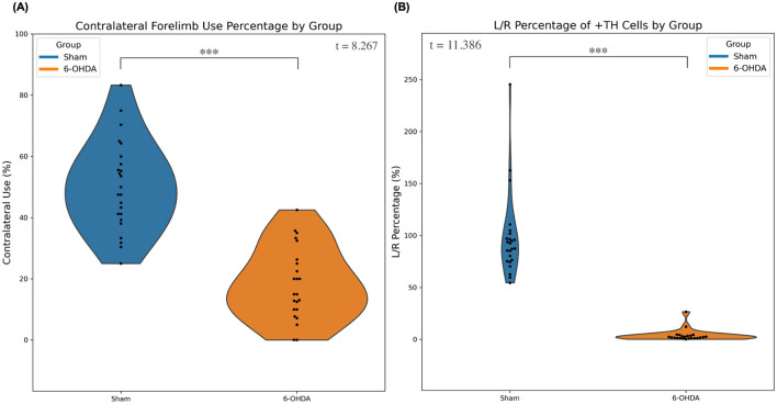
Methods: Behavioral assessments revealed hallmark motor deficits, while MRI was performed to assess complementary functional connectivity and structural connectivity. Post-mortem tyrosine hydroxylase (TH) staining confirmed extensive dopaminergic neuron loss, validating the pathological relevance of the model and ensuring data integrity. MRI data were collected at 7T in 46 male Fischer F344 rats (23 6-OHDA, 23 sham) to characterize functional and structural connectivity differences between cohorts.

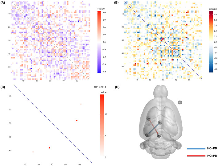
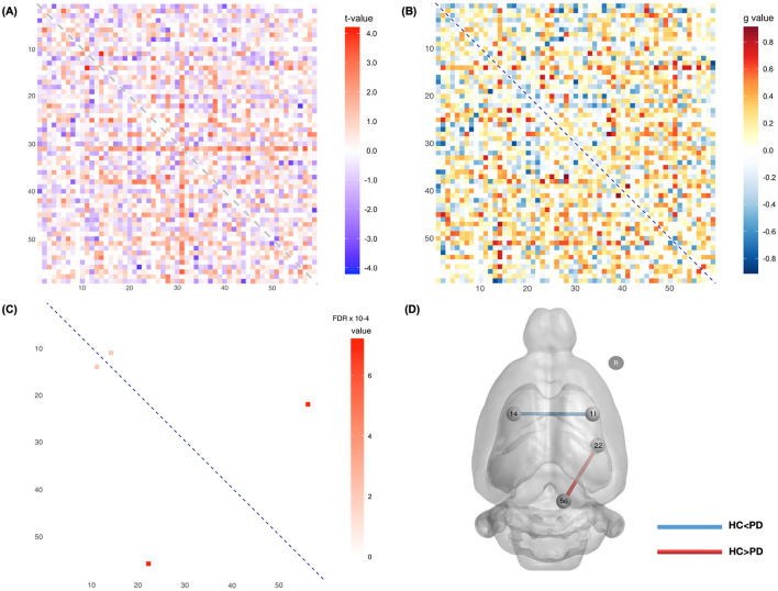
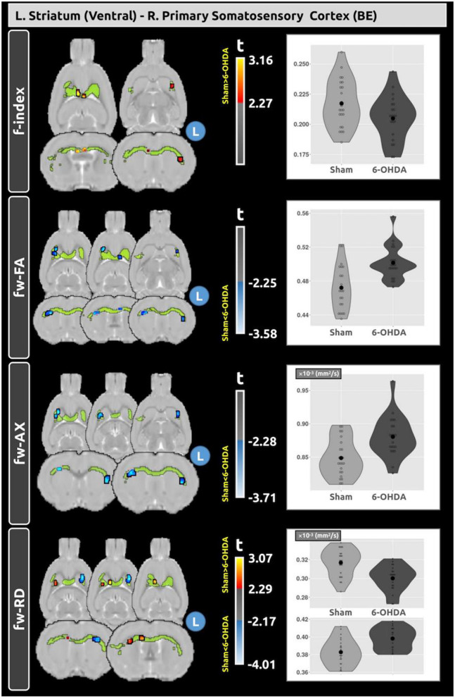
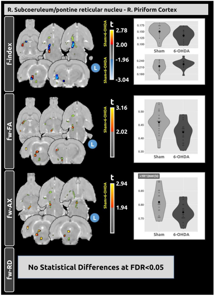
Results: Functionally, decreased connectivity between the retrosplenial and endopiriform cortices in the 6-OHDA model suggests disrupted sensory processing, while increased connectivity between the hippocampus and retrosplenial cortex indicates possible compensatory mechanisms. Structurally, we observed reduced connectivity between the subcoeruleum and piriform cortex in the 6-OHDA model, which may reflect axonal degeneration, and increased connectivity between the ventral striatum and primary somatosensory cortex, which likely reflects compensatory changes to support motor-sensory integration. Diffusion MRI analysis further revealed changes in the white matter tracts connecting these regions, supporting these findings and highlighting adaptive responses to neurodegeneration in PD.

Discussion: These findings demonstrate the utility of combining functional and structural connectivity analyses to capture PD-related network disruptions. These structural connectivity changes were further associated with microstructural alterations. The development of MRI biomarkers for understanding brain connectivity may enhance our understanding of PD pathology and advancing translation of these techniques to clinical applications.

Keywords: 6-hydroxydopamine; Parkinson's disease; diffusion magnetic resonance imaging; free water diffusion tensor imaging; functional connectivity; functional magnetic resonance imaging; structural connectivity.


Conflict of interest statement

The authors declare that the research was conducted in the absence of any commercial or financial relationships that could be construed as a potential conflict of interest.

Figures

Figure 1
Figure 1
Motor impairment and TH+ cell density in 6-OHDA and sham groups. (A) Contralateral forelimb use was significantly reduced in the 6-OHDA group compared to the sham group (p < 0.001, quantified in Table 2). (B) L/R ratio showed a marked reduction in the 6-OHDA group vs. the sham group (p < 0.001, quantified in Table 2). ***Indicates p < 0.001.
Figure 2
Figure 2
Functional connectivity between the sham and 6-OHDA groups. The left hemisphere (L) is defined as the lesion side. (A) Matrix displaying t-values across connections, where blue denotes reduced connectivity and red denotes enhanced connectivity in 6-OHDA rats. (B) Matrix illustrating corresponding effect size across connections. (C) Matrix displaying connections with significant FDR-corrected p-values (p < 0.05). (D) Visualization of the spatial locations of these significant connectivity differences. Specifically, compared to the sham group, a significant increase in functional connectivity was found between the ipsilateral left cornu ammonis and retrosplenial granular cortex in 6-OHDA rats, while a significant decrease was observed between the ipsilateral left retrosplenial cortex and endopiriform cortex. The statistical details for these differences are presented in Table 3. R, right hemisphere.
Figure 3
Figure 3
Structural connectivity between the sham and 6-OHDA groups. (A) Matrix displaying t-values across connections, where blue denotes reduced connectivity and red denotes enhanced connectivity in 6-OHDA rats. (B) Matrix illustrating the variability in effect size across connections. (C) Matrix displaying connections with significant FDR-corrected p-values (p < 0.001). (D) Provides a visualization of the spatial locations of these significant connectivity differences. Specifically, compared to the sham rats, a significant increase in SC in the 6-OHDA rats was found between the left ventral striatum and the right primary somatosensory cortex, while a significant decrease in SC was found between the right subcoeruleum/pontine reticular nucleus and the right piriform cortex. The statistical details for these differences are presented in Table 4. R, right hemisphere.
Figure 4
Figure 4
Voxel-based analysis reveals differences in fw-DTI metrics within the tract connecting the left ventral striatum and the right primary somatosensory cortex between the sham and 6-OHDA groups. A significant increase in structural connectivity was observed in the 6-OHDA group compared to the sham group (t = 4.068; FDR < 0.001). Within this tract, distinct clusters show lower f-index and increased fw-FA and fw-AxD in the 6-OHDA group. Plots depict the average values of the fw diffusion metrics within these significant clusters for both groups.
Figure 5
Figure 5
Voxel-based analysis reveals differences in fw-DTI metrics within the tract connecting the right subcoeruleus/pontine reticular nucleus and the right piriform cortex between the sham and 6-OHDA groups. A significant decrease in structural connectivity was observed in the 6-OHDA group compared with the sham group (t = −3.656; FDR < 0.001). Within this tract, distinct clusters of differences in f-index, fw-FA, and fw-AxD were observed between the groups. No statistical differences were found for fw-RD. Plots depict the average values of the fw-DTI metrics within these significant clusters for both groups.

Similar articles

References

    1. Andersson J. L. R., Sotiropoulos S. N. (2016). An integrated approach to correction for off-resonance effects and subject movement in diffusion MR imaging. Neuroimage 125, 1063–1078. 10.1016/j.neuroimage.2015.10.019 - DOI - PMC - PubMed
    1. Arribarat G., Pasternak O., Barros A. D., Galitzky M., Rascol O., Péran P., et al. (2019). Substantia nigra locations of iron-content, free-water and mean diffusivity abnormalities in moderate stage Parkinson's disease. Parkinsonism Relat. Disord. 65, 146–152. 10.1016/j.parkreldis.2019.05.033 - DOI - PubMed
    1. Avants B. B., Epstein C. L., Grossman M., Gee J. C. (2008). Symmetric diffeomorphic image registration with cross-correlation: evaluating automated labeling of elderly and neurodegenerative brain. Med. Image Anal. 12, 26–41. 10.1016/j.media.2007.06.004 - DOI - PMC - PubMed
    1. Avants B. B., Tustison N. J., Song G., Cook P. A., Klein A., Gee J. C., et al. (2011). A reproducible evaluation of ANTs similarity metric performance in brain image registration. Neuroimage 54, 2033–2044. 10.1016/j.neuroimage.2010.09.025 - DOI - PMC - PubMed
    1. Barbagallo G., Caligiuri M. E., Arabia G., Cherubini A., Lupo A., Nisticò R., et al. (2017). Structural connectivity differences in motor network between tremor-dominant and nontremor Parkinson's disease. Hum. Brain Mapp. 38, 4716–4729. 10.1002/hbm.23697 - DOI - PMC - PubMed

LinkOut - more resources