Review
. 2025 Jun 3:15:1563061.
doi: 10.3389/fcimb.2025.1563061. eCollection 2025.

Microbiota and gastric cancer: from molecular mechanisms to therapeutic strategies

Affiliations
Review

Microbiota and gastric cancer: from molecular mechanisms to therapeutic strategies

Zhou Chen et al. Front Cell Infect Microbiol. .

Abstract

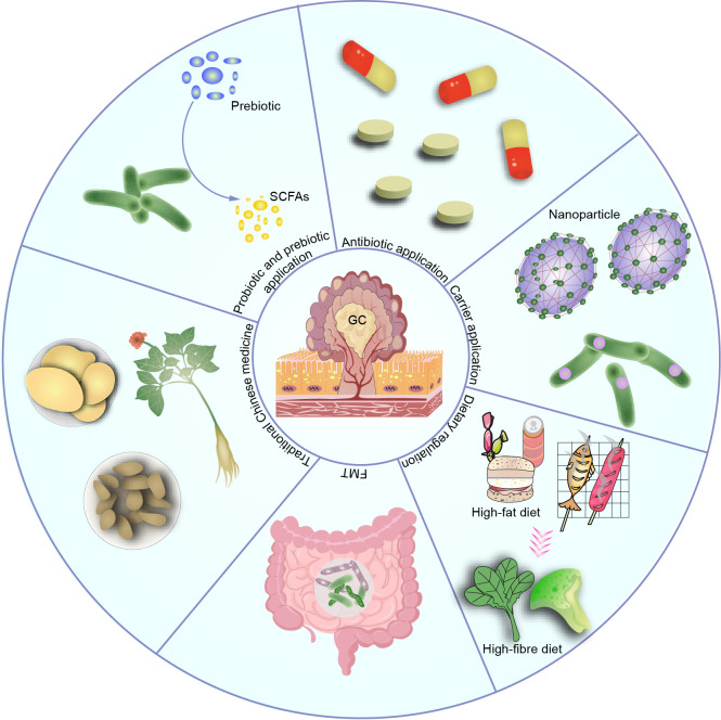
Gastric cancer, a prevalent malignancy globally, is influenced by various factors. The imbalance in the gut microbiome and the existence of particular intratumoural microbiota could have a strong connection with the onset and progression of gastric cancer. High-throughput sequencing technology and bioinformatics analysis have revealed a close correlation between abnormal abundance of specific microbial communities and the risk of gastric cancer. These microbial communities contribute to gastric cancer progression through mechanisms including increasing cellular genomic damage, inhibiting DNA repair, activating abnormal signaling pathways, exacerbating tumor hypoxia, and shaping a tumor immune-suppressive microenvironment. This significantly impacts the efficacy of gastric cancer treatments, including chemotherapy and immunotherapy. Probiotic, prebiotic, antibiotic, carrier-based, dietary interventions, fecal microbiota transplantation, and traditional Chinese medicine show potential applications in gastric cancer treatment. However, the molecular mechanisms regarding dysbiosis of microbiota, including gut microbiota, and intra-tumoral microbiota during the progression of gastric cancer, as well as the therapeutic efficacy of microbiota-related applications, still require extensive exploration through experiments.

Keywords: gastric cancer; microbiota; molecular mechanisms; targeted therapy; tumor microenvironment.


Conflict of interest statement

The authors declare that the research was conducted in the absence of any commercial or financial relationships that could be construed as a potential conflict of interest.

Figures

Figure 1
Figure 1
Microbial adhesion and invasion of gastric epithelial cells. H. pylori can bind to gastric epithelial cells via adhesin HopQ, glycan-modified proteins AlpA/B and BabA/B. H. pylori directly injects a potent virulence protein CagA into epithelial cells via the T4SS. F. nucleatum adheres to gastric epithelial cells through the pre-FadA-mFadA complex and Fap2 galactose-binding lectin, ensuring bacterial invasion of host cells. F. nucleatum produces high levels of H2S, increasing DNA damage. Once internalized by host cells, E. coli secretes genotoxin colibactin, inducing DNA double-strand breaks.
Figure 2
Figure 2
The microbiota increases host cell genomic damage and suppresses genome repair. Primary bile acids, nitrate, and proteins metabolized by certain microbial communities produce substances such as DCA, N-nitrosamines, and heterocyclic amines, leading to DNA damage. F. nucleatum increases the expression of miR-205-5p through the TLR4 and MyD88-dependent innate immune signaling pathway, suppressing the expression of MLH1, MSH2, and MSH6. H. pylori upregulates miR-150-5p, miR-155-5p, and miR-3163 to suppress the expression of MSH2 and MSH3 proteins.
Figure 3
Figure 3
Microbiota dysbiosis alters gastric epithelial cell signaling pathways. H. pylori promotes nuclear accumulation and transcriptional activity of YAP and β-catenin in gastric epithelial cells, leading to activation of target genes CDX2, LGR5, and RUVBL1, facilitating cell proliferation and expansion, ultimately resulting in GC development. H. pylori also induces the expression of IL-11 and cancer-related genes Ptger4 and TGF-β. H. pylori enhances autophagy gene ATG16L1, increasing IL-8 production, driving carcinogenesis. H. pylori induces the expression of G6PD and D-LDH in host cells, facilitating glycolysis, and energy production. F. nucleatum upregulates transcription factor SP1, activates lncRNA ENO1-IT1 transcription, guides KAT7 histone acetyltransferase to modify target gene ENO1, increasing host cell glycolysis. δ-valerobetaine produced by various bacteria inhibits mitochondrial FAO and increases lipid accumulation via transcription factor PPAR-α. SCFAs serve as substrates for lipid synthesis. Additionally, the microbiota can induce sustained inflammatory responses, generate ROS, causing DNA fragmentation, membrane disintegration, and protein misfolding through modification of key substrates such as nucleic acids, lipids, and proproteins, leading to cellular senescence, secretion of SASPs, and accelerated tumor growth. EBV infection can activate the cGAS-STING pathway and upregulate the expression of OLFM4, thereby leading to the activation of YAP in recipient cells.
Figure 4
Figure 4
Microbiota shapes the suppressive immune microenvironment. H. pylori induces expression of the NKG2D ligand in gastric epithelial cells, which is released from the cell surface via protein hydrolysis or extracellular vesicles, leading to decreased expression of the NKG2D receptor on NK cells and cytotoxic granule degranulation, thereby facilitating immune evasion by tumor cells. P. acnes activates the TLR4/PI3K/Akt signaling pathway, inducing M2 TAM polarization, promoting secretion of immunosuppressive factors IL-10 and CCR-2. Butyrate, a derivative of probiotics, negatively regulates the NLRP3-mediated inflammatory signaling pathway, inhibits related macrophage activation, and decreases levels of PD-L1 and IL-10 expression, thereby suppressing tumor growth in mice. H. pylori activates TLR9, promotes expression of negative feedback regulator TRIM30a, downregulates activation of transcription factor IRF3, inhibits the STING signaling pathway, and promotes Th17 inflammatory responses and anti-tumor responses in vivo. H. pylori drives activation of pro-inflammatory T cells, secretes IL-21, induces phosphorylation of STAT3, and induces expression of RORγ-t, promoting Th17 differentiation and IL-17 secretion. H. pylori activates dendritic cells via the TLR2/NLRP3/caspase-1/IL-18 axis to induce Tregs, shaping the immune suppressive microenvironment. H. pylori and Methylobacterium can reduce expression of TGF-β and CD8+ T cell infiltration in a GC mouse model, but their mechanisms remain to be elucidated.
Figure 5
Figure 5
Therapeutic applications based on the microbiota, such as probiotic, prebiotic, antibiotic use, carrier application, dietary modulation, and traditional Chinese medicine, have shown promising efficacy. However, most of these applications are still in the preclinical stage, and their clinical efficacy and potential complications remain to be determined.

Similar articles

References

    1. Abed J., Emgård J. E. M., Zamir G., Faroja M., Almogy G., Grenov A., et al. (2016). Fap2 mediates fusobacterium nucleatum colorectal adenocarcinoma enrichment by binding to tumor-expressed gal-galNAc. Cell Host Microbe 20, 215–225. doi: 10.1016/j.chom.2016.07.006 - DOI - PMC - PubMed
    1. Akira S., Takeda K. (2004). Toll-like receptor signalling. Nat. Rev. Immunol. 4, 499–511. doi: 10.1038/nri1391 - DOI - PubMed
    1. Anthofer M., Windisch M., Haller R., Ehmann S., Wrighton S., Miller M., et al. (2024). Immune evasion by proteolytic shedding of natural killer group 2, member D ligands in Helicobacter pylori infection. Front. In Immunol. 15, 1282680. doi: 10.3389/fimmu.2024.1282680 - DOI - PMC - PubMed
    1. Arita S., Inagaki-Ohara K. (2019). High-fat-diet-induced modulations of leptin signaling and gastric microbiota drive precancerous lesions in the stomach. Nutr. (Burbank Los Angeles County Calif.) 67-68, 110556. doi: 10.1016/j.nut.2019.110556 - DOI - PubMed
    1. Arita S., Ogawa T., Murakami Y., Kinoshita Y., Okazaki M., Inagaki-Ohara K. (2019). Dietary fat-accelerating leptin signaling promotes protumorigenic gastric environment in mice. Nutrients 11, 2127. doi: 10.3390/nu11092127 - DOI - PMC - PubMed

MeSH terms

LinkOut - more resources