. 2025 Jun 3:16:1585953.
doi: 10.3389/fimmu.2025.1585953. eCollection 2025.

Phosphatidylcholine-specific B cells are enriched among atypical CD11chigh and CD21low memory B cells in antiphospholipid syndrome

Affiliations

Phosphatidylcholine-specific B cells are enriched among atypical CD11chigh and CD21low memory B cells in antiphospholipid syndrome

Eduard Nitschke et al. Front Immunol. .

Abstract

Background: Patients with antiphospholipid syndrome (APS) carry an increased risk of thrombosis and adverse pregnancy outcomes due to the presence of antiphospholipid autoantibodies (aPL). However, the pathogenesis of the disease remains incompletely understood regarding various aPL and the role of autoreactive B cells as precursors of antibody-secreting plasma cells (PC).

Objective: To assess B-cell dysregulation in APS, with a focus on the distribution of B cell subsets and phosphatidylcholine (PtC)-specific cells.

Methods: We used flow cytometry to study B cell subsets in peripheral blood mononuclear cells (PBMCs) from 20 healthy controls (HCs), 21 patients with primary APS (pAPS), and 16 patients with secondary APS (sAPS). A novel fluorescent liposome-based method was used to identify PtC-specific B cells in these subsets. Data were analyzed using manual gating and unsupervised clustering. We quantified aPtC antibody serum levels using ELISA and conducted correlation analyses between PtC-specific B cell subsets and aPL titers.

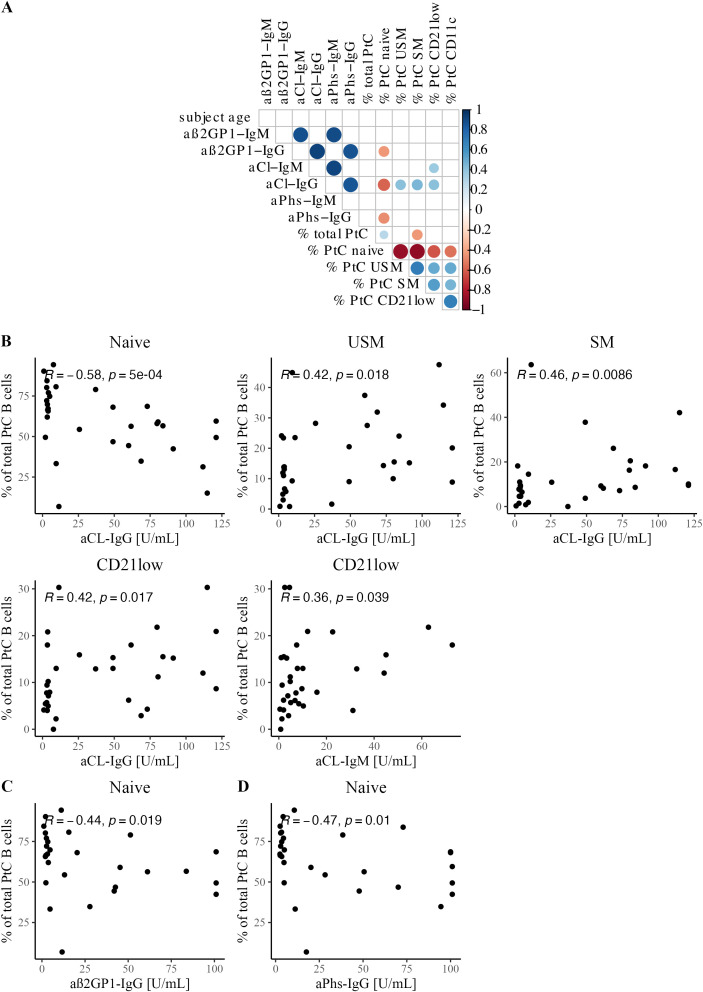
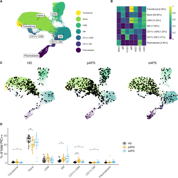
Results: Patients with pAPS and sAPS exhibited significantly increased frequencies of atypical CD21low and CD11chigh B cells, including PtC-specific B cells. Notably, both total and unswitched memory PtC-specific B cells were elevated in pAPS patients and correlated with aPL antibody titers. Unsupervised clustering further highlighted the increased frequencies of PtC-specific CD21lowCD11chigh unswitched and switched memory B cells in both pAPS and sAPS.

Conclusion: The enrichment of PtC-specific B cells among CD21low and CD11chigh atypical memory subsets, along with their correlation with aPL serum levels, suggest a linkage between these atypical memory B cell subsets and autoantibody producing cells in APS.

Keywords: APS - antiphospholipid syndrome; B cells; adaptive immunity; anti-phospholipid antibodies; antigen-specific B cells; primary APS; secondary APS.


Conflict of interest statement

The authors declare that the research was conducted in the absence of any commercial or financial relationships that could be construed as a potential conflict of interest.

Figures

Figure 1
Figure 1
Increased atypical B Cell Frequencies in APS. (A) Assessment of absolute counts for CD19+ B cells and CD4+/CD8+ T cells in HCs, pAPS, and sAPS. (B) B cell subset distribution in APS: categorization according to surface marker expression into transitional (CD24intCD38high), naïve (IgD+CD27-), USM (IgD+CD27+), SM (IgD-CD27+), DN (IgD-CD27-) or plasmablasts (CD27highCD38high). A complete gating strategy is provided in Supplementary Figure 1. (C) Quantification of atypical CD21low and CD11chigh B cells among the cohorts. APS, antiphospholipid syndrome; pAPS, primary APS; sAPS, secondary APS; HC, healthy control; USM, unswitched memory; SM, switched memory; DN, double negative. Statistics: Wilcoxon rank-sum test | reference group: HC | *p<0.05, **p<0.01.
Figure 2
Figure 2
Specific staining and analysis of PtC-Specific B cells in pAPS using PtC liposomes. (A) Overlay of PtC-specific B cells stained with either CF594 labeled PtC liposomes, OG labeled liposomes, non-fluorescent control liposomes or without the addition of any liposomes (FMO). (B) Representative dot plot for the identification of PtC-specific B cells. Only B cells that are positive for both PtC-CF594 and PtC-OG were considered autoantigen-specific. (C) Two representative pseudocolor dot plots of PtC-specific B cells with a complete staining (left) and after blocking of specific binding-sites with non-fluorescent control liposomes (right). (D) Quantification of the relative decrease in PtC-specific B cells in 4 representative samples before- and after blocking with non-fluorescent control liposomes. (E) Frequencies of PtC-specific B cells and their distribution to major B cell subsets. B cell subsets were classified as in Figure 1 . (F) Frequencies of atypical PtC++CD21low and PtC++CD11chigh B cells among the cohorts. PtC, phosphatidylcholine; APS, antiphospholipid syndrome; pAPS, primary APS; sAPS, secondary APS; HC, healthy control; USM, unswitched memory; SM, switched memory; DN, double negative; OG, Oregon-Green. Statistics: Wilcoxon rank-sum testreference group: HC*p<0.05, **p<0.01, ****p<0.0001.
Figure 3
Figure 3
Correlation between PtC-specific B cells and serological features. (A) Correlation matrix shows Spearman correlation coefficients and significant correlations with p<0.05. (B) Scatterplots with Spearman correlation between aCL IgG and/or IgM titer and PtC-specific B cell subsets: naïve, USM, SM, and CD21low. (C) Scatterplot with Spearman correlation for aß2GP1-IgG and naïve PtC-specific B cells. (D) Scatterplot with Spearman correlation for aPhs-IgG and naïve PtC-specific B cells.
Figure 4
Figure 4
B cell clustering analysis in APS. (A, B) UMAP Visualization of B Cells: A representation of 120’000 B cells composed of a random subsample of 40’000 B cells from each group. Different colors represent B cell clusters identified using the FlowSOM algorithm. (B) Surface Marker Heatmap showing the expression of each surface marker within each FlowSOM metacluster. B cell clusters were classified according to certain expression profiles as follows: naïve, USM, SM, CD11c USM, CD11c SM, and plasmablasts. The percentages show the distribution of all B cells used for clustering to different clusters. (C) Individual UMAP graphs: Overlaid black dots represent all PtC-specific B cells within the random 40’000 per group subset. An increase in PtC-specific B cells in pAPS became evident both visually in the UMAP graphs and by cell count data in the subsample of B cells (PtC-specific B cell counts: HD: 347; pAPS: 613; sAPS: 392). (D) Distribution quantification: Analysis of the distribution of PtC-specific B cells to the FlowSOM metaclusters for each subject. UMAP, Uniform Manifold Approximation and Projection; PtC, phosphatidylcholine; PtC++, positive for both phosphatidylcholine liposome colors; APS, antiphospholipid syndrome; pAPS primary APS; sAPS, secondary APS; HC, healthy control; USM, unswitched memory; SM, switched memory; DN, double negative. Statistics: Wilcoxon rank sum test | reference group: HC | p-value adjustment: Bonferroni | *p<0.05, **p<0.01, ***p<0.001.
Figure 5
Figure 5
aPtC IgG, IgM and IgA levels among the groups. PtC, phosphatidylcholine; aPtC, anti-phosphatidylcholine antibodies; APS, antiphospholipid syndrome; pAPS, primary APS; sAPS. secondary APS; HC, healthy control; OD, optical density; ELISA, enzyme-linked immunosorbent assay. Statistics: Wilcoxon rank sum test | reference group: HC | p-value adjustment: Benjamini-Hochberg | *p<0.05, **p<0.01, ****p<0.0001.

Similar articles

References

    1. Tektonidou MG, Andreoli L, Limper M, Amoura Z, Cervera R, Costedoat-Chalumeau N, et al. EULAR recommendations for the management of antiphospholipid syndrome in adults. Ann Rheum Dis Oct. (2019) 78:1296–304. doi: 10.1136/annrheumdis-2019-215213 - DOI - PMC - PubMed
    1. Miyakis S, Lockshin MD, Atsumi T, Branch DW, Brey RL, Cervera R, et al. International consensus statement on an update of the classification criteria for definite antiphospholipid syndrome (APS). J Thromb Haemost. (2006) 4:295–306. doi: 10.1111/j.1538-7836.2006.01753.x - DOI - PubMed
    1. Cervera R, Tektonidou MG, Espinosa G, Cabral AR, González EB, Erkan D, et al. Task Force on Catastrophic Antiphospholipid Syndrome (APS) and Non-criteria APS Manifestations (I): catastrophic APS, APS nephropathy and heart valve lesions. Lupus. (2011) 20:165–73. doi: 10.1177/0961203310395051 - DOI - PubMed
    1. Guédon AF, Catano J, Ricard L, Laurent C, de Moreuil C, Urbanski G, et al. Non-criteria manifestations in primary antiphospholipid syndrome: a French multicenter retrospective cohort study. Arthritis Res Ther. (2022) 24:33. doi: 10.1186/s13075-022-02726-9 - DOI - PMC - PubMed
    1. Barbhaiya M, Zuily S, Naden R, Hendry A, Manneville F, Amigo MC, et al. The 2023 ACR/EULAR antiphospholipid syndrome classification criteria. Arthritis Rheumatol. (2023) 75:1687–702. doi: 10.1002/art.42624 - DOI - PubMed

Substances

LinkOut - more resources