. 2025 May 29:9:100344.
doi: 10.1016/j.ijpx.2025.100344. eCollection 2025 Jun.

Neutrophil-Mimetic oleanolic acid-loaded Liposomes targeted to alleviate oxidative stress for renal ischemia-reperfusion injury treatment

Affiliations

Neutrophil-Mimetic oleanolic acid-loaded Liposomes targeted to alleviate oxidative stress for renal ischemia-reperfusion injury treatment

Qiaojing Chen et al. Int J Pharm X. .

Abstract

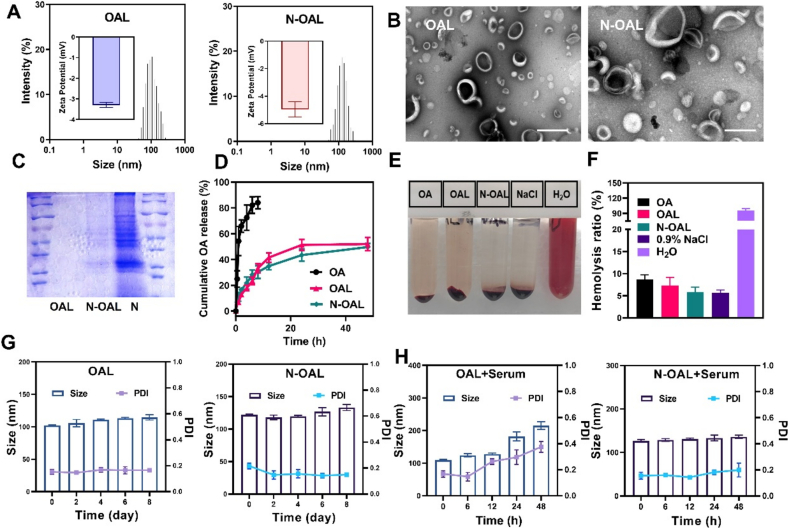
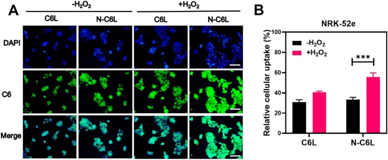
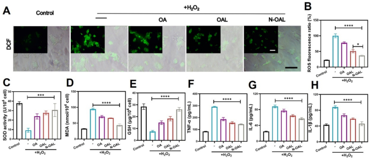
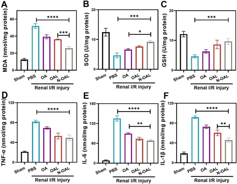
Acute kidney injury (AKI) is a prevalent clinical condition characterized by a sudden decline or loss of renal function, exacerbated by the lack of effective diagnostic and therapeutic tools. Renal ischemia-reperfusion injury serves as the primary cause of AKI, initiating a complex signaling cascade that mediates renal cell necrosis, apoptosis, and inflammation. Oxidative stress plays a crucial role in the pathogenesis and progression of ischemia-reperfusion injury, thus prompting the exploration of antioxidants as potential therapeutic interventions. Oleanolic acid, derived from natural plant extracts, exhibits significant antioxidant and anti-inflammatory properties; however, its clinical application has been hindered by inherent limitations such as poor water solubility and low bioavailability. To address this issue, we developed an innovative approach involving oleanolic acid-loaded liposomes fused with neutrophil membranes, resulting in hybrid liposomes (N-OAL). This strategy aims to enhance the accumulation and retention of N-OAL at inflammatory sites associated with AKI through biomimetic chemotaxis mediated by neutrophil membranes specifically targeting damaged renal tubular epithelial cells. The optimized N-OAL presented a spherical morphology with an average particle size of 125.6 ± 4.9 nm and a surface potential of -4.8 ± 0.3 mV. In addition, N-OAL exhibited favorable sustained release, outstanding stability, and satisfactory biocompatibility. In vitro studies demonstrated that N-OAL effectively attenuated H2O2-induced intracellular reactive oxygen species generation and inflammation while exhibiting superior antioxidant and anti-apoptotic properties. Furthermore, our in vivo results confirmed the remarkable protective effect of N-OAL on oxidative-damaged renal tissue caused by AKI induction. Overall, our study provides novel insights into targeted delivery strategies for oleanolic acid therapy in acute kidney injury.

Keywords: Ischemia/reperfusion injury; Liposome; Neutrophil-mimetic; Oleanolic acid; Oxidative stress.


Conflict of interest statement

The authors declare that they have no known competing financial interests or personal relationships that could have appeared to influence the work reported in this paper.

Figures

Unlabelled Image
Graphical abstract
Fig. 1
Fig. 1
Schematic illustration of N-OAL for AKI management. (A) The synthetic scheme of oleanolic acid neutrophil membrane-hybrid liposome (N-OAL). (B) Schematic illustration of N-OAL targeting renal tubular epithelial cells in ischemia-reperfusion (IR) induced acute kidney injury (AKI) in mice via systemic administration.
Fig. 2
Fig. 2
Characterization of OAL and N-OAL. (A) DLS data and zeta potential for OAL (left) and N-OAL (right). (B) TEM images of OAL (left) and N-OAL (right). Scale bars = 100 nm. (C) SDS-PAGE image of neutrophils, OAL and N-OAL. (D) Cumulative OA release profiles from OAL and N-OAL. (E) Hemolysis analysis of OA, OAL and N-OAL. (F) Relative hemolysis ratio. (G) The variations in size and PDI of OAL (left) and N-OAL (right) in pH 7.4 PBS at 4 °C for a week. (H) The variations in size and PDI of OAL (left) and N-OAL (right) after incubation with culture medium (FBS, 10 %, v/v) at 37 °C for 48 h. Data represent mean ± SD (n = 3).
Fig. 3
Fig. 3
Cellular uptake profiles of N-OAL. (A) Cell uptake efficiency of C6L and N-C6L in untreated or H2O2-treated NRK-52e cells. Scale bar = 100 μm. (B) Mean fluorescence intensity. Data represent mean ± SD. n = 3, ∗∗∗P < 0.001.
Fig. 4
Fig. 4
In vitro antioxidative and anti-inflammatory properties of N-OAL. (A) The levels of cellular ROS measured by DCF staining. Scale bar = 100 μm. (B) Quantitative analysis of intracellular ROS levels. The levels of (C) SOD, (D) MDA, and (E) GSH after various treatments for 24 h. The levels of (F) TNF-α, (G) IL-6, and (H) IL-1β after various treatment for 24 h. Data represent mean ± SD. n = 3, ∗P < 0.05, ∗∗∗P < 0.001, ∗∗∗∗P < 0.0001.
Fig. 5
Fig. 5
In vitro MMP stabilization and inhibition effect against apoptosis of N-OAL. (A) Fluorescence images of JC-1 assay to measure mitochondrial membrane depolarization in H2O2-stimulated cells after various treatments. Scale bar = 100 μm. (B) Quantitative analysis of mitochondrial membrane potential. (C) The apoptosis of NRK-52e cells with N-OAL treatment by flow cytometry after stained with PI and Annexin V-FITC. (D) Statistical diagram of apoptosis. Data represent mean ± SD. n = 3, ∗∗P < 0.01, ∗∗∗P < 0.001.
Fig. 6
Fig. 6
Renal distribution of DiRL and N-DiRL was examined via imaging system. (A) Overview of the in vivo biodistribution experimental design. (B) Representative ex vivo fluorescence images of main organs resected from AKI mice with intravenous injection of DiR, DiRL, and N-DiRL for 10 h. (C) Relative fluorescence intensity of the organs (n = 3).
Fig. 7
Fig. 7
N-OAL attenuated renal I/R injury in AKI mice. (A) Experimental procedure for the treatment of I/R AKI mice. (B) Representative H&E staining images of kidney sections from different groups including sham operated mice, I/R-injured mice, I/R + OA, I/R + OAL, and I/R + N-OAL. (C) Tunel assay indicating apoptosis cells in mice with various treatments. (D) Fluorescence images of 4-HNE (green)- and Hoechst (blue)-stained kidney tissues in I/R AKI mice. The quantitative analysis of (E) Tunel and (F) 4-HNE staining. Scale bar =100 μm. Data represent mean ± SD. n = 3, ∗∗P < 0.01, ∗∗∗P < 0.001, ∗∗∗∗P < 0.0001. (For interpretation of the references to colour in this figure legend, the reader is referred to the web version of this article.)
Fig. 8
Fig. 8
N-OAL alleviated oxidative stress and suppressed inflammation in renal I/R injury. The levels of (A) MDA, (B) SOD, and (C) GSH were determined in I/R injured kidney. Protein levels of (D) TNF-α, (E) IL-6, and (F) IL-1β in the kidney were determined by ELISA. Data represent mean ± SD. n = 5, ∗P < 0.05, ∗∗P < 0.01, ∗∗∗P < 0.001, ∗∗∗∗P < 0.0001.

References

    1. Asati A., Santra S., Kaittanis C., Perez J.M. Surface-charge-dependent cell localization and cytotoxicity of cerium oxide nanoparticles. ACS Nano. 2010;4:5321–5331. - PMC - PubMed
    1. Castaneda M.P., Swiatecka-Urban A., Mitsnefes M.M., Feuerstein D., Kaskel F.J., Tellis V., Devarajan P. Activation of mitochondrial apoptotic pathways in human renal allografts after ischemiareperfusion injury. Transplantation. 2003;76:50–54. - PubMed
    1. Chen S., Li Q., Shi H., Li F., Duan Y., Guo Q. New insights into the role of mitochondrial dynamics in oxidative stress-induced diseases. Biomed. Pharmacother. 2024;178 - PubMed
    1. Chen Y., Zhu M., Huang B., Jiang Y., Su J. Advances in cell membrane-coated nanoparticles and their applications for bone therapy. Biomaterials Advances. 2023;144 - PubMed
    1. Duni A., Liakopoulos V., Roumeliotis S., Peschos D., Dounousi E. Oxidative stress in the Pathogenesis and Evolution of Chronic Kidney Disease: Untangling Ariadne’s Thread. Int. J. Mol. Sci. 2019;20 - PMC - PubMed

LinkOut - more resources