. 2025 Jun 2:12:1580684.
doi: 10.3389/fnut.2025.1580684. eCollection 2025.

Assessing the efficacy of the natural disaccharide trehalose in ameliorating diet-induced obesity and metabolic dysfunction

Affiliations

Assessing the efficacy of the natural disaccharide trehalose in ameliorating diet-induced obesity and metabolic dysfunction

Yu-Sheng Yeh et al. Front Nutr. .

Abstract

Trehalose is a naturally occurring disaccharide with versatile commercial applications and health benefits, including promise as a therapeutic for obesity and diabetes. Although numerous previous reports purport the therapeutic uses of orally ingested trehalose, the abundance of glycosidases in the gastrointestinal tract suggest the potential for significant limitations of oral trehalose that have not been addressed. We first fed mice a high-fat diet (HFD) while providing trehalose by both oral and intraperitoneal routes. This combined strategy was broadly efficacious in reversing HFD-induced weight gain, fat mass, insulin resistance, and the development of hepatosteatosis. In contrast, oral-only trehalose failed to improve HFD-induced obesity and insulin resistance. This was due to trehalase (Treh)-mediated metabolism as blood trehalose levels remained low despite a significant rise in glucose. We next developed systemically deficient Trehalase (Treh-KO) mice to enhance the efficacy of trehalose. Surprisingly, oral trehalose therapy could not be facilitated resulting in neither an increase in serum trehalose levels nor metabolic benefits. Parenteral trehalose resulted in higher trehalose levels with lower serum glucose in Treh-KO mice, yet no additive metabolic benefits were observed. Overall, our findings still support a therapeutic role for trehalose in obesity and metabolic disease but with practical limitations in its delivery by oral route.

Keywords: hepatosteatosis; insulin resistance; obesity; oral vs. parenteral; trehalase; trehalose.


Conflict of interest statement

The authors declare that the research was conducted in the absence of any commercial or financial relationships that could be construed as a potential conflict of interest.

Figures

Figure 1
Figure 1
Combination of oral and intraperitoneal trehalose treatment abrogates high-fat diet-induced obesity. (A) Schematic process of trehalose or sucrose intervention experiment. The mice were treated with saline, sucrose, or trehalose by injecting intraperitoneally (3 times per week) and as a supplement to drinking water. (B) Body weight gain, (C) body composition, and (D) iWAT, eWAT, and BAT weights of mice treated with saline (n = 10), sucrose (n = 15), or trehalose (n = 13) under HFD condition were measured after 16 weeks of HFD feeding. (E) Schematic illustration of trehalose treatment time course experiment. The mice were fed HFD for 16 weeks, during which time mice also received trehalose for 0, 1, 2, or 3 months (0 M, 1 M, 2 M or 3 M) both orally and intraperitoneally. (F) Body weight variation, and (G) Tissue mass of iWAT, eWAT, and liver in the mice after 16 weeks of HFD treatment, along with trehalose added to drinking water for 0 M, 1 M, 2 M, and 3 M (n = 4). All mice were male and fed HFD. Values are presented as mean ± SE. Significant differences were determined by Student’s t-test, with comparison to Saline or 0 M group: *p < 0.05.
Figure 2
Figure 2
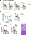
Combination of oral and intraperitoneal trehalose treatment improved high-fat diet-induced insulin resistance and fatty liver. (A) Serum glucose, triglyceride, free fatty acid, and cholesterol levels of mice receiving saline (n = 10), sucrose (n = 15), or trehalose (n = 13) were measured at 16 weeks of HFD feeding. (B) Insulin tolerance test was performed at 15 weeks of HFD-fed mice treated with saline (n = 10), sucrose (n = 15), or trehalose (n = 13). Results were shown as a time-dependent graph (left panel) or as the area (%*min) under the curve (right panel). (C) Liver mass, (D) triglyceride content, and (E) histological analysis with H&E staining were performed. Liver samples from 16 weeks HFD-fed mice treated with saline (n = 10), sucrose (n = 15), or trehalose (n = 13). Scale bars, 200 μm. All mice were male and fed HFD. Values are presented as mean ± SE. Significant differences were determined by Student’s t-test, as compared with Saline group: *p < 0.05.
Figure 3
Figure 3
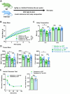
Oral trehalose is therapeutically ineffective. (A) Schematic representation of trehalose or vehicle intervention experiment. Treatments were provided as supplementation to drinking water. (B) Body weights were measured in mice provided drinking water with or without 3% w/v trehalose (n = 15). (C) Body composition, and (D) tissue weights of iWAT, eWAT, and liver were measured after 16 weeks of HFD feeding with or without 3% w/v trehalose as a supplement in drinking water (n = 15). (E) Insulin tolerance test was performed at 15 weeks HFD-fed mice treated with or without 3% w/v trehalose, provided as a supplement in drinking water (n = 15). Results were shown as a time-dependent graph (left panel) or as the area (%*min) under the curve (right panel). All mice were male and fed HFD. Values are presented as mean ± SE.
Figure 4
Figure 4
Oral trehalose treatment enhanced blood glucose level but only show a minor effect on serum trehalose. (A) Trehalose tolerance test was performed based on oral (Oral) and intraperitoneal (IP) treatment (3 g/Kg body weight) and the serum trehalose level was measured at indicated time points (n = 4). (B) Schematic image of the metabolism of trehalose through trehalose (Treh) to glucose. (C) Glucose and trehalose levels were measured in serum and portal circulation 30 min after oral trehalose treatment (n = 3). (D) Tissue mRNA expression and (E) activity of Treh were measured in the duodenum, kidney, colon, liver, and quadriceps of 8-week-old male C57BL/6 J mice (n = 4). (F) Trehalose tolerance test was performed based on oral (Oral) and intraperitoneal (IP) treatment (3 g/Kg body weight) and the serum glucose level was measured at indicated time points (n = 4). All mice were male and fed a normal chow diet. Values are presented as mean ± SE. Significant differences were determined by Student’s t-test compared with IP group: *p < 0.05, **p < 0.01, ***p < 0.001.
Figure 5
Figure 5
Systemic trehalase-deficient mice showed no metabolic changes at baseline. (A) Indicated dose of validamycin (Trehehalase inhibitor) was used for mice treated with trehalose (3 g/Kg body weight), after 6 h fasting. Blood glucose levels were measured at indicated time points (left panel), and area under the curve (%*min) was calculated (right panel) (n = 3). (B) Illustration of strategy used to establish a systemic trehalase (Treh) knockout (Treh KO) mouse model. Treh DNA genotyping was performed on mouse tail clippings from heterozygous (Het), wildtype (WT) and Treh KO mice (Embedded panel). (C) mRNA and (D) protein expression levels of Treh in the kidney and duodenum of 8-week-old WT and Treh KO male mice (n = 3). (E) Body weight, and (F) serum glucose, triglyceride, free fatty acid, and cholesterol levels were measured in 8-week-old female WT (n = 9) and Treh KO (n = 6) mice. All mice were fed a normal chow diet. Values are presented as mean ± SE. Significant differences were determined by Student’s t-test in comparison with vehicle or WT group: *p < 0.05, **p < 0.01.
Figure 6
Figure 6
Systemic trehalase deficiency delayed serum trehalose clearance but did not improve trehalose oral absorption. (A) Schematic illustration of the experimental process employed. WT and Treh KO mice were tested for trehalose tolerance test after oral or i.p. trehalose administration. Serum glucose levels were quantified at indicated time points (left panel) after (B) i.p. (WT and Treh KO, n = 3 and 5) or (C) oral (n = 4) trehalose treatment (3 g/Kg body weight) and area under the curve calculated (mg/dL*min, right panel). Serum trehalose levels were quantified at indicated time points (left panel) after (D) i.p. (WT and Treh KO, n = 3 and 5) or (E) oral (n = 4) trehalose treatment (3 g/Kg body weight), and area under the curve calculated (mg/dL*min, right panel). All mice were male and fed a normal chow diet. Values are presented as mean ± SE. Significant differences were determined by Student’s t-test, as compared with WT group: *p < 0.05, **p < 0.01.
Figure 7
Figure 7
I.P. trehalose alone effectively reduced obesity and trehalase deficiency did not improve efficacy. (A) Schematic representation of trehalose or vehicle intervention experiment. WT and Treh KO mice were treated with vehicle or trehalose by i.p. injection. (B) Body weight, (C) body composition, and (D) tissue mass of iWAT (left panel), eWAT (middle panel), and liver (right panel) were measured in WT and Treh KO mice fed a HFD and treated by i.p. injection of trehalose or vehicle (3 g/Kg body weight, 3 times per week) for 16 weeks (Vehicle i.p. WT, Trehalose i.p. WT, Vehicle i.p. Treh KO, and Trehalose i.p. Treh KO, n = 13, 14, 11, and 18). (E) Insulin tolerance test was performed in WT (n = 4) and Treh KO (n = 5) mice i.p. injected with trehalose (3 g/Kg body weight, 3 times per week) for 15 weeks. Blood glucose level were measured at indicated time points (left panel), and area under the curve was calculated (mg/dL*min, right panel). All mice were male and fed HFD. Values are presented as mean ± SE. Significant differences were determined by Student’s t-test compared with indicated groups: *p < 0.05.

References

    1. Rakhra V. Obesity and the Western diet: how we got Here. Mo Med. (2020) 117:536–8. - PMC - PubMed
    1. Gonzalez-Muniesa P. Obesity. Nat Rev Dis Primers. (2017) 3:17034. doi: 10.1038/nrdp.2017.34, PMID: - DOI - PubMed
    1. Kahn CR, Wang G, Lee KY. Altered adipose tissue and adipocyte function in the pathogenesis of metabolic syndrome. J Clin Invest. (2019) 129:3990–4000. doi: 10.1172/JCI129187, PMID: - DOI - PMC - PubMed
    1. Ahima RS, Lazar MA. Adipokines and the peripheral and neural control of energy balance. Mol Endocrinol. (2008) 22:1023–31. doi: 10.1210/me.2007-0529, PMID: - DOI - PMC - PubMed
    1. Srivastava G, Apovian CM. Current pharmacotherapy for obesity. Nat Rev Endocrinol. (2018) 14:12–24. doi: 10.1038/nrendo.2017.122, PMID: - DOI - PubMed

LinkOut - more resources