. 2025 Jun 3:16:1566622.
doi: 10.3389/fphar.2025.1566622. eCollection 2025.

Trapidil attenuates diabetic cardiomyopathy via GPX3/Nrf2-mediated inhibition of myocardial pyroptosis

Affiliations

Trapidil attenuates diabetic cardiomyopathy via GPX3/Nrf2-mediated inhibition of myocardial pyroptosis

Zihao Wang et al. Front Pharmacol. .

Abstract

Background: Currently, there is a paucity of clinically effective medications for the treatment of diabetic cardiomyopathy (DCM), while the strategy of drug repurposing offers a promising avenue for advancing therapeutic development.

Methods: The investigation explored the ameliorative effects and uncovered underlying mechanisms of trapidil (TRA), a drug commonly employed in the management of coronary heart disease, on DCM by inhibiting myocardial pyroptosis. Type 1 DCM models were established utilizing C57BL/6 mice and primary neonatal mouse cardiomyocytes (NMCMs), which were subsequently treated with TRA.

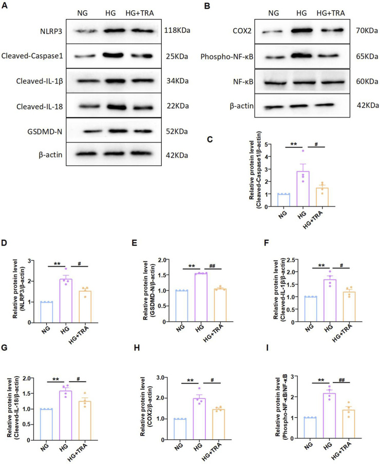
Results: Results demonstrated that in DCM mice, TRA significantly enhanced cardiac function, effectively alleviated pathological changes in myocardial tissue, reversed ultrastructural alterations, and reduced pyroptosome formation in myocardial cells. TRA significantly increased the body weight of the mice in the DCM model group, whereas there was no significant alteration in blood glucose levels following TRA treatment. In the myocardial tissue of DCM mice and high-glucose (HG)-treated NMCMs, TRA was found to correct the aberrant expression of key proteins involved in pyroptosis, including cleaved-caspase1, NLRP3, phospho-NF-κB cyclooxygenase-2, interleukin Cleaved-IL-1β, Cleaved-IL-18, and gasdermin D. Furthermore, TRA effectively curtailed the excessive production of ROS and augmented the mitochondrial membrane potential in NMCMs under the HG environment. Proteomics analysis identified 90 differentially expressed proteins between DCM mice and TRA-treated mice, with glutathione peroxidase 3 (GPX3) emerging as a standout due to its critical role in the cellular antioxidant defense system. Further investigations revealed that the protein and mRNA levels of GPX3, as well as the activated Nrf2 protein levels, were significantly downregulated in the myocardial tissue of DCM mice and HG-treated NMCMs cells. However, these levels were notably upregulated following TRA treatment. Upon knocking down GPX3 mRNA expression using siRNA technology, the anti-pyroptotic effect of TRA in cardiomyocytes was markedly diminished, and the level of activated Nrf2 protein also significantly decreased.

Conclusion: In conclusion, TRA holds potential for improving DCM, with the inhibition of myocardial pyroptosis via the GPX3/Nrf2 pathway playing a pivotal role. HG-induced Downregulation of the GPX3/Nrf2 pathway is a critical mechanism underlying pyroptosis in DCM. This pathway can be targeted for the design of DCM-related therapeutics, utilizing the aforementioned signaling mechanisms.

Keywords: GPX3; Nrf2; diabetic cardiomyopathy; pyroptosis; trapidil.


Conflict of interest statement

The authors declare that the research was conducted in the absence of any commercial or financial relationships that could be construed as a potential conflict of interest.

Figures

FIGURE 1
FIGURE 1
Trapidil ameliorates cardiac function in C57BL/6 diabetic cardiomyopathy mice. (A) Mouse modeling flow chart. (B) Representative M--mode echocardiography of mouse hearts in each group. (C–F) Quantitative analysis of LVIDd (mm), LVIDs (mm), EF (%) and FS (%). (G, H) Changes of body weight and blood glucose in each group. Data are presented as mean ± standard error, n = 8. CON: control group, DCM: model group, DCM+TRA: treatment group. The data passed the Shapiro-Wilk test, indicating a normal distribution (p > 0.05), except for the blood glucose data in the sixth week (p < 0.05). For the remaining data, the Levene’s test showed homogeneity of variances (p > 0.05). A ordinary one-way ANOVA analysis was performed, and the multiple comparisons were adjusted using the Tukey’s method. However, for the blood glucose data in the sixth week, Welch’s (p < 0.05) correction was applied. *p < 0.05, **p < 0.01, compared with the CON group; #p < 0.05, ##p < 0.01, compared with the DCM group.
FIGURE 2
FIGURE 2
Trapidil ameliorates pathological injury of myocardial tissue in C57BL/6 diabetic cardiomyopathy mice. (A) H&E staining results of the left ventricle of mice from each group (scale bar, 100 µm). (B) Masson trichromatic staining results of the left ventricle of mice from each group (scale bar, 100 µm). (C) Quantitative statistical analysis of perivascular collagen volume fraction (CVF) in mice. Data are presented as mean ± standard error, n = 4. CON: control group, DCM: model group, DCM+TRA: treatment group. Since the data were normalized and there was at least one group of non-normal data, a non-parametric test was directly selected. Specifically, the Kruskal–Wallis test (p < 0.05) was used, and the post-hoc analysis was adjusted by the Dunn-Bonferroni method. **p < 0.01, compared with the CON group; ##p < 0.01, compared with the DCM group.
FIGURE 3
FIGURE 3
Trapidil ameliorates pyroptosis in myocardial tissue of C57BL/6 mice. (A–I) Representative Western blot images of cleaved-caspase1, NLRP3, phospho-NF-κB, COX2, Cleaved-IL-1β, Cleaved-IL-18, and GSDMD-N in the hearts of mice from each group. (J) Ultrastructure of the hearts of mice from each group observed by transmission electron microscopy (scale bar, 2 µm). Data are presented as mean ± standard error, n = 4. CON: control group, DCM: model group, DCM+TRA: treatment group.Since the data were normalized and there was at least one group of non-normal data, a non-parametric test was directly selected. Specifically, the Kruskal–Wallis test (p < 0.05) was used, and the post-hoc analysis was adjusted by the Dunn-Bonferroni method. **p < 0.01, compared with the CON group; #p < 0.05, ##p < 0.01, compared with the DCM group.
FIGURE 4
FIGURE 4
Cell viability of neonatal mouse cardiomyocytes cells in each group measured using methylthiazolyldiphenyl-tetrazolium bromide assay. The data are represented as mean ± standard error, n = 4. NG: normal glucose, HG: high glucose, HG+TRA: treatment group. Since the data were normalized and there was at least one group of non-normal data, a non-parametric test was directly selected. Specifically, the Kruskal–Wallis test (p < 0.05) was used, and the post-hoc analysis was adjusted by the Dunn-Bonferroni method. **p < 0.01, compared with the NG group; ##p < 0.01, compared with the HG group.
FIGURE 5
FIGURE 5
Trapidil inhibits pyroptosis in neonatal mouse cardiomyocytes cells. (A–I) Representative Western blot images of cleaved-caspase1, NLRP3, phospho-NF-κB, COX2, Cleaved-IL-1β, Cleaved-IL-18, and GSDMD-N in each group of NMCMs. Data are presented as mean ± standard error, n = 4. NG: normal glucose, HG: high glucose, HG+TRA: treatment group. Since the data were normalized and there was at least one group of non-normal data, a non-parametric test was directly selected. Specifically, the Kruskal–Wallis test (p < 0.05) was used, and the post - hoc analysis was adjusted by the Dunn-Bonferroni method. **p < 0.01, compared with the NG group; #p < 0.05, ##p < 0.01, compared with the HG group.
FIGURE 6
FIGURE 6
Trapidil inhibits oxidative stress occurring in neonatal mouse cardiomyocytes cells. (A, B) ROS levels in NMCMs of each group (scale bar, 20 µm). (C, D) Mitochondrial membrane potential levels in NMCMs of each group (scale bar, 50 µm). Data are presented as mean ± standard error, n = 4. NG: normal glucose, HG: high glucose, HG+TRA: treatment group. Since the data were normalized and there was at least one group of non-normal data, a non-parametric test was directly selected. Specifically, the Kruskal–Wallis test (p < 0.05) was used, and the post - hoc analysis was adjusted by the Dunn-Bonferroni method. **p < 0.01, compared with the NG group; ##p < 0.01, compared with the HG group.
FIGURE 7
FIGURE 7
Proteomic analysis based on cardiac tissue from mice in each group. (A) Schematic diagram of volcanic plot results. (B) Cluster diagram of histone differences between DCM and DCM+TRA groups. (C) GO enrichment analysis results. n = 3. The p-value of the significance test was calculated using the T-test. Differential protein screening was performed under the condition of a 1.2-fold change (FC) and p < 0.05 threshold. Proteins with FC ≥ 1.2 and p < 0.05 were considered upregulated, and those with FC ≤ 0.833 and p < 0.05 were considered downregulated.
FIGURE 8
FIGURE 8
Trapidil upregulated the GPX3/Nrf2 pathway. (A–D) Representative Western blot images of GPX3 in each group. (E, F) mRNA expression of GPX3 in each group. (G–J) representative Western blot images of Nrf2 in each group. Data are presented as mean ± standard error, n = 4. CON and NG: control groups; normal glucose, DCM and HG: model groups; high glucose, DCM + TRA and HG + TRA: treatment groups. *p < 0.05, **p < 0.01, compared with the CON or NG groups; Since the data were normalized and there was at least one group of non-normal data, a non-parametric test was directly selected. Specifically, the Kruskal–Wallis test (p < 0.05) was used, and the post-hoc analysis was adjusted by the Dunn-Bonferroni method. #p < 0.05, ##p < 0.01, compared with the DCM or HG groups.
FIGURE 9
FIGURE 9
TRA inhibits pyroptosis in cardiomyocytes by regulating the GPX3/Nrf2 pathway. (A–E) Representative Western blot images of cleaved-caspase1, NLRP3, Nrf2, and GPX3 in NMCMs from each group; (F, G) The ROS fluorescence intensity in NMCM of each group. Data are presented as mean ± standard error, n = 4. NG: control group, GPX3 siRNA: GPX3 siRNA treatment group, HG: model group, HG+TRA: treatment group, HG+TRA+GPX3 siRNA: HG+TRA+GPX3 siRNA treatment group. **p < 0.01, compared with the NG group; Since the data were normalized and there was at least one group of non-normal data, a non-parametric test was directly selected. Specifically, the Kruskal–Wallis test (p < 0.05) was used, and the post-hoc analysis was adjusted by the Dunn-Bonferroni method. #p < 0.05, ##p < 0.01, compared with the HG group; &p < 0.05, &&p < 0.01, compared with the HG+TRA group.
FIGURE 10
FIGURE 10
Trapidil inhibits myocardial pyroptosis in DCM via the GPX3/Nrf2 pathway. In vivo and in vitro studies revealed that TRA activates the expression of GPX3 and Nrf2, thereby decreasing the levels of downstream signaling molecules, including phospho-NF-κB, COX2, NLRP3, cleaved-caspase1, IL-1β, IL-18, and GSDMD. This cascade of effects aids in protecting cardiac function and maintaining mitochondrial homeostasis, while alleviating oxidative stress and pyroptosis in cardiomyocytes. TRA: Trapidil; GPX3: glutathione peroxidase 3; Nrf2: nuclear factor E2 related factor 2; ROS: Oxidative stress; NF-κB: nuclear factor-κB; COX2: cyclooxygenase 2; NLRP3: Nucleic acid-bound oligomerized domain-like receptor protein 3; Caspase1: aspartate proteolytic enzyme 1; ASC: GSDMD: gasdermin D.

Similar articles

References

    1. Bellezza I., Giambanco I., Minelli A., Donato R. (2018). Nrf2-Keap1 signaling in oxidative and reductive stress. Biochim. Biophys. Acta Mol. Cell Res. 1865 (5), 721–733. 10.1016/j.bbamcr.2018.02.010 - DOI - PubMed
    1. Berndt A., Looby M., Pönicke K., Zipprich B., Weiss M. (1996). Pharmacokinetics of trapidil in patients with chronic liver disease. Clin. Pharmacol. 36 (10), 897–902. 10.1002/j.1552-4604.1996.tb04756.x - DOI - PubMed
    1. Bierl C., Voetsch B., Jin R. C., Handy D. E., Loscalzo J. (2004). Determinants of human plasma glutathione peroxidase (GPx-3) expression. J. Biol. Chem. 279 (26), 26839–26845. 10.1074/jbc.M401907200 - DOI - PubMed
    1. Boudina S., Abel E. D. (2010). Diabetic cardiomyopathy, causes and effects. Rev. Endocr. & metabolic Disord. 11 (1), 31–39. 10.1007/s11154-010-9131-7 - DOI - PMC - PubMed
    1. Burk R. F., Olson G. E., Winfrey V. P., Hill K. E., Yin D. (2011). Glutathione peroxidase-3 produced by the kidney binds to a population of basement membranes in the gastrointestinal tract and in other tissues. Am. J. physiology. Gastrointest. liver physiology 301 (1), G32–G38. 10.1152/ajpgi.00064.2011 - DOI - PMC - PubMed

LinkOut - more resources