Review
. 2025 Jun 2;129(23):10378-10414.
doi: 10.1021/acs.jpcc.5c01343. eCollection 2025 Jun 12.

A Review on Pulsed Laser-Based Synthesis of Carbon and Graphene Quantum Dots in Liquids: From Fundamentals, Chemistry to Bio Applications and Beyond

Affiliations
Review

A Review on Pulsed Laser-Based Synthesis of Carbon and Graphene Quantum Dots in Liquids: From Fundamentals, Chemistry to Bio Applications and Beyond

Francis Rey U Cortes et al. J Phys Chem C Nanomater Interfaces. .

Abstract

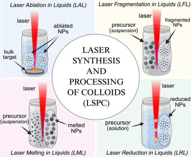
The constantly growing interest in zero-dimensional carbon-based nanomaterials such as carbon quantum dots (CQDs) and graphene quantum dots (GQDs) as the vital components in advancing various bio-related, catalysis, and energy-relevant applications has inspired nanotechnology research centered mainly on their synthesis and modifications for the betterment of their exceptional features including their strong and tunable fluorescence. Among the multitude of synthesis approaches in fabricating CQDs and GQDs, laser-based synthesis in liquids, such as pulsed laser ablation in liquids (PLAL) and pulsed laser fragmentation in liquids (PLFL), has emerged as a more beneficial technique owing to its versatility, flexibility, green synthesis process, and ease of scalability. With the modern trend of employing this method for CQDs and GQDs synthesis, this review article will revisit the foundation of laser synthesis in liquids, starting from its fundamental mechanism of nanoparticle formation to the effect of different variables such as laser parameters (e.g., laser energy, laser wavelength, frequency), chosen liquids, and the starting carbon material to the final attributes (morphological, optical, and surface) of CQDs and GQDs. In this paper, we will also address the different post-laser treatments, such as modifications and conjugation, and how they affect the properties of CQDs and GQDs. We will also emphasize the diverse applications of laser-synthesized CQDs and GQDs, ranging from bioapplications to beyond bio-related applications. This article hoped to provide practical insights for researchers to develop further laser synthesis in liquids to produce carbon-based nanomaterials such as CQDs and GQDs and their applicability to other applications.


Figures

1
1
General techniques of the LSPC-based synthesis of nanomaterials.
2
2
(a) Historical timeline and development of LSPC-based synthesis of CQDs and GQDs starting from the pioneering works of Patil et al. and Fotjik and Henglein. Figures from Sun et. al. were reprinted with permission from ref . Copyright 2006 American Chemical Society. Figure from Santiago et. al. was reprinted with permission from ref . Copyright 2016 Royal Society of Chemistry. Figure from Habiba et. al. was reprinted with permission from ref . Copyright 2013 Elsevier Ltd. Figure from Li et. al. was reprinted with permission from ref . Copyright 2025 Elsevier B.V. The whole timeline and schematic figure from Hu et. al. were created in BioRender.com. (b) Number of published papers on CQDs and GQDs and (c) Comparison of published papers on laser synthesis of CQDs and GQDs to other conventional approaches based on the Scopus database between 2010 and 2024.
3
3
(a) CDs can be classified as GQDs, CQDs, or CPDs and structural representation of CQDs or GQDs when viewed at the (b) top and (c) side. Created in BioRender.com.
4
4
(a) Illustration of CDs fluorescence originating from the quantum confinement effect (Created in BioRender.com), (b) Fluorescence of prepared CQDs with increasing size and their corresponding fluorescence spectra (Reprinted with permission from ref . Copyright 2016 John Wiley and Sons, Inc.), (c) CQDs under ambient light and UV lamp with their (d) corresponding fluorescence spectra, and (e) band gap relation to their size (Reprinted with permission from ref . Copyright 2010 John Wiley and Sons, Inc.).
5
5
(a) Illustration of CDs fluorescence originating from the surface state effect (Created in BioRender.com), (b) Fluorescence of prepared CQDs at different solvent and their corresponding fluorescence spectra (Reprinted with permission from ref . Copyright 2022 Elsevier B.V.), (c) CQDs under UV lamp and the band gap illustration from the degree of surface oxidation (Reprinted with permission from ref . Copyright 2016 American Chemical Society), and (d) synergistic effect of carbon core and surface state emission (Reprinted with permission from ref . Copyright 2023 John Wiley & Sons, Inc.).
6
6
Top-down and bottom-up synthesis routes for CDs. Created in BioRender.com.
7
7
(a) Classic PLAL experimental setups together with the incorporation of translational, rotational stage, (b) ultrasonication bath and electric field, (c) PLFL experimental setups (horizontal and vertical beam orientation), and (d) flow-jet configuration technique for CDs synthesis (Created in BioRender.com). (e) SSTF technique for NPs synthesis (Reprinted with permission from ref . Copyright 2019 Optica Publishing Group).
8
8
Stages of nanoparticles formation via PLAL technique: (a) laser-matter interaction stage, (b) plasma formation stage, (c) cavitation bubble formation stage (primary nanoparticle ejection), (d) expansion of cavitation bubble and secondary nanoparticles formation stage, (e) release of secondary nanoparticles stage to liquid, and (f) further reirradiation of formed nanoparticles. Created in BioRender.com.
9
9
Nonlinear ionization processes: (a) Multiphoton ionization and (b) electron impact ionization (avalanche ionization). Created in BioRender.com
10
10
PLFL mechanism for nanoparticle synthesis: (a) thermal ablation and (b) Coulomb explosion mechanism. Created in BioRender.com.
11
11
TEM images of CDs synthesized at different laser fluence: (a) 150 J/cm2, (b) 350 J/cm2, and (c) 750 J/cm2 (Reprinted with permission from ref . Copyright 2015 American Institute of Physics Publishing). (d) Fluorescence spectra of CDs synthesized at different fluences as indicated: (a) 0.17, (b) 0.42, (c) 0.70, (d) 0.90, and (e) 1.0 J/cm2, where the asterisk (*) represents the equipment lamp. Inset: Corresponding fluorescence of CDs at different fluences under UV lamp. (Reprinted with permission from ref . Copyright 2015 Elsevier Ltd.).
12
12
TEM images of CDs synthesized at different laser wavelengths: (a) 355 nm, (b) 532 nm, and (c) 1064 nm. (d) Fluorescence emission of CDs synthesized at different laser wavelengths with varying irradiation time under UV lamp (Reprinted with permission from ref . Copyright 2016 Springer Nature). (e) Fluorescence emission of CDs synthesized at different wavelengths under violet laser pointer and their (f) corresponding fluorescence spectra (Reprinted with permission from ref . Copyright 2017 American Institute of Physics Publishing). (g) Schematic representation of the type of GQDs produced from MWCNT ablated at different wavelengths and the XPS spectra of (h) GOQDs and (i) GQDs, respectively (Reprinted with permission from ref . Copyright 2019 Royal Society of Chemistry).
13
13
(a) Schematic representation of the product yield at different pulse durations from pure toluene molecules (Reprinted with permission from ref . Copyright 2023 Peter the Great St. Petersburg Polytechnic University); (b) Fluorescence emission of CDs synthesized directly from different organic solvents at two pulse durations (30 fs and 4 ps) under 365 nm illumination (Reprinted with permission from ref . Copyright 2023 American Chemical Society); (c) Fluorescence spectra of laser-synthesized CDs at different laser pulse duration (Reprinted with permission from ref . Copyright 2011 Springer Nature).
14
14
TEM images of laser modification of graphene oxide dispersion in water at different irradiation times: (a) 5 min, (b) 15 min, and (c) 30 min. (Reprinted with permission from ref . Copyright 2016 Royal Society of Chemistry). (d) Fluorescence emission of laser-synthesized GQDs at different irradiation times as indicated and (e) their corresponding fluorescence spectra. (f) Proposed mechanism of the origin of blue and green emission (Reprinted with permission from ref . Copyright 2016 Royal Society of Chemistry).
15
15
HR-TEM images of CDs synthesized in different liquid environments: (a) water, (b) diethylenetriaminepentaacetic (DTPA) aqueous solution, and (c) ethanol. Scale bars are 2 nm (Reprinted with permission from ref . Copyright 2017 John Wiley & Sons, Inc.). (d) Proposed structure of GQDs synthesized at different nitrogen-containing solvents. (e) Content of different nitrogen configurations in GQDs synthesized in ammonia (NA-GQDs), ethylenediamine (NE-GQDs), and pyridine (NP-GQDs) (Reprinted with permission from ref . Copyright 2019 American Chemical Society). (f) GQDs synthesized in (i) ethanol (e-GQDs) and (ii) hexane (h-GQDs) under ambient light (left) and 360 nm lamp (right). (Reprinted with permission from ref . Copyright 2016 Springer Nature.) (g) Fluorescence of CQDs synthesized in DMF, EDA, and DMSO using a laser pointer of 405 nm wavelength.
16
16
(a) TEM image of CDs synthesized from charcoal powder and their (b) corresponding fluorescence spectra at different excitation wavelengths (Reprinted with permission from ref . Copyright 2021 Elsevier Ltd.). TEM images of CDs from (c) graphite flakes and (d) carbon black powder. Inset: HR-TEM of CDs; (e) Raman spectra of CDs from graphite flakes (S1) and from carbon black powder (S2) (Reprinted with permission from ref . Copyright 2012 Royal Society of Chemistry).
17
17
(a) Process of Bougainvillea alba flower extract for laser irradiation. (b) HR-TEM of CDs produced from flower extract with amorphous and crystalline features. Inset: Closer look to the crystalline structure of CDs (Reprinted with permission from ref . Copyright 2019 Springer Nature). (c) CDs produced from lysine under ambient light (left) and under UV lamp (right) and their (d) corresponding TEM image. Inset: HR-TEM of CDs produced from amino acids (Reprinted with permission from ref . Copyright 2024 American Chemical Society).
18
18
(a) Fluorescence emission of CQDs passivated with PEG1500N at different excitation wavelengths as indicated. (b) Laser-synthesized CDs after passivated with PEG1500N resulting to emission (Reprinted with permission from ref . Copyright 2006 American Chemical Society). (c) Fluorescence emission of CQDs under UV lamp before (left) and after passivated with PEG400N (right); (d) GQDs before and after solvothermal treatment in DMF; and (e) fluorescence spectra of GQDs subjected to solvothermal at different temperatures: 65 °C, 90 °C, and 120 °C (Reprinted with permission from ref . Copyright 2019 Elsevier Ltd.). (f) SEC purification of CQDs conjugated with boronic acid via amidation reaction (Reprinted with permission from ref . Copyright 2024 Elsevier B.V.). (g) Removal of by-products from carbon nanodots (CNDs) solution through dialysis method. (Reprinted with permission from ref . Copyright 2022 American Chemical Society).
19
19
(a) Cell viability test of DMSO-CQDs at different concentration in μg/mL; (b) Bright-field and the corresponding overlay images of HeLa cells incubated with CQDs from confocal microscope (Reprinted with permission from ref .Copyright 2020 Elsevier Ltd.); (c) Confocal microscope image of A549 cells after addition of Lys-CDs (Reprinted with permission from ref . Copyright 2024 American Chemical Society); Confocal fluorescence (left) and bright-field images (right) of (d) OEC and (e) HT-29 cells incubated with CQDs (Reprinted with permission from ref . Copyright 2018 American Chemical Society); (f) In vivo imaging of a euthanized mouse implanted with GQDs-acrylamide gel (red) in the thoracic region as indicated by the white circle (Reprinted with permission from ref .Copyright 2017 Royal Society of Chemistry); (g) Different region of CDs injection to a zebrafish; (h) Blue luminescence of CDs located in the eye of a zebrafish after (i) gill, (ii) intestinal, (iii) dorsal, and (iv) tail injection. (Reprinted with permission from ref .Copyright 2023 Jurnal Ilmu Fisika).
20
20
(a) Fluorescence spectra of FGQDs at increasing Fe3+ concentration centered at 450 nm; (b) FGQDs fluorescence emission under UV lamp with the addition of different metal ion species as indicated (Reprinted with permission from ref .Copyright 2022 American Chemical Society); (c) Fluorescence spectra of (1) N-GQDs, (2) N-GQDs after addition of Fe3+ ions, and the (3) fluorescence recovery after addition of ascorbic acid and (d) their corresponding fluorescence emission under UV lamp (Reprinted with permission from ref .Copyright 2019 Elsevier Ltd.); (e) Fluorescence spectra of CQDs-APBA after addition of glucose at increasing concentration (Reprinted with permission from ref .Copyright 2024 Elsevier B.V.); (f) Fluorescence spectra of N-doped CDs with two maximum emissions (402 and 518 nm) at different pH (Reprinted with permission from ref .Copyright 2017 Elsevier B.V.); (g) Fluorescence spectra of laser-synthesized CDs with two maximum emissions (400 and 465 nm) at increasing temperature (°C) and (h) their corresponding linear relationship curve (Reprinted with permission from ref .Copyright 2017 Elsevier B.V.).
21
21
(a) Kirby Bauer diffusion test results of (iii) laser-synthesized CDs in EDA solution in comparison to (i) DMSO only as negative control, (ii) CDs synthesized in deionized water, and (iv) terramycin only as positive control; (b) Kirby Bauer diffusion test results of (iii) laser-synthesized CDs in chitosan solution in comparison to (i) DMSO only as negative control, (ii) CDs synthesized in deionized water, and (iv) terramycin only as positive control (Reprinted with permission from ref .Copyright 2023 Elsevier B.V.); Agar well diffusion assay results of CDs synthesized at different fluences: (B) 60 mJ, (C) 80 mJ, (D) 160 mJ, and (E) 220 mJ with (A) deionized water as the control, against (c) S. aureus and (d) E. coli bacteria (Reprinted with permission from ref .Copyright 2022 John Wiley & Sons, Inc.); (e) Microscopy images of MCF-7 cells: (i) untreated and treated with CDs at different concentrations: (ii) 400 μg/mL and (iii) 800 μg/mL and their (iv) corresponding viability assay result; (f) MCF-7 cells (i-ii) untreated and treated with CDs of two concentrations: (iii-iv) 400 μg/mL and (v-vi) 800 μg/mL, stained with AO/EtBr (left) and DAPI (right). Scale bar 10 μm (Reprinted with permission from ref .Copyright 2020 Institute of Physics Publishing Vietnam Academy of Science & Technology)
22
22
(a) SEM images of worn surfaces lubricated by (i) PAO only, (ii) N-doped CDs + PAO, (iii) S-doped CDs + PAO, and (iv) N,S-doped CDs + PAO (Reprinted with permission from ref .Copyright 2024 John Wiley & Sons, Inc.); (b) PCL (i) without and wet with CDs for (ii) 30 mins and (iii) 60 mins (Reprinted with permission from ref .Copyright 2024 Multidisciplinary Digital Publishing Institute); (c) TEM images of β-amyloid peptides (Aβ42) (i) alone and incubated with (ii) 40 μg/mL of CQDs and ThS fluorescence images of Caenorhabditis elegans nematodes (iii) untreated and (iv) treated with CQDs. Note: White arrows indicate Aβ plaques in nematodes. Scale bars are 20 μm. (Reprinted with permission from ref .Copyright 2022 Elsevier B.V.).

Similar articles

Cited by

References

    1. Das S., Mondal S., Ghosh D.. Carbon Quantum Dots in Bioimaging and Biomedicines. Front Bioeng Biotechnol. 2024;11:1333752. doi: 10.3389/fbioe.2023.1333752. - DOI - PMC - PubMed
    1. Paulo S., Palomares E., Martinez-Ferrero E.. Graphene and Carbon Quantum Dot-Based Materials in Photovoltaic Devices: From Synthesis to Applications. Nanomaterials. 2016;6(9):157. doi: 10.3390/nano6090157. - DOI - PMC - PubMed
    1. Walther B. K., Dinu C. Z., Guldi D. M., Sergeyev V. G., Creager S. E., Cooke J. P., Guiseppi-Elie A.. Nanobiosensing with Graphene and Carbon Quantum Dots: Recent Advances. Mater. Today. 2020;39:23–46. doi: 10.1016/j.mattod.2020.04.008. - DOI - PMC - PubMed
    1. Li M., Chen T., Gooding J. J., Liu J.. Review of Carbon and Graphene Quantum Dots for Sensing. ACS Sens. 2019;4(7):1732–1748. doi: 10.1021/acssensors.9b00514. - DOI - PubMed
    1. Alaghmandfard A., Sedighi O., Tabatabaei Rezaei N., Abedini A. A., Malek Khachatourian A., Toprak M. S., Seifalian A.. Recent Advances in the Modification of Carbon-Based Quantum Dots for Biomedical Applications. Materials Science and Engineering: C. 2021;120:111756. doi: 10.1016/j.msec.2020.111756. - DOI - PubMed

LinkOut - more resources