Same-side recurrence of unilateral multiple evanescent white dot syndrome following the thermal laser photocoagulation for inflammatory macular neovascularization
- PMID: 40529803
- PMCID: PMC12171975
- DOI: 10.3205/oc000250
Same-side recurrence of unilateral multiple evanescent white dot syndrome following the thermal laser photocoagulation for inflammatory macular neovascularization
Abstract
Purpose: We report the same side recurrence of multiple evanescent white dot syndrome (MEWDS) subsequent to 532 nm laser treatment for the macular neovascularization (MNV) associated with the first MEWDS episode.
Method: Retrospective case documentation with the multimodal imaging.
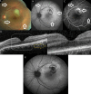
Result: A 24-year-old otherwise healthy woman who was diagnosed as having left MEWDS four years ago was re-examined for a visual disturbance of the duration of one month in the same eye. Fundus evaluation led us to the diagnosis of left extrafoveal inflammatory MNV. Surprisingly, she developed further visual deterioration a month following the uneventful 532 nm laser photocoagulation in her left eye. Fundus examination and multimodal imaging tests confirmed the recurrent MEWDS after full negative laboratory work-up. Visual acuity and fundus changes were improved with the help of a short course oral steroid therapy.
Conclusion: MEWDS can very rarely recur and thermal laser photocoagulation may be a possible triggering factor.
Keywords: choriodal neovascularization; fundus autofluorescence; laser photocoagulation; macular neovascularization; multifocal ERG; multiple evanescent white dot syndrome.
Copyright © 2025 Ünlü et al.
Conflict of interest statement
The authors declare that they have no competing interests.
Figures


Similar articles
-
Multiple evanescent white dot syndrome associated with focal scleral nodule: a case report and literature review.BMC Ophthalmol. 2025 Jul 16;25(1):413. doi: 10.1186/s12886-025-04231-4. BMC Ophthalmol. 2025. PMID: 40671005 Free PMC article. Review.
-
Contralateral Recurrences of Post-vaccination Multiple Evanescent White Dot Syndrome.Cureus. 2022 Dec 7;14(12):e32300. doi: 10.7759/cureus.32300. eCollection 2022 Dec. Cureus. 2022. PMID: 36628035 Free PMC article.
-
A Multiple Evanescent White Dot Syndrome-like Reaction to Concurrent Retinal Insults.Ophthalmol Retina. 2021 Oct;5(10):1017-1026. doi: 10.1016/j.oret.2020.12.007. Epub 2020 Dec 22. Ophthalmol Retina. 2021. PMID: 33348087 Review.
-
Multiple evanescent white dot syndrome in highly myopic eye in which fundus autofluorescence was diagnostically useful: A case report.Medicine (Baltimore). 2023 Feb 3;102(5):e32713. doi: 10.1097/MD.0000000000032713. Medicine (Baltimore). 2023. PMID: 36749227 Free PMC article.
-
Case report: Visual snow as the presenting symptom in multiple evanescent white dot syndrome. Two case reports and literature review.Front Neurol. 2022 Oct 6;13:972943. doi: 10.3389/fneur.2022.972943. eCollection 2022. Front Neurol. 2022. PMID: 36277919 Free PMC article.
Cited by
-
Multiple evanescent white dot syndrome associated with focal scleral nodule: a case report and literature review.BMC Ophthalmol. 2025 Jul 16;25(1):413. doi: 10.1186/s12886-025-04231-4. BMC Ophthalmol. 2025. PMID: 40671005 Free PMC article. Review.
References
-
- Ramirez Marquez E, Ayala Rodríguez SC, Rivera L, Pappaterra-Rodriguez MC, Requejo-Figueroa GA, Rios R, Rivera-Grana E, Rodríguez-García EJ, Oliver AL. Contralateral Recurrences of Post-vaccination Multiple Evanescent White Dot Syndrome. Cureus. 2022 Dec;14(12):e32300. doi: 10.7759/cureus.32300. - DOI - PMC - PubMed
Publication types
LinkOut - more resources
Full Text Sources

