. 2025 Jun 5:53:63-81.
doi: 10.1016/j.jot.2025.04.015. eCollection 2025 Jul.

Regulating inflammation microenvironment and tenogenic differentiation as sequential therapy promotes tendon healing in diabetic rats

Affiliations

Regulating inflammation microenvironment and tenogenic differentiation as sequential therapy promotes tendon healing in diabetic rats

Jingyi Dang et al. J Orthop Translat. .

Abstract

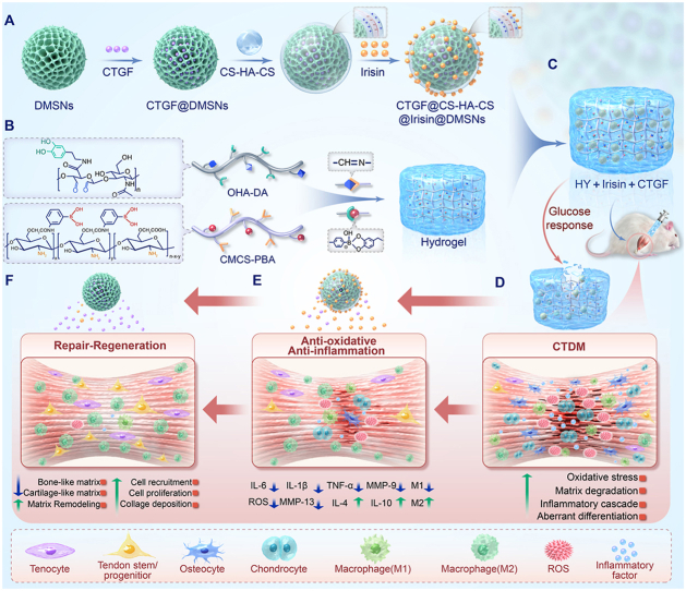
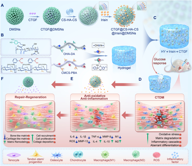
Background: Chronic tendinopathy with diabetes mellitus (CTDM) poses significant therapeutic challenges due to persistent inflammation and impaired tenogenesis. While the supplementation of tendon stem/progenitor cells (TSPCs) has the potential to facilitate tenogenesis, premature recruitment and proliferation in inflammatory microenvironments risks fibrosis or heterotopic ossification (HO). Consequently, balancing inflammation regulation and tenogenic differentiation is critical for effective healing.

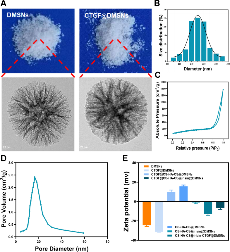
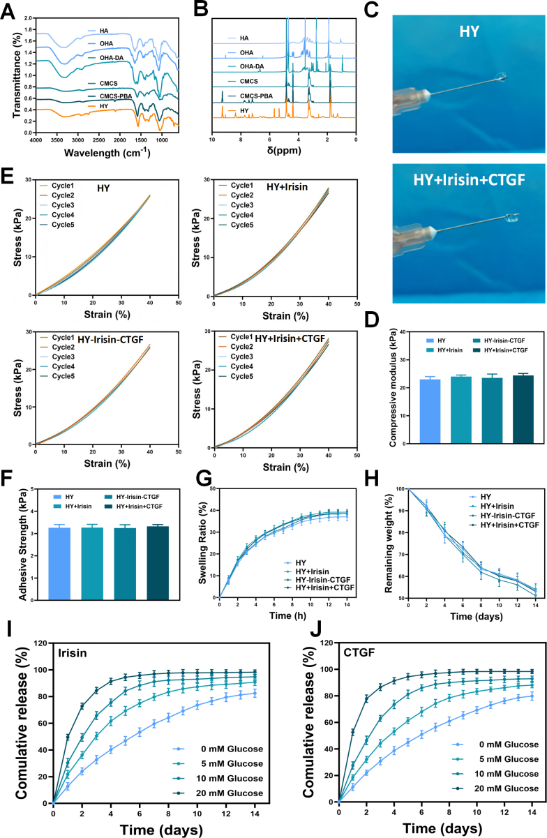
Methods: An injectable glucose-responsive dual-drug-sequential delivery hydrogel (GDSH) was developed utilizing oxidized hyaluronic acid-modified dopamine and phenylboronic acid-functionalized carboxymethyl chitosan. Dendritic mesoporous silica nanospheres (DMSNs) encapsulating irisin and connective tissue growth factor (CTGF) were incorporated into the GDSH matrix. A comprehensive characterization of the hydrogel's properties, including rheological, mechanical, adhesive, swelling/degradation, and drug release behaviors, was conducted. In vitro assessments were performed to evaluate cytocompatibility, as well as antioxidant and anti-inflammatory effects, alongside the migration, proliferation, and differentiation of TSPCs. The therapeutic efficacy was further investigated using a collagenase type I/streptozotocin-induced CTDM model in rats, with analyses conducted through histological, biomechanical, and micro-CT methods. Transcriptome sequencing and Western blot analyses were employed to elucidate the involvement of specific signaling pathways in the tissue repair process.

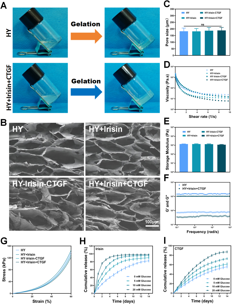
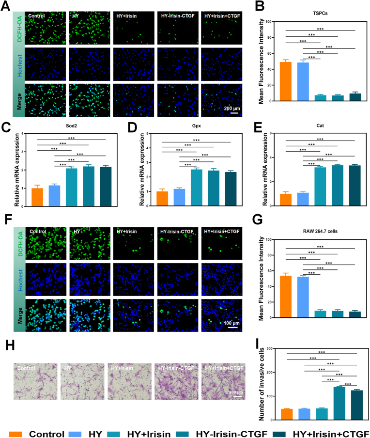
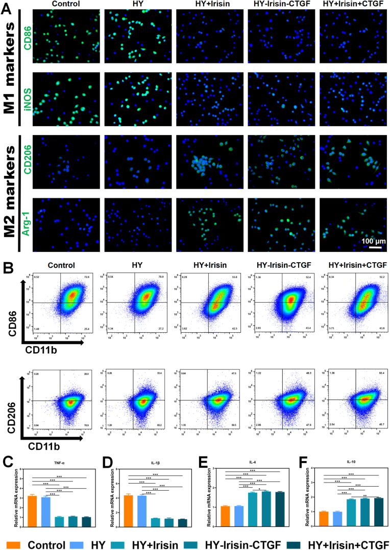
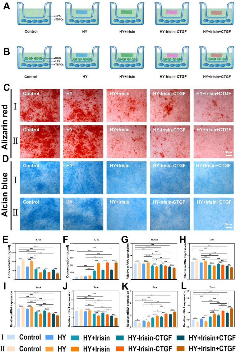
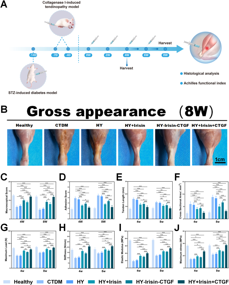
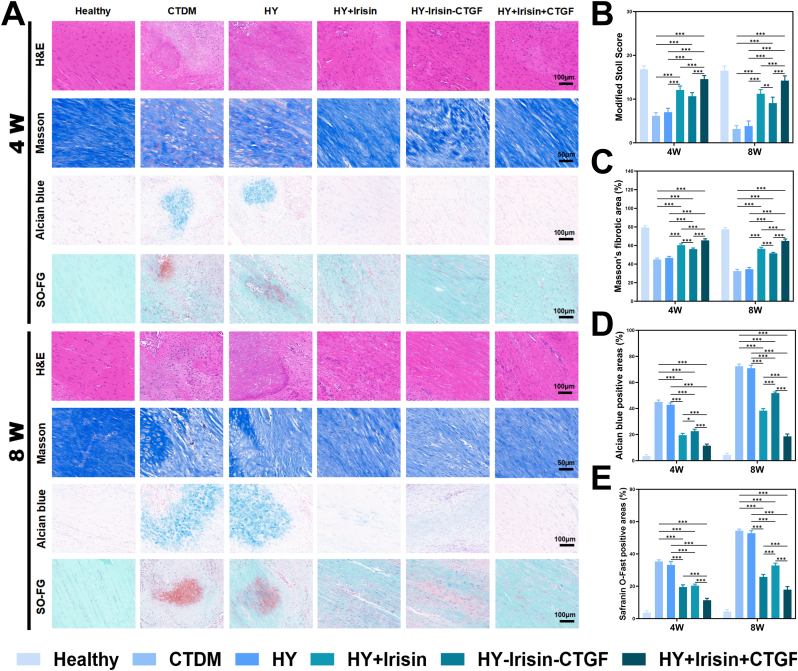
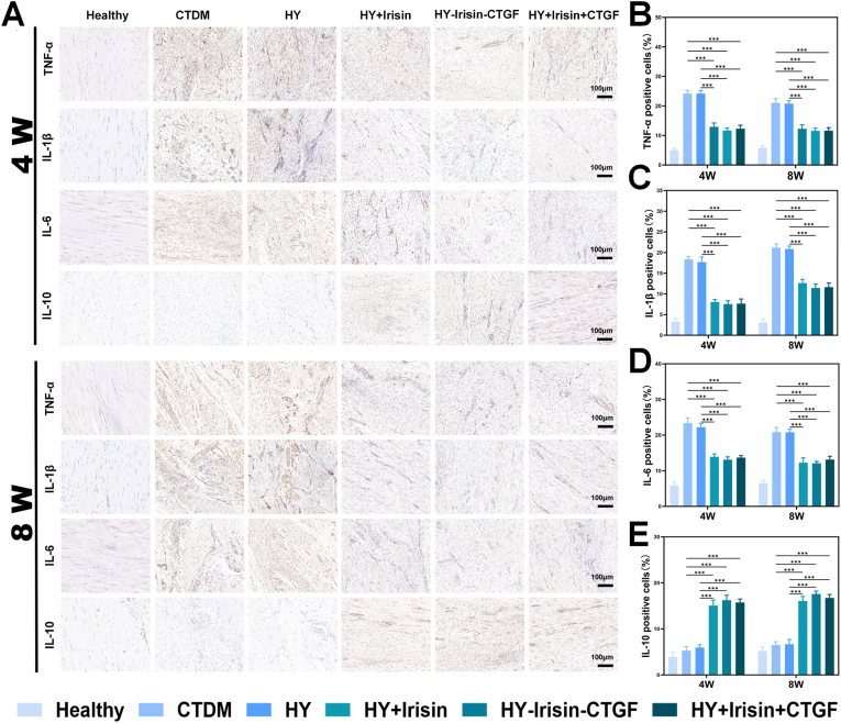
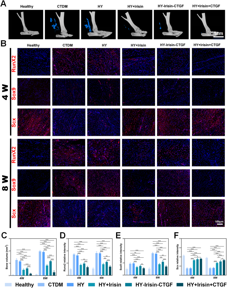
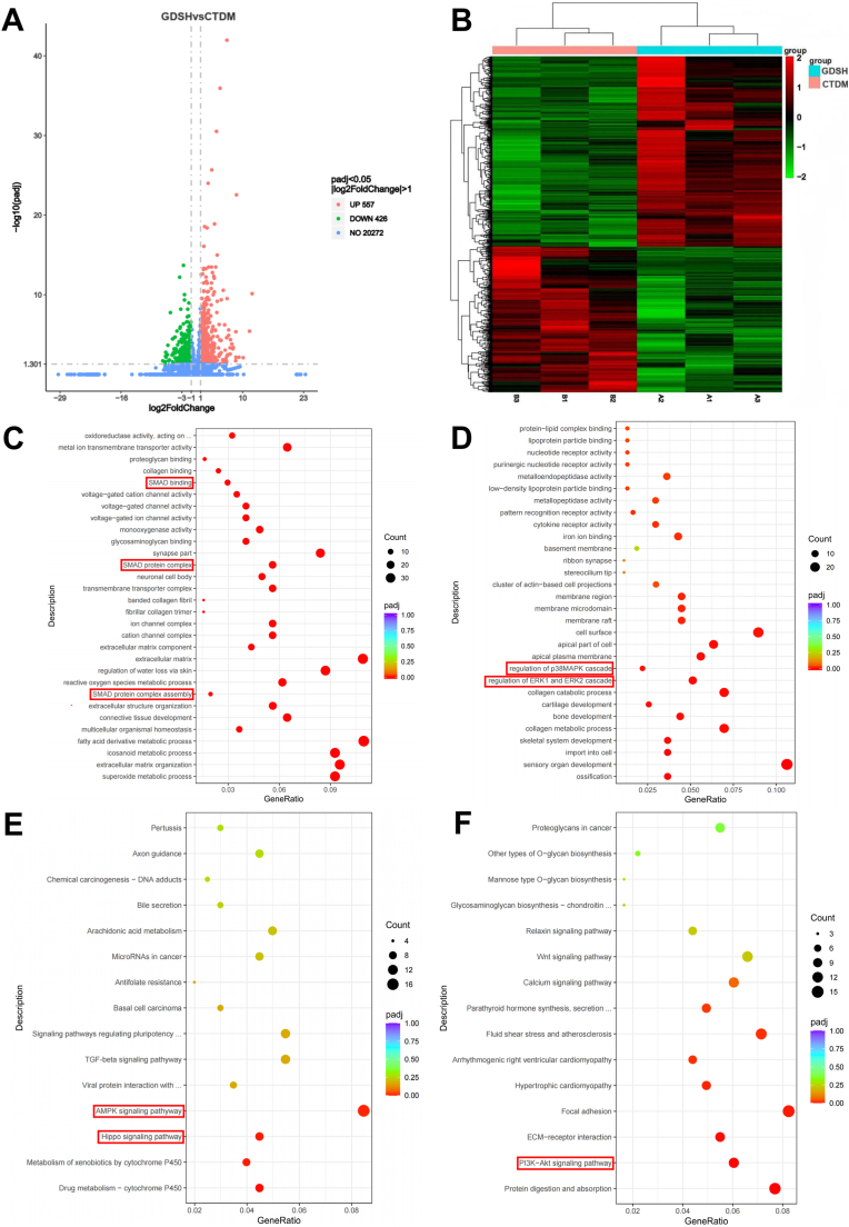
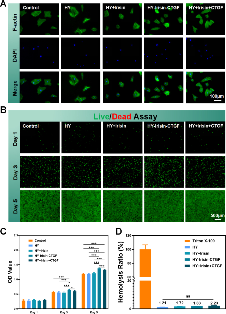
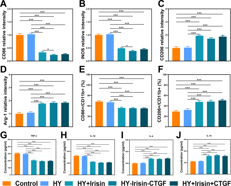
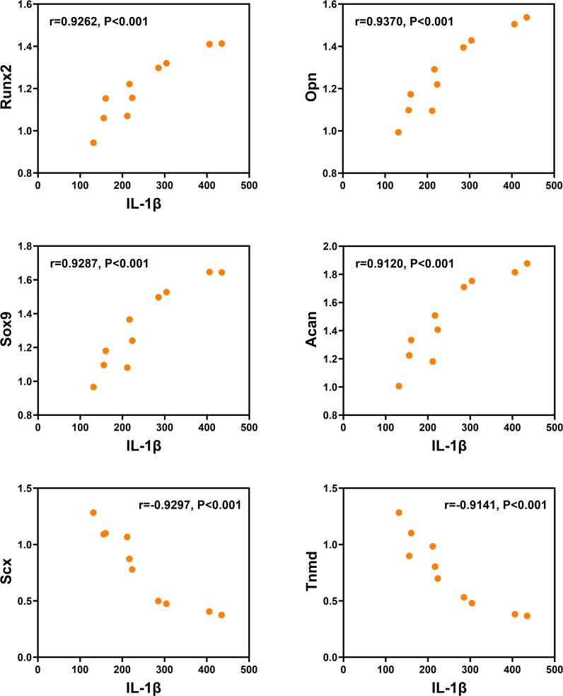
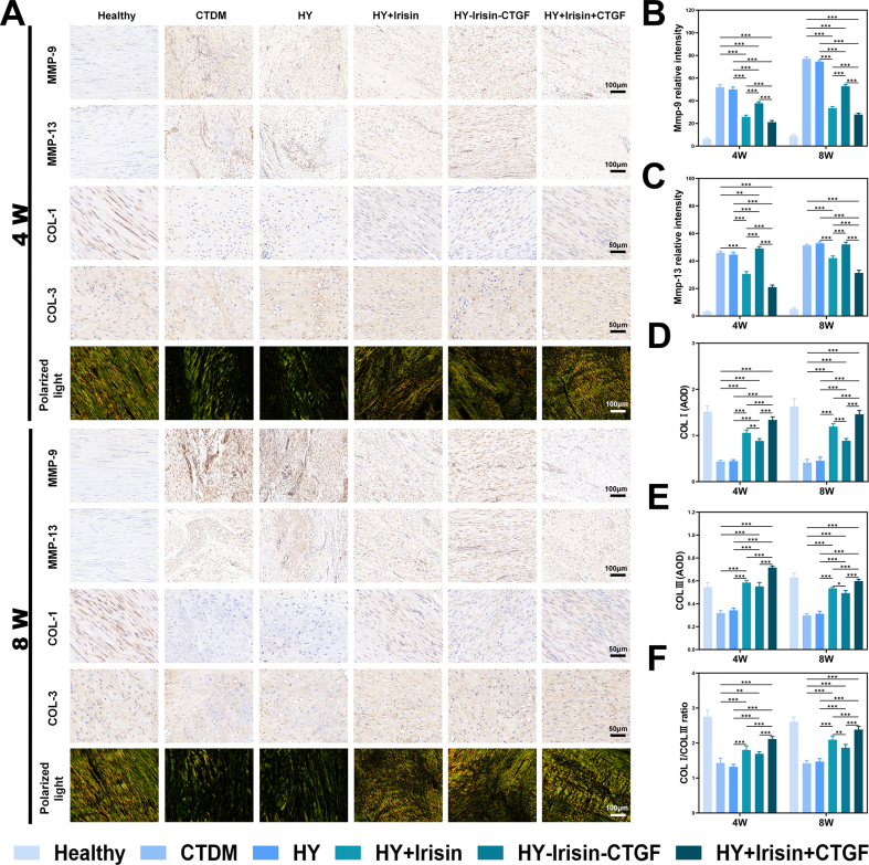
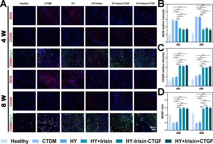
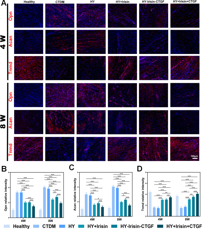
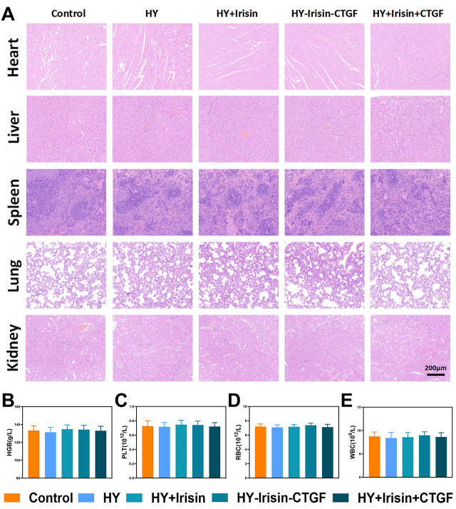
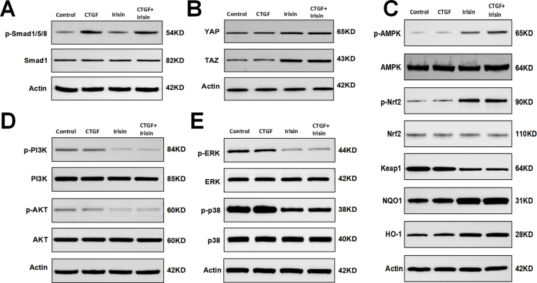
Results: The GDSH composite hydrogels possess a range of advantageous properties, including exceptional mechanical strength, optimal adhesiveness, superior biocompatibility, and appropriate swelling and degradation rates, in addition to controllable and sequential drug release capabilities. In vitro investigations revealed that these composite hydrogels exhibit antioxidant and anti-inflammatory effects, while also promoting cell proliferation and migration. Furthermore, they facilitate tenogenic differentiation and simultaneously inhibit the aberrant differentiation of TSPCs. In vivo studies demonstrated that the composite hydrogels significantly improved the morphological and biomechanical properties of injured tendons, reduced inflammation, corrected abnormal differentiation, and displayed favorable biosafety profiles. Transcriptome sequencing and Western blotting analysis indicated that the composite hydrogels repaired CTDM through the MAPK, AMPK, Smad, Hippo and PI3K/AKT signaling pathways.

Conclusion: GDSH achieves spatiotemporal control of inflammation resolution and tenogenesis via glucose-responsive sequential delivery of irisin and CTGF. This strategy restores tendon microstructure, biomechanics, and redox homeostasis in CTDM, offering a translatable platform for diabetic tendon regeneration.

The translational potential of this article: This study presents a glucose-responsive dual-drug-sequential delivery hydrogel (GDSH) designed for the treatment of chronic tendinopathy with diabetes mellitus (CTDM). This innovative approach aims to balance the regulation of inflammation and promote tenogenic differentiation. The sequential release of irisin and connective tissue growth factor (CTGF) effectively addresses the dual challenges posed by oxidative stress/inflammation and aberrant differentiation during tendon repair. The hydrogel's demonstrated biocompatibility, controlled drug release, and efficacy in restoring tendon structure and function highlight its potential for clinical translation. This platform represents a safer and more effective alternative to conventional treatments. Future research should focus on scaling up production, assessing long-term safety, and facilitating the translation of this technology into human clinical trials for the management of tendon injuries in diabetic patients.

Keywords: Chronic tendinopathy; Controllable release; Diabetes mellitus; Glucose-responsive; Inflammation microenvironment; Sequential delivery; Tenogenic differentiation.


Conflict of interest statement

We would like to submit a revised original article entitled“Regulating inflammation microenvironment and tenogenic differentiation as sequential therapy promotes tendon healing in diabetic rats”, which we wish to be considered for publication in Journal of Orthopaedic Translation. No conflict of interest exits in the submission of this manuscript, and manuscript is approved by all authors for publication. I would like to declare on behalf of my co-authors that the work described was original research that has not been published previously, and not under consideration for publication elsewhere, in whole or in part. All the authors listed have approved the manuscript that is enclosed. In this work, we demonstrated the advantages of sequential release of irisin and CTGF in the treatment of tendon injuries, highlighting the benefits of balancing inflammation and tenogenesis.

Figures

Image 1
Graphical abstract
Scheme 1
Scheme 1
Schematic illustration of the GDSH. (A). Fabrication of CTGF@CS-HA-CS@ Irisin@DMSNs; (B). Synthesis of OHA-DA/CMCS-PBA hydrogel; (C). Application of GDSH; (D–F). Schematic illustration of CTDM repair through the regulation of the repair microenvironment and the promotion of tenogenic differentiation.
Fig. 1
Fig. 1
Gross morphology and characterizations of the composite hydrogels. (A). Optical photographs of the OHA-DA/CMCS-PBA mixture solution and the formed hydrogel with or without CTGF and irisin. (B). Representative SEM images of composite hydrogels.Scale bar: 100 μm. (C). Pore sizes of composite hydrogels. (D). The viscosity of hydrogels with the shear rate from 0.1 to 10 1/s. (E). Storage moduli of the hydrogels at 10 rad s−1 frequency and 1 % strain. (F). Variations of storage and loss moduli (G′ and G″) versus angular frequency (0.1–100 rad s−1). (G).Typical stress–strain curves. The releasing kinetics of irisin (H) and CTGF (I).
Fig. 2
Fig. 2
Antioxidant properties of composite hydrogels and TSPCs migratory abilities. (A). The DCFH-DA staining results of TSPCs. Scale bar: 200 μm. (B). Image J analysis of the DCFH-DA staining in TSPCs. (C–E). qPCR analysis of relative gene expression. (F). The DCFH-DA staining results of macrophages. Scale bar: 100 μm. (G). Image J analysis of the DCFH-DA staining in macrophages. (H). Representative light photomicrographs of migrated TSPCs. Scale bar: 200 μm. (I). Quantitative analysis of migrated TSPCs density.
Fig. 3
Fig. 3
Anti-inflammatory properties of composite hydrogels. (A). Immunostaining of CD86, iNOS, CD206, Arg-1(green) and nuclei (blue). Scale bar: 100 μm. (B). Cytometry analysis of CD86 and CD206 expression in macrophages. (C–F). The mRNA expression of pro-infammatory genes (TNF-α and IL-1β) and anti-infammatory genes (IL-4 and IL-10).
Fig. 4
Fig. 4
GDSH inhibited aberrant differentiation and promoted tenogenic differentiatition in vitro. (A). Schematic illustration of the first condition. (B). Schematic illustration of the second condition. (C). ARS staining. Scale bar: 100 μm. (D). Alcian blue staining. Scale bar: 100 μm. ELISA results of cytokines IL-1β (E) and IL-10 (F). (G–L). The mRNA expression of markers of osteogenic differentiation (Runx2, Opn), chondrogenic differentiation (Sox9, Acan) and tenogenic differentiation (Scx, Tnmd).
Fig. 5
Fig. 5
Efficacy assessment of GDSH by morphological and biomechanical properties. (A). The timeline of the in vivo experiment. (B). Gross appearance of achilles tendon on week 8. Scale bar: 1 cm. (C). Macroscopical score of achilles tendon. (D). Adhesion score of achilles tendon. (E). Achilles tendon length. (F). Achilles tendon CSA. (G). Maximum Load. (H). Stiffness. (I). Elastic Modulus. (J). Maximun stress (n = 5).
Fig. 6
Fig. 6
Efficacy assessment of GDSH by histological staining. (A). H&E, Masson, Alcian blue and Safranin O-Fast green staining at 4 and 8 weeks postoperatively. Scale bar: 50 or 100 μm. (B). Modified stoll score. (C). Quantitative analysis of percentage of Masson's fibrotic area. (D). Quantitative analysis of percentage of Alcian blue positive area. (E). Quantitative analysis of percentage of Safranin O-Fast green positive area.
Fig. 7
Fig. 7
GDSH alleviated the inflammatory in vivo. (A). Immunohistochemical staining against TNF-α, IL-1β, IL-6, IL-10. Scale bar:100 μm. (B–E). Percentage of Immunohistochemical positive staining against TNF-α, IL-1β, IL-6, IL-10.
Fig. 8
Fig. 8
GDSH attenuated tendon heterotopic ossification in vivo. (A). Micro-CT images of repaired tendons after treatment at post-operative week 8. Scale bar:1 cm. (B). Immunofluo-rescence staining of Runx2, Sox9 and Scx in repaired tendons. Scale bar:100 μm. (C). Statistical analysis of bone volume measurement of the ectopic bone. (D–F). The quantifcation results of Runx2, Sox9, Scx.
Fig. 9
Fig. 9
Mechanism analysis of GDSH treated CTDM. (A). Volcano plots of differently expressed genes. (B). Heatmap of differently expressed genes. (C). GO analysis of upregulated genes in GDSH versus CTDM samples. (D). GO analysis of downregulated genes in GDSH versus CTDM samples. (E). KEGG analysis of upregulated genes in GDSH versus CTDM samples. (F). KEGG analysis of downregulated genes in GDSH versus CTDM samples.
Fig. S1
Fig. S1
Characterization of the DMSNs. (A). Gross morphology and TEM images of DMSNs and CTGF@DMSNs. Scale bar: 20 nm (B). Diameter distribution of DMSNs (n = 100). (C). Nitrogen sorption isotherms of DMSNs. (D). Pore size distribution of the DMSNs. (E). Zeta potential of DMSNs and modified DMSNs.
Fig. S2
Fig. S2
Characterization of the composite hydrogels. (A). FTIR spectrum of HA, OHA, OHA-DA, CMCS, CMCS-PBA, OHA-DA/CMCS-PBA hydrogel. (B). 1H NMR spectrum of HA, OHA, OHA-DA, CMCS, CMCS-PBA, OHA-DA/CMCS-PBA hydrogel. (C). Photographs of the injectability of the hydrogel through the needle. (D). Compression modulus (n = 3). (E). Cyclic compression stress–strain curve. (F). Adhesion strength of hydrogels (n = 3). (G). Swelling ratio of hydrogels. (H). Residual weight of hydrogels. The releasing kinetics of irisin (I) and CTGF (J) in HY-Irisin-CTGF.
Fig. S3
Fig. S3
The biocompatibility of the hydrogels. (A). Cytoskeleton staining images of TSPCs. Scale bar: 100 μm (B). Live/Dead staining of TSPCs after incubated with the hydrogels for 1, 3 and 5 days. Scale bar: 500 μm. (C). CCK-8 assays of TSPCs at 1, 3, and 5 days. (D). Hemolytic percentage of the hydrogels.
Fig. S4
Fig. S4
Anti-inflammatory properties of composite hydrogels. (A-D).The quantifcation results of M1-related markers (CD86, iNOS) and M2-related markers (CD206, Arg-1) in immuno-fluorescence images(n = 3). (E-F). Quantitative analysis of CD86/CD11b and CD206/CD11b in flow cytometry (n = 3).(G-J). ELISA results of cytokines TNF-α, IL-6, IL-1β and IL-10 (n = 3).
Fig. S5
Fig. S5
Correlation analysis between heterotopic ossification and inflammation.
Fig. S6
Fig. S6
GDSH promoted Collagen remodeling in vivo. (A). Immunohistochemical staining against MMP-9, MMP-13, COL I, COL III and polarized light of achilles tendon. Scale bar: 50 or 100 μm. (B-E). Percentage of positive staining for the MMP-9, MMP-13, COL Ⅰ, COL Ⅲ. AOD, average optical density. (F). COL I/COL III ratio of different groups.
Fig. S7
Fig. S7
GDSH alleviated the inflammatory in vivo.(A). Immunofuorescent images of sections in diferent groups: red (iNOS, CD86, CD206), green (Arg-1). Scale bar:100 μm. (B). The quantifcation results of iNOS. (C). The quantifcation results of CD206. (D). M2/M1 ratio.
Fig. S8
Fig. S8
GDSH attenuated tendon heterotopic ossification in vivo. (A).Immunofluorescence staining of Opn, Acan and Tnmd in repaired tendons, Scale bar: 100 μm. (B-D).The quantifcation results of Opn, Acan, Tnmd.
Fig. S9
Fig. S9
In vivo biosafety evaluation. (A) H&E stained histological images of the main organs in rats from different treatment groups. Scale bar: 200 μm. Hematological examination in hemoglobin (HGB) (B) and the counts of platelet (PLT) (C), red blood cell (RBC) (D), and white blood cell (WBC) (E) after the subcutaneous injection of hydrogels for 3 days.
Fig. S10
Fig. S10
Mechanism analysis of Irisin and CTGF treatment. (A).The protein expressions of key markers in Smad signaling pathway. (B).The protein expressions of key markers in Hippo signaling pathway. (C).The protein expressions of key markers in AMPK signaling pathway. (D).The protein expressions of key markers in PI3K/AKT signaling pathway. (E).The protein expressions of key markers in MAPK signaling pathway.
Table S1
Table S1
The premiers used in this study.
Table S2
Table S2
Details of primary and secondary antibodies.
Table S3
Table S3
(A). Irisin loading efficacy. (B). The detailed information of CTGF loading efficacy. (C). CTGF loading efficacy.

Similar articles

References

    1. Lui P.P.Y. Tendinopathy in diabetes mellitus patients-Epidemiology, pathogenesis, and management. Scand J Med Sci Sports. 2017;27(8):776–787. doi: 10.1111/sms.12824. - DOI - PubMed
    1. Patel S.H., Mendias C.L., Carroll C.C. Descriptive transcriptome analysis of tendon derived fibroblasts following in-vitro exposure to advanced glycation end products. PLoS One. 2022;17(7) doi: 10.1371/journal.pone.0271770. - DOI - PMC - PubMed
    1. Ackerman J.E., Geary M.B., Orner C.A., Bawany F., Loiselle A.E. Obesity/Type II diabetes alters macrophage polarization resulting in a fibrotic tendon healing response. PLoS One. 2017;12(7) doi: 10.1371/journal.pone.0181127. - DOI - PMC - PubMed
    1. Korntner S., Kunkel N., Lehner C., Gehwolf R., Wagner A., Augat P., et al. A high-glucose diet affects Achilles tendon healing in rats. Sci Rep. 2017;7(1):780. doi: 10.1038/s41598-017-00700-z. - DOI - PMC - PubMed
    1. Vvaidya R., Lake S.P., Zellers J.A. Effect of diabetes on tendon structure and function: not limited to collagen crosslinking. J Diabetes Sci Technol. 2023;17(1):89–98. doi: 10.1177/19322968221100842. - DOI - PMC - PubMed

LinkOut - more resources