

Multiparametric MRI in Diagnosis of Parotid Gland Tumor: An Observational Study in 3-T MRI
- PMID: 40529971
- PMCID: PMC12169940
- DOI: 10.1055/s-0044-1800861
Multiparametric MRI in Diagnosis of Parotid Gland Tumor: An Observational Study in 3-T MRI
Abstract
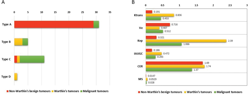
Background Preoperative magnetic resonance imaging (MRI) has an important role in the management and prognostication of parotid gland tumors. We aim to evaluate the role of multiparametric MRI in differentiating the major subgroup of parotid tumors. Materials and Methods Multiparametric MRI: T1-weighted imaging (T1WI), T2WI, diffusion-weighted imaging (DWI), pseudo-continuous arterial spin labeling (ASL) and dynamic contrast-enhanced (DCE) imaging were acquired in all patients. Apparent diffusion coefficient (ADC) values and tumor blood flow (TBF) were calculated from DWI and ASL, respectively. Ktrans, Kep, Ve, initial area under the gadolinium enhancement concentration curve (IAUGC), maximum slope, and contrast enhancement ratio (CER) were calculated from DCE-MRI perfusion. The above parameters were compared between three major subgroups of parotid gland tumors, such as non-Warthin benign tumors (NWBT), Warthin's tumors (WT), and malignant parotid tumors (MT). Results The mean ADC of MT ( n = 13), WT ( n = 5), and NWBT ( n = 29) was 1.03 × 10 -3 mm 2 /s, 0.97 × 10 -3 mm 2 /s, and 1.89 × 10 -3 mm 2 /s, respectively. The mean TBF (in mL/100 g/min) was the highest MT (70.33), followed by WT (62.04) and NWBT (21.99). A cutoff of 40.51 mL/100 g/min showed a sensitivity of 96.6% and specificity of 77.8% for predicting NWBT. In DCE-MRI, 96.6% of the NWBT showed a type A time-signal intensity curve. Although the majority of MT and WT had type C and B curves, respectively, there was overlapping. Among the quantitative DCE parameters, Ktrans and Kep were highly sensitive for differentiating NWBT, WT, and MT. Ktrans, Kep, IAUGC, and MS were maximum in WT, followed by MT and NWBT. Kep and Ktrans both had an accuracy of 84.7% for predicting pleomorphic adenoma. Conclusion Multiparametric MRI is useful for differentiating NWBT, WT, and MT. DCE-MRI helps in differentiating benign from malignant tumors. DWI and ASL are useful in differentiating NWBT from parotid malignancy.
Keywords: arterial spin labeling; diffusion-weighted MRI; dynamic contrast enhanced MRI; parotid tumor; salivary gland tumor.
Indian Radiological Association. This is an open access article published by Thieme under the terms of the Creative Commons Attribution-NonDerivative-NonCommercial License, permitting copying and reproduction so long as the original work is given appropriate credit. Contents may not be used for commercial purposes, or adapted, remixed, transformed or built upon. ( https://creativecommons.org/licenses/by-nc-nd/4.0/ ).
Conflict of interest statement
Conflict of Interest None declared.
Figures


References
-
- Stenner M, Klussmann J P. Current update on established and novel biomarkers in salivary gland carcinoma pathology and the molecular pathways involved. Eur Arch Otorhinolaryngol. 2009;266(03):333–341. - PubMed
-
- Perkins C, Toll E, Reece P. Fine-needle aspiration cytology and radiological imaging in parotid gland tumours: our experience in 103 patients. Clin Otolaryngol. 2019;44(06):1124–1127. - PubMed
-
- Yabuuchi H, Fukuya T, Tajima T, Hachitanda Y, Tomita K, Koga M. Salivary gland tumors: diagnostic value of gadolinium-enhanced dynamic MR imaging with histopathologic correlation. Radiology. 2003;226(02):345–354. - PubMed
-
- Coudert H, Mirafzal S, Dissard A, Boyer L, Montoriol P F. Multiparametric magnetic resonance imaging of parotid tumors: a systematic review. Diagn Interv Imaging. 2021;102(03):121–130. - PubMed
LinkOut - more resources
Full Text Sources