Review
. 2025 Feb 13;35(3):374-386.
doi: 10.1055/s-0044-1801268. eCollection 2025 Jul.

Multimodality Imaging in Wrist Fractures and Dislocations

Affiliations
Review

Multimodality Imaging in Wrist Fractures and Dislocations

Madhurima Sharma et al. Indian J Radiol Imaging. .

Abstract

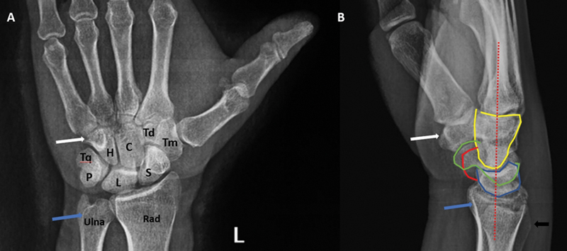
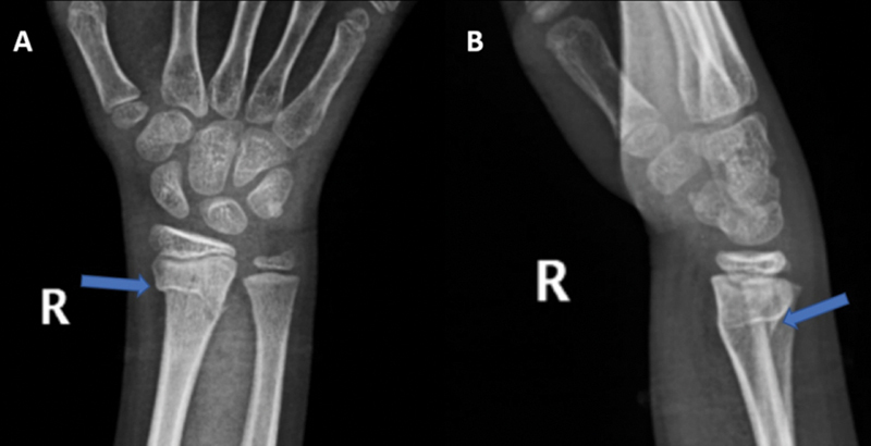
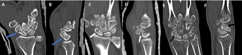
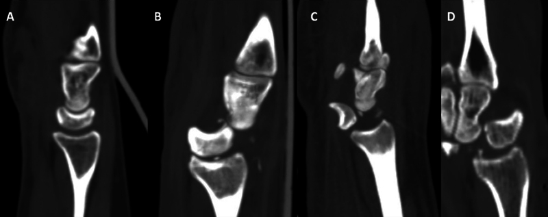
Wrist fractures and dislocations are frequently encountered in the emergency department and can cause significant long-term disability. Imaging plays a crucial role in the evaluation of wrist injuries, with conventional radiography being the first imaging investigation. Cross-sectional imaging is playing an increasingly important role in management of wrist injuries. Computed tomography with 3D and multiplanar reformatting capabilities is in particular useful for detailed evaluation of bony injuries and can provide vital information to orthopaedic surgeons for adequate surgical planning. In this article, we provide a brief review of the normal wrist anatomy, imaging appearance, and various patterns of fractures and dislocations commonly encountered in the emergency department.

Keywords: computed tomography; radiography; radius fractures; wrist injuries; wrist trauma.


Conflict of interest statement

Conflict of Interest None declared.

Figures

Fig. 1
Fig. 1
( A ) Volume rendered computed tomography (CT) image showing the normal bony anatomy of the wrist. C, capitate; H, hamate; L, lunate; P, pisiform; Rad, radius; S, scaphoid; Td, trapezoid; Tm, trapezium; Tq, triquetral. ( B ) Volume-rendered CT image showing various joints of the wrist: distal radioulnar joint ( blue ), radiocarpal joint ( green ), intercarpal joints ( purple ), and carpometacarpal joints ( red ).
Fig. 2
Fig. 2
Posteroanterior radiograph of the wrist demonstrating the Gilula arcs. Arc I is drawn through the proximal articular surface of the scaphoid, lunate, and triquetral; arc II is drawn through the distal articular surface of the scaphoid, lunate, and triquetral; and arc III is drawn through the proximal articular surface of the capitate and hamate in the distal carpal row. Note that the pisiform, trapezium, and trapezoid are not a part of the Gilula arcs.
Fig. 3
Fig. 3
( A, B ) Scapholunate and lunotriquetral ligaments ( green ) with volar, dorsal, and interosseous components. ( C ) Various components of the triangular fibrocartilaginous complex: articular disk with radial and ulnar attachments ( yellow ); volar ( blue ) and dorsal ( purple ) radioulnar ligaments; ulnolunate ( dark green ) and ulnotriquetral ( light green ) ligaments; ulnar collateral ligament ( gray ) and tendon sheath of the extensor carpi ulnaris tendon ( orange ).
Fig. 4
Fig. 4
Standard radiographic views of the wrist: ( A ) posteroanterior, ( B ) lateral, and ( C ) oblique views.
Fig. 5
Fig. 5
Normal anatomy on posteroanterior (PA) and lateral views. ( A ) PA radiograph showing all the carpal bones, distal radius, and ulna. Also note the hook of the hamate ( white arrow ), which is seen as a ring-like structure on the PA view. In the optimal PA view, the groove for the extensor carpi ulnaris tendon should be seen in the profile ( blue arrow ). ( B ) Lateral view showing the capitate ( yellow ), lunate ( blue ), scaphoid ( green ), pisiform ( red ), trapezium ( white arrow ), distal radius ( blue arrow ), and ulna ( black arrow ). In the true lateral view, the palmar cortex of the pisiform should lie between the palmar cortices of the scaphoid and the capitate. Also note the normal relationship between the capitate, lunate, and radius ( dashed red line ).
Fig. 6
Fig. 6
Posteroanterior and lateral radiographs of the wrist in different patients demonstrating ( A, B ) Colle's fracture ( blue arrow ), ( C, D ) Smith's fracture ( blue arrow ), and ( E, F ) volar Barton's fracture ( blue arrow ). ( G ) Oblique radiograph of the wrist demonstrating a fracture of the radial styloid ( blue arrow ) suggestive of a Hutchinson fracture. Also note the coexisting fractures of the ulnar styloid ( white arrow ) and the hamate ( black arrow ).
Fig. 7
Fig. 7
( A ) Lateral radiograph of the wrist showing a normal pronator quadratus fat stripe ( white arrow ). ( B ) Extra-articular fracture of the distal radius with obliteration of the pronator quadratus fat stripe.
Fig. 8
Fig. 8
Distal radius measurements on radiograph. ( A ) On posteroanterior view: radial length is the distance between the tip of the radial styloid and the ulnar articular surface ( white arrow ). Radial inclination is the angle between two lines drawn through the distal articular surface of the radius and a line drawn perpendicular to the long axis of the radius. ( B ) On lateral view: radial tilt is the angle between lines drawn through the distal articular surface and the line drawn perpendicular to mid-shaft of the radius.
Fig. 9
Fig. 9
Posteroanterior radiographs of the wrist in different patients showing ( A ) positive, ( B ) neutral, and ( C ) negative ulnar variance.
Fig. 10
Fig. 10
( A ) Posteroanterior (PA) radiograph of the wrist demonstrating a comminuted fracture of the distal radius ( blue arrow ) with intra-articular extension ( white arrow ) and loss of the radial length and inclination. ( B ) PA radiograph in another patient demonstrating a comminuted fracture of the distal radius ( blue arrow ) with intra-articular extension ( black arrow ). The radial length and inclination are, however, preserved.
Fig. 11
Fig. 11
( A ) Posteroanterior radiograph of the wrist demonstrating a comminuted fracture of the distal radius ( blue arrow ) with suspicious intra-articular extension. Also note the associated fracture of the ulnar styloid (white arrow). ( B ) Coronal computed tomography image showing an extensively comminuted intra-articular fracture of the distal radius with articular step-off ( black arrow ), which was not seen on radiograph. ( C ) Also note the associated fracture of the triquetral ( black arrow ) missed on radiograph.
Fig. 12
Fig. 12
( A ) T1-weighted (T1W) coronal, ( B ) T2W coronal fat-saturated, and ( C ) proton density fat-saturated sagittal images demonstrating a hypointense fracture line in the distal radius ( blue arrow in A ) with adjacent marrow edema ( arrows in B and C ). The fracture was occult on radiograph.
Fig. 13
Fig. 13
( A ) Posteroanterior and ( B ) lateral radiographs of the wrist in a child demonstrating a torus fracture of the distal radius ( blue arrow ) with involvement of the lateral cortex.
Fig. 14
Fig. 14
( A, B ) Fracture of the tip of the ulnar styloid. Coronal reformatted computed tomography (CT) image showing a fracture of the tip of the ulnar styloid ( blue arrow ), which was not appreciated on radiograph ( A ). Also note the associated distal radial fracture ( black arrow ). ( C ) Fracture base of the ulnar styloid. PA radiograph of the wrist showing fracture of base of the ulnar styloid ( white arrow ) with an associated comminuted fracture of the distal radius ( black arrow ). Also note the poorly visualized hook of the hamate with fractures of the fourth and fifth metacarpals. The fracture of the hook of the hamate was present on CT (not shown here).
Fig. 15
Fig. 15
Epicenter method for the distal radioulnar joint (DRUJ) instability in ( A ) a stable and ( B ) subluxated DRUJ. A line is drawn through the center of the ulnar head and the styloid ( yellow line ), and another line through the sigmoid notch ( green line ). A line perpendicular to the sigmoid notch ( dashed red line ) is then drawn from the center of this line. In a stable DRUJ, this perpendicular lies in the middle of the sigmoid notch ( A ).
Fig. 16
Fig. 16
Posteroanterior radiographs of the wrist in three different patients demonstrating fractures of the scaphoid through ( A ) the proximal pole ( blue arrow ), ( B ) waist ( blue arrow ), and ( C ) distal pole ( blue arrow ).
Fig. 17
Fig. 17
Coronal and sagittal reformatted images in three different patients depicting ( A, B ) a minimally displaced fracture through the waist of the scaphoid without significant angulation ( blue arrow ), ( C, D ) displaced fracture through the scaphoid waist with volar angulation of the proximal fracture fragment ( white arrow ), and ( E, F ) displaced fracture through the scaphoid waist with significant volar angulation of the proximal and distal fracture fragments resulting in humpback deformity ( black arrow ).
Fig. 18
Fig. 18
( A ) T1-weighted (T1W) coronal, ( B ) T2W coronal fat-saturated, and ( C ) proton density fat-saturated sagittal images demonstrating a hypointense fracture line through the waist of the scaphoid ( blue arrow ) with an adjacent marrow edema.
Fig. 19
Fig. 19
( A ) Coronal reformatted and ( B ) axial computed tomography images show fractures of the triquetrum ( blue arrow ) and hamate ( black arrow ). Also note the associated fractures of the base of the fourth and fifth metacarpals ( white arrow ).
Fig. 20
Fig. 20
( A ) Axial, ( B ) coronal, and ( C ) sagittal reformatted images demonstrate a linear undisplaced fracture of the lunate (blue arrow). Also note the comminuted fracture involving the distal radius ( white arrow ).
Fig. 21
Fig. 21
( A ) The hook of the hamate is not visualized on the posteroanterior radiograph of the wrist ( blue arrow ). ( B ) Axial and ( C ) sagittal reformatted computed tomography (CT) images demonstrate a fracture of the hook of the hamate ( blue arrow ). Also note the fracture dislocations involving the proximal carpal row ( white arrow in A ) and undisplaced fracture involving the trapezoid ( black arrow in B ). On CT, fractures were also seen involving the triquetrum and the scaphoid with an associated midcarpal dislocation (not shown here).
Fig. 22
Fig. 22
( A ) Oblique radiograph, ( B ) sagittal reformatted, and ( C ) axial computed tomography images showing a fracture through the dorsal surface of the hamate (blue arrow).
Fig. 23
Fig. 23
( A ) Posteroanterior radiograph and ( A ) coronal computed tomography image showing a fracture of the pisiform ( blue arrow ). Also note the presence of the comminuted fractures of the distal radius ( white arrow ) and ulna ( black arrow ).
Fig. 24
Fig. 24
( A ) Coronal and ( B ) sagittal reformatted images showing a comminuted fracture of the capitate ( blue arrow ). Also note the comminuted fractures of the distal radius ( white arrow ) and ulna ( black arrow ).
Fig. 25
Fig. 25
Sagittal reformatted computed tomography images showing alignment of the capitate, lunate, and radius in ( A ) normal case, ( B ) perilunate dislocation, ( C ) lunate dislocation, and ( D ) midcarpal dislocation. In the perilunate dislocation, alignment between the radius and the lunate is maintained, while in the lunate dislocation ( B ), alignment between the radius and the capitate is maintained ( C ). In the midcarpal dislocation, alignment between all three bones is lost ( D ).
Fig. 26
Fig. 26
Trans-scaphoid fracture dislocation. ( A ) Posteroanterior radiograph of the wrist showing a displaced fracture through the waist of the scaphoid (blue arrow). ( B ) Lateral radiograph demonstrating a perilunate dislocation where alignment of the radius with the lunate is maintained (white line) with a posterior dislocation of the capitate (*). Coronal ( C ) and Sagittal ( D ) reformatted CT images confirm the findings. Also note the undisplaced fracture through the radial styloid (white arrow).
Fig. 27
Fig. 27
( A ) Lateral radiograph and ( B ) sagittal reformatted computed tomography image showing a lunate dislocation ( blue arrow ) with a “spilled tea-cup” sign.
Fig. 28
Fig. 28
Lateral radiograph of the wrist showing a midcarpal dislocation with complete loss of alignment between the distal radius (*), lunate ( blue arrow ), and capitate (*).

Similar articles

References

    1. Squires J H, England E, Mehta K, Wissman R D. The role of imaging in diagnosing diseases of the distal radioulnar joint, triangular fibrocartilage complex, and distal ulna. AJR Am J Roentgenol. 2014;203(01):146–153. - PubMed
    1. van der Post A S, Jens S, Smithuis F F, Obdeijn M C, Oostra R J, Maas M. The triangular fibrocartilage complex on high-resolution 3 T MRI in healthy adolescents: the thin line between asymptomatic findings and pathology. Skeletal Radiol. 2021;50(11):2195–2204. - PMC - PubMed
    1. Kaewlai R, Avery L L, Asrani A V, Abujudeh H H, Sacknoff R, Novelline R A. Multidetector CT of carpal injuries: anatomy, fractures, and fracture-dislocations. Radiographics. 2008;28(06):1771–1784. - PubMed
    1. Bateni C P, Bartolotta R J, Richardson M L, Mulcahy H, Allan C H. Imaging key wrist ligaments: what the surgeon needs the radiologist to know. AJR Am J Roentgenol. 2013;200(05):1089–1095. - PubMed
    1. Gilbert T J, Cohen M. Imaging of acute injuries to the wrist and hand. Radiol Clin North Am. 1997;35(03):701–725. - PubMed

LinkOut - more resources