Sphenoidal Meningoencephalocele Secondary to a Persistent Sternberg Canal: A Case Report
- PMID: 40530234
- PMCID: PMC12172811
- DOI: 10.7759/cureus.84332
Sphenoidal Meningoencephalocele Secondary to a Persistent Sternberg Canal: A Case Report
Abstract
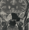
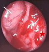
The Sternberg canal represents a congenital defect of the lateral wall of the sphenoid sinus, predisposing to spontaneous cerebrospinal fluid (CSF) leakage and the formation of meningoencephaloceles. We report the case of a 54-year-old patient presenting with a four-month history of anterior rhinorrhea in the absence of any other associated symptoms. High-resolution computed tomography and magnetic resonance imaging identified an osteomeningeal defect in the lateral sphenoid sinus wall, associated with a meningoencephalocele. The patient underwent a right transsphenoidal sphenoidotomy with multilayer reconstruction utilizing abdominal fat, conchal cartilage, and biological adhesive. Favorable clinical outcomes were achieved, with no recurrence noted during a nine-month follow-up period. Surgical repair of CSF leaks through the Sternberg canal remains technically demanding, primarily due to the anatomical complexity and restricted accessibility of the lateral sphenoid recess.
Keywords: cerebrospinal fluid; lateral craniopharyngeal canal; meningoencephalocele; rhinorrhea; sternberg canal.
Copyright © 2025, Ben Ameur El Youbi et al.
Conflict of interest statement
Human subjects: Consent for treatment and open access publication was obtained or waived by all participants in this study. Conflicts of interest: In compliance with the ICMJE uniform disclosure form, all authors declare the following: Payment/services info: All authors have declared that no financial support was received from any organization for the submitted work. Financial relationships: All authors have declared that they have no financial relationships at present or within the previous three years with any organizations that might have an interest in the submitted work. Other relationships: All authors have declared that there are no other relationships or activities that could appear to have influenced the submitted work.
Figures


Similar articles
-
Nerve-Sparing Approach to the Lateral Sphenoid Sinus Recess in a Patient With Multiple Sphenoid Encephaloceles.Ear Nose Throat J. 2025 Jul 1:1455613251353646. doi: 10.1177/01455613251353646. Online ahead of print. Ear Nose Throat J. 2025. PMID: 40596760
-
Endoscopic transpterygoid approach and skull base repair after sphenoid meningoencephalocele resection. Our experience.Acta Otorrinolaringol Esp. 2015 Jan-Feb;66(1):1-7. doi: 10.1016/j.otorri.2014.03.008. Epub 2014 Jul 20. Acta Otorrinolaringol Esp. 2015. PMID: 25052487 English, Spanish.
-
Analysis of the Causes and Experience in the Diagnosis and Treatment of Meningocele Caused by Sternberg's Canal of the Sphenoid Sinus: Two Case Reports and a Review of the Literature.Curr Med Imaging. 2023;19(9):1063-1070. doi: 10.2174/1573405619666230206103036. Curr Med Imaging. 2023. PMID: 36748216 Review.
-
Endoscopic endonasal repair of temporal lobe meningoencephalocele in the lateral recess of the sphenoid sinus, complicated by intracerebral hematoma: illustrative case.J Neurosurg Case Lessons. 2023 Dec 25;6(26):CASE23575. doi: 10.3171/CASE23575. Print 2023 Dec 25. J Neurosurg Case Lessons. 2023. PMID: 38145563 Free PMC article.
-
Transcranial approach for spontaneous CSF rhinorrhea due to Sternberg's canal intrasphenoidal meningoencephalocele: case report and review of the literature.Turk Neurosurg. 2012;22(2):242-5. doi: 10.5137/1019-5149.JTN.2902-10.1. Turk Neurosurg. 2012. PMID: 22437302 Review.
References
-
- A retrospective analysis of spontaneous sphenoid sinus fistula: MR and CT findings. Shetty PG, Shroff MM, Fatterpekar GM, Sahani DV, Kirtane MV. https://pubmed.ncbi.nlm.nih.gov/10696020/ AJNR Am J Neuroradiol. 2000;21:337–342. - PMC - PubMed
-
- A previously undescribed canal in the sphenoid bone of humans (Article in German) Sternberg M. Anat Anz. 1888;3:784–785.
-
- Sternberg's canal--cause of congenital sphenoidal meningocele. Schick B, Brors D, Prescher A. Eur Arch Otorhinolaryngol. 2000;257:430–432. - PubMed
Publication types
LinkOut - more resources
Full Text Sources
