. 2025 Jun 3:13:1611226.
doi: 10.3389/fcell.2025.1611226. eCollection 2025.

Induced neural stem cells ameliorate blood-brain barrier injury by modulating the calcium signaling pathway of astrocyte in cerebral ischemia-reperfusion rats

Affiliations

Induced neural stem cells ameliorate blood-brain barrier injury by modulating the calcium signaling pathway of astrocyte in cerebral ischemia-reperfusion rats

Xueyun Liang et al. Front Cell Dev Biol. .

Abstract

Background: Neural stem cells offer new hope for ischemic stroke patients on the basis of their potential to reverse neurological sequelae, but it is still difficult to obtain sufficient neural stem cells in the clinic. We induced human placental mesenchymal stem cells to neural stem cells (iNSCs), the therapeutic effects and possible mechanisms of iNSCs in ischemic stroke were observed in this study.

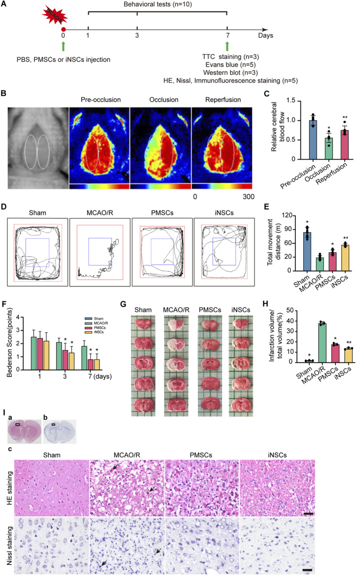
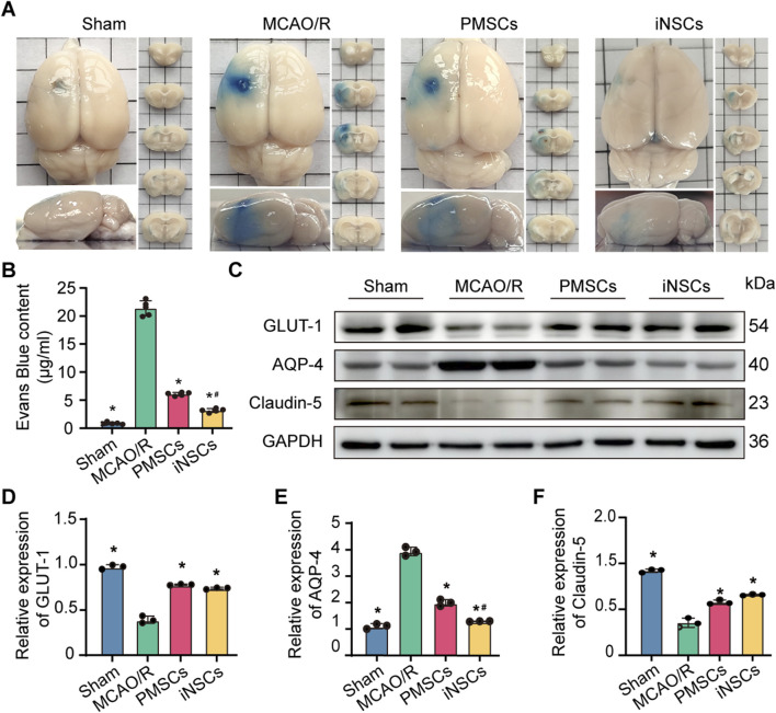
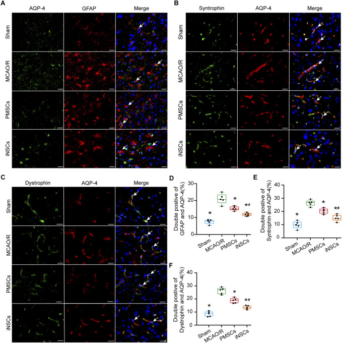
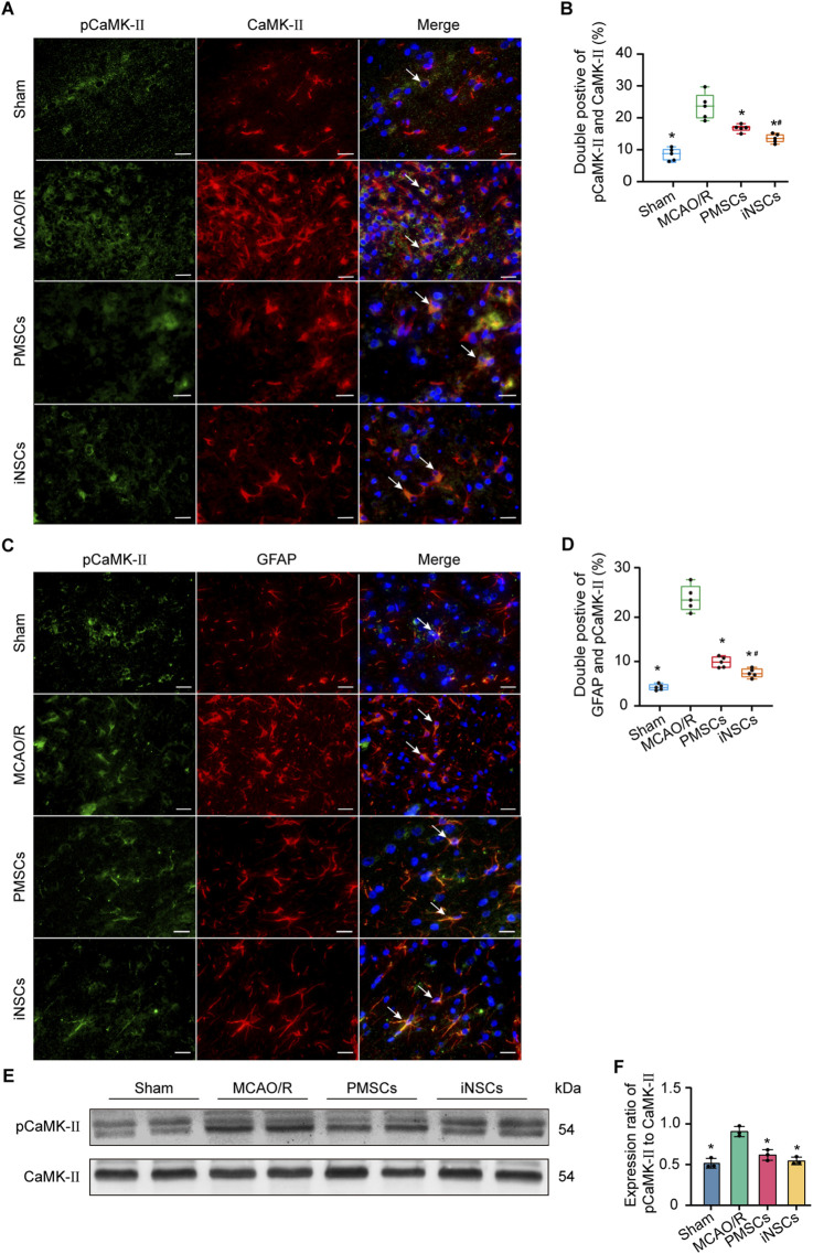
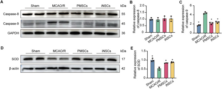
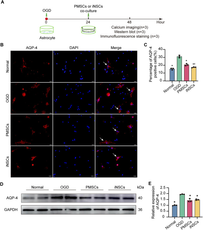
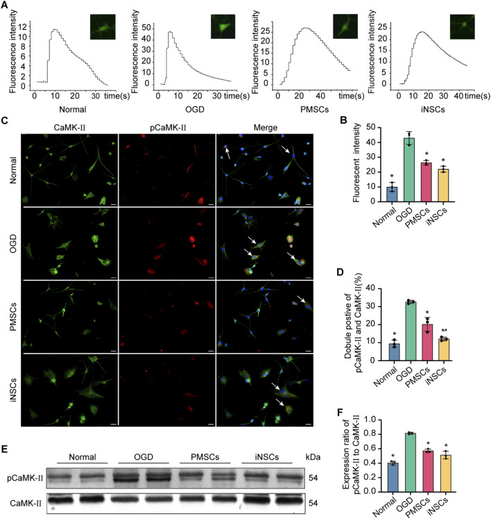
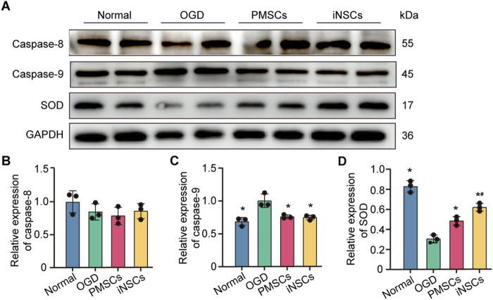
Results: Transplanted iNSCs improved neurological deficits, increased the integrity of blood-brain barrier (BBB) structure and its related proteins expression level in middle cerebral artery occlusion/reperfusion (MCAO/R) rats. The in vitro study demonstrated that iNSCs treatment inhibited Ca2+ influx in oxygen-glucose deprived (OGD)-damaged astrocytes. Additionally, iNSCs downregulated the expression level of pCaMK-II, increased the expression level of superoxide dismutase, and inhibited the expression of caspase 9 in both brain of MCAO/R rats and OGD-damaged astrocytes.

Conclusion: iNSCs transplantation improved BBB function by modulating calcium signaling pathway of astrocyte in MCAO/R rats, which proved iNSCs may be a new promising neural stem cells origin for the treatment of cerebral ischemia-reperfusion injury.

Keywords: BBB; astrocytes; calcium signaling pathways; cerebral ischemia-reperfusion; induced neural stem cells.


Conflict of interest statement

The authors declare that the research was conducted in the absence of any commercial or financial relationships that could be construed as a potential conflict of interest.

Figures

FIGURE 1
FIGURE 1
Effects of iNSCs transplantation on MCAO/R rats. (A) Schematic drawing about the design of the animal experiments. (B) Representative laser scatter images of cerebral blood flow during the entire procedure of the MCAO/R model establishment. (C) Quantitative analysis of the cerebral blood flow level during the procedure of the MCAO/R model establishment. The data are expressed as the means ± SDs, (n = 6). (D) Representative images of the open field test. (E) Quantitative analysis of the movement distance of the open field test in each group. The data are expressed as the means ± SDs, (n = 10). (F) Quantitative analysis of the Bederson score test; behavioral evaluation was performed on the 1st, 3rd, and 7th days after cells transplantation in each group. The data are expressed as the means ± SDs, (n = 10). (G) Representative images of TTC staining in each group on day 7 after cells transplantation. (H) Quantitative analysis of the infarction volume of brain on the 7th days after cells’ transplantation in each group. The data are expressed as the means ± SDs, (n = 3). (I) Representative images of HE staining and Nissl staining in each group after cells transplantation. Scale bar = 20 μm (n = 5). Comparisons of means among multiple groups were performed via one-way ANOVA followed by Tukey’s post hoc test, and *P < 0.05, compared with the MCAO/R group; # P < 0.05, compared with the PMSCs group.
FIGURE 2
FIGURE 2
Effects of iNSCs transplantation on BBB function in MCAO/R rats. (A) Representative images of Evans blue stained samples from each group. (B) Quantitative analysis of the Evans blue content in each group. The data are expressed as the means ± SDs, (n = 5). (C) Representative Western blot images of BBB structure-related protein expression in each group. (D–F) Results of quantitative analysis of the proteins expression levels of GLUT-1, AQP-4, and Claudin-5, respectively, and were normalized to that of GAPDH. The data are expressed as the means ± SDs, (n = 3). Comparisons of means among multiple groups were performed via one-way ANOVA followed by Tukey’s post hoc test, and *P < 0.05, compared with the MCAO/R group; # P < 0.05, compared with the PMSCs group.
FIGURE 3
FIGURE 3
Effects of iNSCs transplantation on AQP4 expressions in MCAO/R rats. (A–C) Representative images of immunofluorescence double staining of AQP-4 with either GFAP, Syntrophin or Dystrophin in each group. Scale bar = 20 μm. (D–F) The results of quantitative analysis results of double positive cells of AQP-4 with either GFAP, Syntrophin or Dystrophin in each group. The data are expressed as the means ± SDs, (n = 5). Comparisons of means among multiple groups were performed via one-way ANOVA followed by Tukey’s post hoc test, and *P < 0.05, compared with the MCAO/R group; # P < 0.05, compared with the PMSCs group.
FIGURE 4
FIGURE 4
Effects of iNSCs transplantation on pCaMK-II expression in MCAO/R rats. (A) Representative images of immunofluorescence double staining of pCaMK-II with CaMK-II in each group. Scale bar = 20 μm. (B) Quantitative analysis results of pCaMK-II and CaMK-II double positive cells in each group. The data are expressed as the means ± SDs, (n = 5). (C) The representative images of immunofluorescence of pCaMK-II with GFAP double staining in each group. Scale bar = 20 μm. (D) Results of Quantitative analysis of the double positive cells of pCaMK-II with GFAP in each group. The data are expressed as the means ± SDs, (n = 5). (E) Representative Western blot images of pCaMK-II and CaMK-II expression in each group. (F) Results of quantitative analysis of the proteins expression ratio of pCaMK-II to CaMK-II, and normalization to one of the values from the MCAO/R group. The data are expressed as the means ± SDs, (n = 3). Comparisons of means among multiple groups were performed via one-way ANOVA followed by Tukey’s post hoc test, and *P < 0.05, compared with the MCAO/R group; # P < 0.05, compared with the PMSCs group.
FIGURE 5
FIGURE 5
Effects of iNSCs transplantation on mitochondrial function in MCAO/R rats. (A) Representative Western blot images of Caspase 8 and Caspase 9 expression in each group. (B,C) Quantitative analysis of the expression level of Caspase 8 and Caspase 9, respectively, and normalized to GAPDH. (D) Representative Western blot images of SOD expression in each group. (E) Quantitative analysis of the expression level of SOD, which was normalized to β-actin. The data are expressed as the means ± SDs, (n = 3). Comparisons of means among multiple groups were performed via one-way ANOVA followed by Tukey’s post hoc test, and *P < 0.05, compared with the MCAO/R group; # P < 0.05, compared with the PMSCs group.
FIGURE 6
FIGURE 6
Effects of iNSCs treatment on AQP-4 expression in OGD-damaged astrocytes. (A) Schematic drawing of the design of the in vivo experiments. (B) Representative images of immunofluorescence staining of AQP-4 in astrocytes from each group. Scale bar = 20 μm. (C) Quantitative analysis of the percentage of AQP-4 positive astrocytes in each group. The data are expressed as the means ± SDs, (n = 3). (D) Representative Western blot images of AQP-4 expression in each group. (E) Quantitative analysis of the expression level of AQP-4, normalized to that of GAPDH. The data are expressed as the means ± SDs, (n = 3). Comparisons of means among multiple groups were performed via one-way ANOVA followed by Tukey’s post hoc test, and *P < 0.05, compared with the OGD group; # P < 0.05, compared with the PMSCs group.
FIGURE 7
FIGURE 7
Effects of iNSCs treatment on the calcium signaling pathway in OGD damaged astrocytes. (A,B) Representative images of calcium imaging in astrocytes from each group and quantitative analysis of the intracellular calcium in astrocytes from each group. (C,D) Representative images of immunofluorescence double staining of pCaMK-II and CaMK-II in astrocytes from each group and quantitative analysis of the percentage of pCaMK-II and CaMK-II positive cells in each group. The arrows indicate co-labeled positive cells, scale bar = 20 μm. (E) Representative Western blot images of pCaMK-II and CaMK-II expression in each group. (F) Quantitative analysis of the expression level of pCaMK-II, which was normalized to that of CaMK-II. The data are expressed as the means ± SDs, (n = 3). Comparisons of means among multiple groups were performed via one-way ANOVA followed by Tukey’s post hoc test, and *P < 0.05, compared with the OGD group; #P < 0.05, compared with the PMSCs group.
FIGURE 8
FIGURE 8
Effects of iNSCs treatment on mitochondrial function in OGD damaged astrocytes. (A) Representative Western blot images of Caspase 8, Caspase 9 and SOD expression in each group. (B–D) Quantitative analysis of the expression levesl of Caspase 8, Caspase 9 and SOD, respectively, and normalization to the level of GAPDH. The data are expressed as the means ± SDs, (n = 3). Comparison of means among multiple groups was performed using one-way ANOVA followed by Tukey’s post hoc test, and *P < 0.05, compared with the OGD group; # P < 0.05, compared with the PMSCs group.

Similar articles

References

    1. Awasthi V. A., Dhankar V., Singh S. (2024). Novel therapeutic targets for reperfusion injury in ischemic stroke: understanding the role of mitochondria, excitotoxicity and ferroptosis. Vasc. Pharmacol. 156, 107413. 10.1016/j.vph.2024.107413 - DOI - PubMed
    1. Ballabh P., Braun A., Nedergaard M. (2004). The blood-brain barrier: an overview: structure, regulation, and clinical implications. Neurobiol. Dis. 16 (1), 1–13. 10.1016/j.nbd.2003.12.016 - DOI - PubMed
    1. Bederson J. B., Pitts L. H., Germano S. M., Nishimura M. C., Davis R. L., Bartkowski H. M. (1986). Evaluation of 2,3,5-triphenyltetrazolium chloride as a stain for detection and quantification of experimental cerebral infarction in rats. Stroke 17 (6), 1304–1308. 10.1161/01.str.17.6.1304 - DOI - PubMed
    1. Buchlak Q. D., Esmaili N., Moore J. (2024). Opportunities for developing neural stem cell treatments for acute ischemic stroke: a systematic review and gap analysis. J. Clin. Neurosci. 120, 64–75. 10.1016/j.jocn.2023.12.021 - DOI - PubMed
    1. Cao G., Guo J., Yang K., Xu R., Jia X., Wang X. (2024). DCPIB attenuates ischemia-reperfusion injury by regulating microglial M1/M2 polarization and oxidative stress. Neuroscience 551, 119–131. 10.1016/j.neuroscience.2024.05.008 - DOI - PubMed

LinkOut - more resources