. 2025 Aug 6;36(11):609-622.
doi: 10.1097/WNR.0000000000002184. Epub 2025 Jun 13.

Knockdown of miR-204-5p promotes nerve regeneration and functional recovery after hypoxic-ischemic brain damage in neonatal rats via the Wnt2/Ephrin-A2/EphA7 pathway

Affiliations

Knockdown of miR-204-5p promotes nerve regeneration and functional recovery after hypoxic-ischemic brain damage in neonatal rats via the Wnt2/Ephrin-A2/EphA7 pathway

Mengzao He et al. Neuroreport. .

Abstract

Objective: Neonatal hypoxic-ischemic brain damage (HIBD) can cause short- and long-term neurological damage. MicroRNA (miR)-204-5p is closely associated with nerve injury caused by brain injury, but its mechanism in HIBD is not very clear.

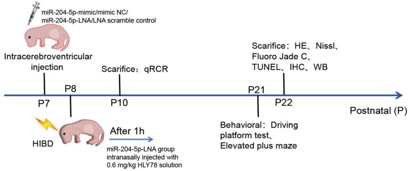
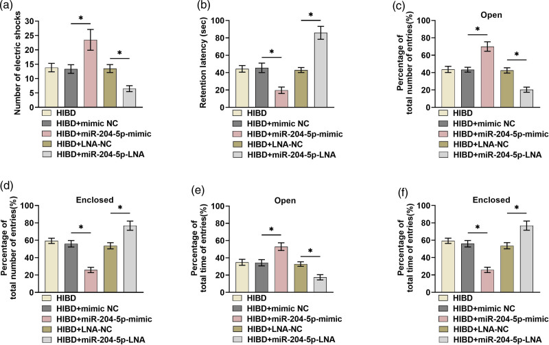
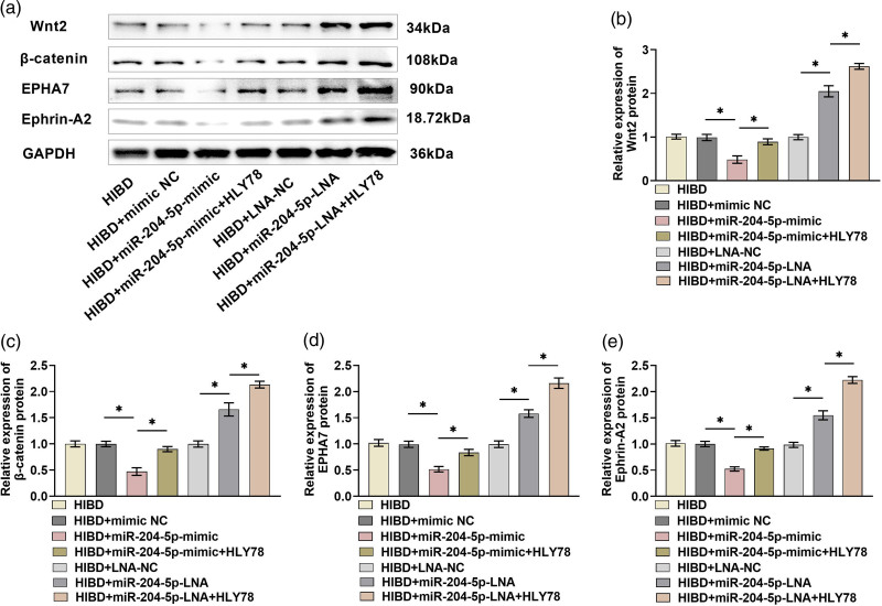
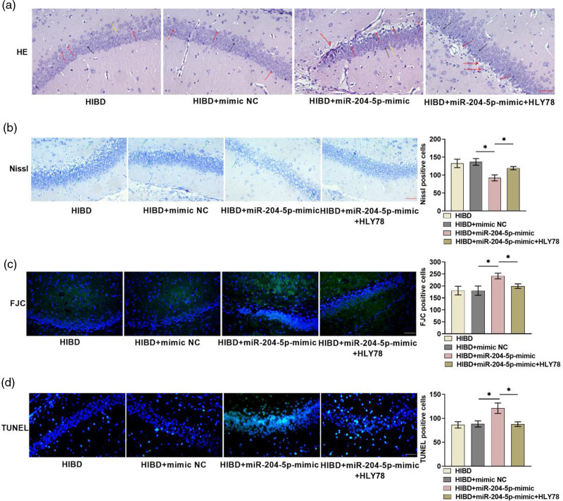
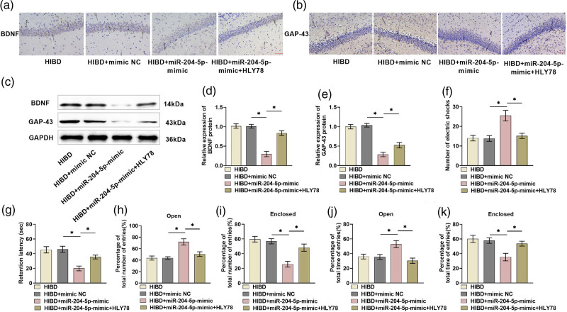
Methods: The neonatal rat's HIBD model was constructed by the modified Rice-Vannucci method, and the expression of miR-204-5p was detected. After overexpression or knockdown of miR-204-5p and application of Wnt2 activator HLY78, the histopathological changes and neuronal degeneration in the hippocampal CA1 region were observed with pathological staining. The neurological function was assessed with a diving platform test and elevated plus-maze test. Nerve regeneration-related protein and Wnt2/Ephrin-A2 (Eph receptor-interacting proteins)/EphA7 (erythropoi-etin-producing hepatomocellular receptor) signaling pathway protein levels were detected by immunohistochemistry and western blot, respectively.

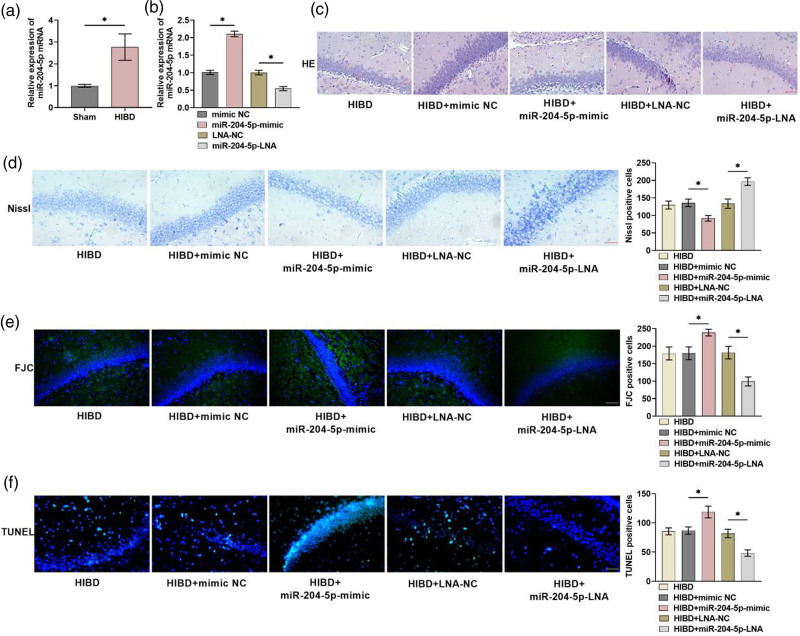
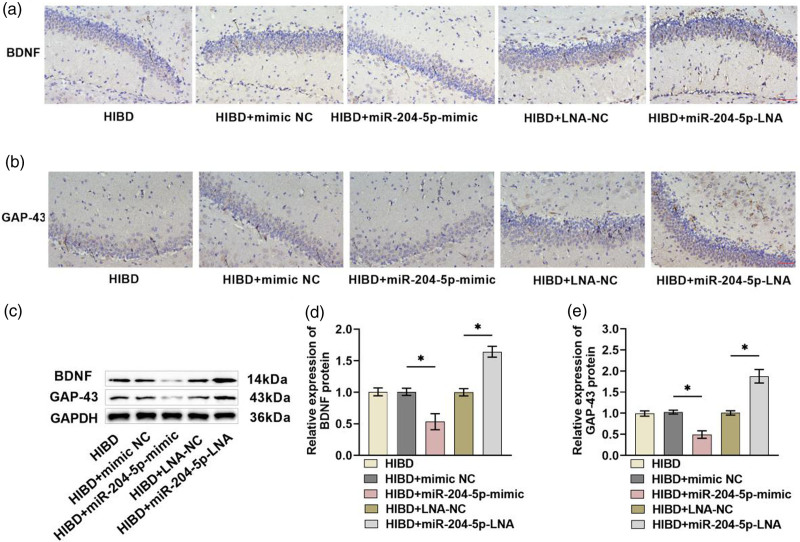
Results: miR-204-5p was highly expressed in HIBD. When miR-204-5p was knocked down, the morphology of nerve cells and Nissl bodies was notably improved, Fluoro-Jade C and terminal deoxynucleotidyl transferase-mediated dUTP nick end labeling-positive cells number was significantly reduced. The levels of brain-derived neurotrophic factor and growth-associated protein 43 were significantly increased, and the behavioral indicators of the diving platform and elevated plus-maze test were significantly alleviated. The nerve injury was repaired, and the Wnt2/Ephrin-A2/EphA7 signaling pathway protein was notably elevated. The overexpressed miR-204-5p aggravated the nerve injury in HIBD rats. After the application of HLY78, the neuropathological damage of HIBD rats was further repaired, and the nerve regeneration and function were also significantly improved.

Conclusion: Knockdown of miR-204-5p can improve HIBD in neonatal rats by activating the Wnt2/Ephrin-A2/EphA7 signaling pathway to encourage nerve regeneration and functional recovery.

Keywords: HIBD; Wnt2/Ephrin-A2/EphA7 pathway; miR-204-5p; nerve regeneration.


Conflict of interest statement

There are no conflicts of interest.

Figures

Fig. 1
Fig. 1
Flow chart of in-vivo experimental design. HE, hematoxylin and eosin; HIBD, hypoxic–ischemic brain damage; IHC, immunohistochemistry; miR, microRNA; NC, negative control; TUNEL, terminal deoxynucleotidyl transferase-mediated dUTP nick end labeling; WB, western blot.
Fig. 2
Fig. 2
The expression of miR-204-5p is up-regulated in HIBD, and the knockdown of miR-204-5p can improve the pathological damage of the hippocampus in HIBD. (a) miR-204-5p expression was tested by qRT-PCR at 48 h after HIBD, and the miR-204-5p level was significantly boosted (n = 4). (b) The overexpression or knockdown efficiency of miR-204-5p was tested by qRT-PCR, and it was effectively overexpressed or knocked down (n = 4). (c) After HE staining of brain tissue sections, nerve cell size, and shape were relatively normal and the arrangement was more regular when miR-204-5p knockdown. After overexpressing miR-204-5p, nerve cells were swollen and irregular in shape (×40, 50 μm). (d) Nissl staining was performed on brain tissue sections, and it was found that after miR-204-5p knockdown, the structure of Nissl bodies was relatively complete and the number was large; after overexpressed miR-204-5p, Nissl bodies morphology was disordered and the number was significantly reduced (×40, 50 μm). (e) FJC staining disclosed that miR-204-5p knockdown alleviated neuronal degeneration, while miR-204-5p overexpression was the opposite (×40, 50 μm). (f) TUNEL staining of brain tissue sections revealed that miR-204-5p knockdown shrunk neuronal apoptosis, while its overexpression was the opposite (×40, 50 μm) (n = 6, *P < 0.05). The black arrow indicates normal neurons; the red arrows indicate damaged neurons with swelling, vacuolation, and nuclear shrinkage; the yellow arrow indicates a low density of neurons in HE staining results. The green arrow indicates the Nissl body in Nissl staining results. FJC, Fluoro-Jade C; HE, hematoxylin and eosin; HIBD, hypoxic–ischemic brain damage; miR, microRNA; NC, negative control; qRT-PCR, quantitative reverse transcription PCR; TUNEL, terminal deoxynucleotidyl transferase-mediated dUTP nick end labeling.
Fig. 3
Fig. 3
Knockdown of miR-204-5p promotes nerve regeneration after HIBD in neonatal rats. (a and b) BDNF and GAP-43 expression were detected by immunohistochemistry. Brown positive cell number elevated significantly after miR-204-5p knockdown and decreased significantly after miR-204-5p overexpression (×40, 50 μm). (c–e) BDNF and GAP-43 levels were tested by WB. They were significantly increased after miR-204-5p knockdown and markedly declined after miR-204-5p overexpression (n = 6, *P < 0.05). The black arrows indicate the location of the protein that is positively expressed in the cell in IHC staining results. BDNF, brain-derived neurotrophic factor; FJC, Fluoro-Jade C; GAP-43, growth-associated protein 43; HIBD, hypoxic–ischemic brain damage; IHC, immunohistochemistry; miR, microRNA; NC, negative control; TUNEL, terminal deoxynucleotidyl transferase-mediated dUTP nick end labeling; WB, western blot.
Fig. 4
Fig. 4
Knockdown of miR-204-5p promotes neurological recovery after HIBD in neonatal rats. (a and b) The results of the diving platform test showed that the error number was obviously reduced after miR-204-5p knockdown, the latency was significantly increased. After miR-204-5p overexpression, the error number was markedly increased, and the latency was significantly reduced. (c–f) The results of the elevated plus-maze test showed that rats entering the open arm number and time were notably reduced after miR-204-5p knockdown, rats entering the closed arm number and time were markedly raised. After miR-204-5p overexpression, rats entering the open arm number and time were obviously increased, rats entering the closed arm number and time were remarkably reduced (n = 6, *P < 0.05). HIBD, hypoxic–ischemic brain damage; miR, microRNA; NC, negative control.
Fig. 5
Fig. 5
miR-204-5p can regulate Wnt2/Ephrin-A2/EphA7 pathway. (a–e) Wnt2/Ephrin-A2/EphA7 pathway protein expression in the hippocampus was detected by western blot. Wnt2, β-catenin, Ephrin-A2, and EphA7 levels were notably decreased after overexpressed miR-204-5p, and significantly raised after miR-204-5p knockdown. The levels were increased after the application of HLY78 on the basis of overexpression or knockdown of miR-204-5p (n = 6, *P < 0.05). HIBD, hypoxic–ischemic brain damage; miR, microRNA; NC, negative control.
Fig. 6
Fig. 6
HLY78 can partially restore miR-204-5p to promote the pathological damage of hypoxic–ischemic brain tissue in neonatal rats. (a) HE staining of brain tissue sections showed that after the application of HLY78, the size and morphology of nerve cells were restored compared with the overexpressed miR-204-5p, the arrangement was more regular (×40, 50 μm). (b) Nissl staining of brain tissue sections showed that compared with miR-204-5p overexpression, Nissl bodies structure was restored and the number was significantly increased after the application of HLY78 (×40, 50 μm). (c) FJC staining of brain tissue sections showed that the application of HLY78 alleviated neuronal degeneration caused by miR-204-5p overexpression (×40, 50 μm). (d) TUNEL staining of brain tissue sections showed that after the application of HLY78, neuronal apoptosis was obviously reduced (×40, 50 μm) (n = 6, *P < 0.05). The black arrow indicates normal neurons; the red arrows indicate damaged neurons with swelling, vacuolation, and nuclear shrinkage; the yellow arrow indicates a low density of neurons in HE staining results. The green arrow indicates the Nissl body in Nissl staining results. FJC, Fluoro-Jade C; HE, hematoxylin and eosin; HIBD, hypoxic–ischemic brain damage; miR, microRNA; NC, negative control; TUNEL, terminal deoxynucleotidyl transferase-mediated dUTP nick end labeling.
Fig. 7
Fig. 7
HLY78 can partially restore the phenomenon that miR-204-5p inhibits nerve regeneration and functional recovery after HIBD in neonatal rats. (a–e) BDNF and GAP-43 levels were tested by immunohistochemistry and WB. The number of brown–yellow positive cells and protein levels were significantly raised after the application of HLY78 relative to overexpressed miR-204-5p (×40, 50 μm). (f and g) The results of the diving platform test showed that the error number was remarkably reduced and the latency was notably rebounded after the application of HLY78. (h–k) The elevated plus-maze test showed that rats entering the open arm number and time were significantly reduced after the application of HLY78, entering the closed arm number and time were significantly increased (n = 6, *P < 0.05). The black arrows indicate the location of the protein that is positively expressed in the cell in IHC staining results. BDNF, brain-derived neurotrophic factor; GAP-43, growth-associated protein 43; HIBD, hypoxic–ischemic brain damage; IHC, immunohistochemistry; miR, microRNA; WB, western blot.

Similar articles

References

    1. Tetorou K, Sisa C, Iqbal A, Dhillon K, Hristova M. Current therapies for neonatal hypoxic-ischaemic and infection-sensitised hypoxic-ischaemic brain damage. Front Synaptic Neurosci 2021; 13:709301. - PMC - PubMed
    1. Greco P, Nencini G, Piva I, Scioscia M, Volta CA, Spadaro S, et al. Pathophysiology of hypoxic-ischemic encephalopathy: a review of the past and a view on the future. Acta Neurol Belg 2020; 120:277–288. - PubMed
    1. Yao HW, Kuan CY. Early neutrophil infiltration is critical for inflammation-sensitized hypoxic-ischemic brain injury in newborns. J Cereb Blood Flow Metab 2020; 40:2188–2200. - PMC - PubMed
    1. Zhu K, Zhu X, Liu S, Yu J, Wu S, Hei M. Glycyrrhizin attenuates hypoxic-ischemic brain damage by inhibiting ferroptosis and neuroinflammation in neonatal rats via the HMGB1/GPX4 pathway. Oxid Med Cell Longevity 2022; 2022:8438528. - PMC - PubMed
    1. Gou Z, Su X, Hu X, Zhou Y, Huang L, Fan Y, et al. Melatonin improves hypoxic-ischemic brain damage through the Akt/Nrf2/Gpx4 signaling pathway. Brain Res Bull 2020; 163:40–48. - PubMed

LinkOut - more resources