. 2025 Sep;37(5):786-799.
doi: 10.1177/10406387251346909. Epub 2025 Jun 18.

Efficient blocking ELISA for bovine alphaherpesvirus 1 using a gB epitope-specific monoclonal antibody

Affiliations

Efficient blocking ELISA for bovine alphaherpesvirus 1 using a gB epitope-specific monoclonal antibody

Shuang-Yan Xiao et al. J Vet Diagn Invest. 2025 Sep.

Abstract

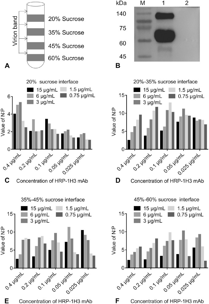
Infectious bovine rhinotracheitis (IBR) is an infectious respiratory disease in cattle that is caused by bovine alphaherpesvirus 1 (BoAHV1). We immunized BALB/c mice with inactivated and purified BoAHV1 to prepare hybridoma cells. After the successful establishment of a positive hybridoma cell line, co-immunoprecipitation coupled with mass spectrometry unveiled the predominant targeting of glycoprotein B (gB) by the hybridoma cells. Through bioinformatics analysis and Western blot techniques, we identified the epitope of the monoclonal antibody (mAb) against gB to amino acids 1-170. Subsequently, the 1H3 mAb was leveraged for the development of a gB blocking ELISA (gB-bELISA), utilizing inactivated BoAHV1 virions as the coating antigen. The optimized protocol involved diluting samples 2-fold with 1% fish gelatin, followed by incubation periods of 120 min for samples, 30 min for HRP-conjugated 1H3 mAb, and 15 min for the TMB substrate. We validated our assay using 268 bovine serum samples with clear backgrounds and established the cutoff value of 43.8% through ROC analysis. Additionally, we tested 256 clinical bovine serum samples using both our gB-bELISA and a virus neutralization test, achieving a concordance rate of 95.3%. Based on testing 495 randomly selected sera from 18 counties for BoAHV1 antibodies with our gB-bELISA, the seroprevalence of IBR in the Central China region was 22.0% (95% CI: 18.4, 25.7). Our gB-bELISA could be a valuable tool for the clinical detection of IBR, supporting disease control and eradication efforts.

Keywords: antigen epitope; blocking ELISA; bovine alphaherpesvirus 1; glycoprotein B; infectious bovine rhinotracheitis.


Conflict of interest statement

Declaration of conflicting interestsThe authors declared no potential conflicts of interest with respect to the research, authorship, and/or publication of this article.

Figures

Figure 1.
Figure 1.
Identification of 1H3 monoclonal antibody (mAb). A. Isotype identification of 1H3 mAb. B. Determination of the 1H3 mAb titer by indirect ELISA. The dotted line indicates an OD450 value of 1.0. C. Analysis of 1H3 mAb purity by SDS-PAGE; lane 1 was purified 1H3 mAb. M = protein ladder. D. Reactivity of 1H3 mAb was analyzed with immunofluorescence assay on MDBK cells infected with bovine alphaherpesvirus 1 (BoAHV1), pseudorabies virus (PRV), and bovine viral diarrhea virus (BVDV). The cells were incubated with 1H3 mAb, with Sp2/0 cell supernatant used as negative control. Bars = 10 µm.
Figure 2.
Figure 2.
Characterization of 1H3 monoclonal antibody (mAb). A. Immunoprecipitation–mass spectrometry analysis identified the interaction protein and binding sites of 1H3 mAb, which reacted with peptides at 7 different sites on bovine alphaherpesvirus 1 (BoAHV1) gB. The black box indicates full-length BoAHV1 gB. B, C. Western blot analysis demonstrating the specificity of 1H3 mAb for eukaryotically expressed gB. B. Membrane incubated with HRP-conjugated mouse anti-His tag antibody (1:5,000 dilution). C. Membrane incubated with HRP-conjugated 1H3 mAb (1:10,000 dilution): lane 1 was the eukaryotically expressed gB; lane 2 was HEK293T cells transfected with pcDNA3.1. D. Neutralizing activity of 1H3 mAb against BoAHV1 was evaluated by plaque reduction neutralization assay. The dotted line indicates a percentage of inhibition value of 50%.
Figure 3.
Figure 3.
Epitope mapping using truncated gB fusion proteins. A. Schematic representation of bovine alphaherpesvirus 1 gB and its truncated derivatives. The black box is the general graphical representation of the gB precursor, with arrowheads indicating signal peptides, extracellular regions, transmembrane regions, and intracellular regions; gray boxes are the mature forms of gB truncation mutants. B. Expression analysis of 8 recombinant gB truncated proteins and full-length gB. All 9 fragments were cloned into pcDNA3.1 and were detected with anti-His antibodies, and each recombinant protein (gB-I to gB-VIII) was analyzed in duplicate. C. Reactivity of full-length and 8 truncated gB fusion proteins (gB-I to gB-VIII) with 1H3 monoclonal antibody; each recombinant protein was tested in duplicate.
Figure 4.
Figure 4.
Antigen selection. A. Distribution of bovine alphaherpesvirus 1 (BoAHV1) following sucrose density gradient ultracentrifugation. B. Reactivity of 1H3 monoclonal antibody (mAb) with BoAHV1 was detected by Western blot; membrane was incubated with HRP-conjugated 1H3 mAb (1:10,000 dilution): lane 1 was the purified BoAHV1; lane 2 was MDBK cells (negative control). C–F. Determination of the optimal concentration of coating antigen and antibody through checkerboard titration; viral fractions were collected from 4 sucrose density interfaces (20%, 20%-35%, 35%-45%, and 45%-60%) and tested with HRP-conjugated 1H3 mAb. C. Virions from 20% sucrose interface. D. Virions from 20%–35% sucrose interface. E. Virions from 35%-45% sucrose interface. F. Virions from 45%-60% sucrose interface. N:P = negative control OD450:positive control OD450 ratio.
Figure 5.
Figure 5.
Establishment of blocking ELISA. A–F. A single-factor assay was performed to determine the optimal conditions for each parameter. A. Blocking buffer [0.1%, 0.2%, or 0.5% bovine serum albumin (BSA), or 5% skim milk]. B. Sample diluents (1% PVP10000, 1% PVP40000, 1% fish gelatin, 1% porcine gelatin, or PBS). C. Serum diluted factors (bovine serum was diluted 0-, 2-, 4-, 6-, and 10-fold with 1% fish gelatin). D. Sample incubation time (30, 45, 60, 90, 120, or 150 min at 37°C). E. HRP-conjugated 1H3 mAb incubation time (15, 30, 45, or 60 min at 37°C). F. TMB substrate incubation conditions (20–25°C for 10 min and 15 min, 37°C for 10, 15, and 20 min). N = negative control OD450; N:P = negative control OD450:positive control OD450 ratio; P = positive control OD450.
Figure 6.
Figure 6.
Analysis of gB blocking ELISA (gB-bELISA) characterization. A, B. Determination of criteria for bELISA: 268 bovine serum samples with known backgrounds [determined by virus neutralization test (VNT)] were used to standardize the threshold for established gB-bELISA. A. The detection results of serum samples with the gB-bELISA. B. ROC analysis of the data. C. The specificity of the gB-bELISA: 8 serum samples were tested, including bovine alphaherpesvirus 1 (BoAHV1)-, BoAHV5-, bovine viral diarrhea virus (BVDV)-, foot-and-mouth disease virus (FMDV)-, Mycobacterium bovis–, Brucella-positive sera, as well as BoAHV1-negative sera and MDBK cell immune sera. D. Limit of detection analysis: high-, medium-, and weak-positive sera were selected from clinical bovine sera previously tested by the VNT; 2-fold gradient dilutions were prepared and tested by the gB-bELISA. E. We analyzed 212 bovine serum samples by the gB-bELISA. F. The within-run CV was calculated between replicates I–II, and the between-run CV was determined between replicates I–IV; the mean value and 95% CIs were calculated for each reactivity level and overall. Means are the dots and squares in each CI. The dotted line indicates CV% value of 0%.

Similar articles

References

    1. Bender FC, et al. Antigenic and mutational analyses of herpes simplex virus glycoprotein B reveal four functional regions. J Virol 2007;81:3827–3841. - PMC - PubMed
    1. Bitsch V. The P 37/24 modification of the infectious bovine rhinotracheitis virus-serum neutralization test. Acta Vet Scand 1978;19:497–505. - PMC - PubMed
    1. Deregt D, et al. A comparative evaluation of two sensitive serum neutralization tests for bovine herpesvirus-1 antibodies. Can J Vet Res 1993;57:56–59. - PMC - PubMed
    1. El-Kholy AA, et al. Baculovirus expression and diagnostic utility of the glycoprotein E of bovine herpesvirus-1.1 Egyptian strain “Abu-Hammad”. J Virol Methods 2013;191:33–40. - PubMed
    1. Esteves PA, et al. An indirect ELISA to detect antibodies to the gC of bovine alphaherpesvirus 1 (BoAHV1) displaying no crossreactivity with antibodies induced by bovine alphaherpesvirus 5 (BoAHV5). J Virol Methods 2023;320:114785. - PubMed

MeSH terms

LinkOut - more resources