. 2025 Jun 6;53(11):gkaf517.
doi: 10.1093/nar/gkaf517.

Non-coding Y RNA fragments in a complex with YBX1 modulate PARP1 residency at DNA double strand breaks

Affiliations

Non-coding Y RNA fragments in a complex with YBX1 modulate PARP1 residency at DNA double strand breaks

Annabelle Shaw et al. Nucleic Acids Res. .

Abstract

To protect genome integrity from pervasive threats of damage and prevent diseases like cancer, cells employ an integrated network of signalling pathways called the DNA damage response. These pathways involve both protein and RNA components, which can act within the damaged cell or be transferred intercellularly to influence population-wide responses to damage. Here, we show that radioprotection can be conferred by damage-derived exosomes and is dependent on YBX1-packaged Y3-derived ysRNA. In recipient cells, ysRNAs are methylated on cytosine and bound by m5C reader, YBX1. YBX1/ysRNA localise at double strand break (DSB) sites to promote efficient DNA repair and cell survival through complex formation with PARP1. YBX1 modulates PARP1 auto-modification by facilitating ysRNA ADP-ribosylation, promoting increased PARP1 residency at DSBs. Our data highlight an unprecedented role for these under-studied species of small non-coding RNAs, identifying them as a novel substrate for PARP1 mediated ADP-ribosylation with a function in DNA repair.


Conflict of interest statement

The authors declare no competing interests.

Figures

Graphical Abstract
Graphical Abstract
Figure 1.
Figure 1.
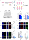
Exosomes derived from damaged cells mediate protective YBX1-dependent bystander effect. (A) Schematic overview of exosome transfer experiment, generated in Biorender. NDD EVs = non-damage-derived exosomes, DD EVs = damage-derived exosomes, IR = ionising radiation. (B) Representative Western blot showing expression levels of pCHK1 S317 and γH2AX, with CHK1 and GAPDH as loading controls, following exosome transfer (24 h) and irradiation (10 Gy, 2 h) of HEK293T cells. CM = clear media only control. (C) Bar chart showing the average fold change in γH2AX and pCHK1 expression levels in IR-treated HEK293T cells upon PBS/exosome transfer. Error bars, mean ± SEM. N ≥ 3, independent experiments. Statistical significance was determined using one-way ANOVA with Tukey’s multiple comparison, *P ≤ 0.05, **P ≤ 0.01, ***P ≤ 0.001. (D) Left panel shows representative immunofluorescence images of γH2AX and pCHK1 expression levels upon transfer of NDD EVs, DD EVs, or a PBS control (24 h) in IR-treated (10 Gy, 2 h) and untreated HEK293T cells. Right panel shows quantification from independent experiments where N ≥ 100. Statistical significance was determined using Kruskall-Wallis test with Dunn’s multiple comparison., ****P ≤ 0.0001. (E) Top panel shows representative immunofluorescence images of γH2AX and pCHK1 expression levels upon transfer of DD EVs (24 h) from wild type (WT) or YBX1 knockout (YBX1-/-) donor HEK293T cells in IR-treated (10 Gy, 2 h) recipient HEK293T cells. Bottom panel shows quantification from independent experiments where N ≥ 100. Statistical significance was determined using Kruskall-Wallis test with Dunn’s multiple comparison., ****P ≤ 0.0001.
Figure 2.
Figure 2.
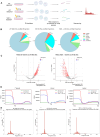
Y RNA derived fragments in exosomes are responsive to DNA damage and YBX1 knockout. (A) Schematic overview of method followed to obtain RNA from exosomes for small RNA sequencing, generated in Biorender. (B) Pie charts showing the proportion of reads corresponding to different classes of sncRNA obtained from wild-type non-damage-derived exosomes (WT NDD EVs), wild-type damage-derived exosomes (WT DD EVs) and YBX1 knockout damage-derived exosomes (YBX1-/- DD EVs). (C) Volcano plots showing differentially expressed Y RNA in exosomes upon damage (left) and YBX1 knockout (right). Coloured points show up- and downregulated Y RNA-corresponding reads with log2FC > 1 and log2FC < −1 and P-adj < 0.001, respectively. Y1 and Y3 associated transcripts are shown in red and blue, respectively. (D) Metagene distribution of commonly differentially expressed Y RNAs, Y1 RNA, and Y3 RNA reads mapped along normalised gene length, in relation to transcription start and end sites (TSS and TES, respectively). (E) Size distribution of reads corresponding to Y RNA from WT NDD EVs, WT DD EVs, and YBX1-/- DD EVs (left to right, respectively).
Figure 3.
Figure 3.
YsRNAs are nuclear, modified by NSUN2 and interact with YBX1 at DSBs. (A) Representation of synthetic ysRNA oligonucleotide sequences used for subsequent experiments. (B) Representative images (left) and quantification (right) of nuclear localisation of AlexaFluor488-labelled ysRNA oligonucleotides following transfection into HEK293T cells and IR-treatment (10 Gy, 1 h). Mean ± SEM. Quantification was carried out using ImageJ and represented as the percentage of cells per frame with nuclear fluorescent signal (N ≥ 6 frames, at least 70 cells per condition). Statistical significance was determined using two-way ANOVA, *P ≤ 0.05, ***P ≤ 0.001. (C) Schematic overview of biotinylated RNA immunoprecipitation, generated in Biorender. (D) Co-immunoprecipitation of YBX1 with biotinylated Y3 5′ or Y1 5′ ysRNA oligonucleotide, or beads only control (no RNA) from HEK293T cell nuclear lysates. Numbers represent fold change in band intensity compared with beads only control, normalised to input. Quantified using ImageJ. (E) Slot blot for m5C modification (left) and total RNA stain (right) following pulldown of biotinylated Y3 5′ ysRNA oligonucleotide after incubation with cytoplasmic (Cyt) and nuclear (Nuc) fractions from IR-treated (10 Gy, 10 min, +IR) and untreated (-IR) cells. Numbers represent signal intensity of m5C in Y3 5′ pulldown, normalised to corresponding total RNA signal. Quantified using ImageJ. (F) Representative images (left) and quantification (right) of nuclear localisation of AlexaFluor488-labelled Y3 5′ oligonucleotides following transfection into wild type (WT) or YBX1 knockout (YBX1-/-) HEK293T cells and IR-treatment (10 Gy, 1 h). Mean ± SEM. Quantification was carried out using ImageJ and represented as the percentage of cells per frame with nuclear fluorescent signal (N ≥ 3, at least 40 cells analysed). Statistical significance was determined using Student’s t-test, *P ≤ 0.05. (G) Representative images (left) and quantification (right) of nuclear localisation of AlexaFluor488-labelled Y3 5′ oligonucleotides following transfection into HEK293T cells treated with negative control siRNA (siNeg) or siRNA against NSUN2 (siNSUN2) (60 nM, 48 h) and IR-treatment (10 Gy, 1 h). Mean ± SEM. Quantification was carried out using ImageJ and represented as the percentage of cells per frame with nuclear fluorescent signal (N ≥ 6 frames, at least 80 cells analysed). Statistical significance was determined using one-way ANOVA with Tukey’s multiple comparison, **P ≤ 0.01. (H) Representative images (top and bottom left) and quantification (bottom right) of PLA between YBX1 and γH2AX, combined with transfection of AlexaFluor488-labelled synthetic ysRNA oligonucleotides (Y1 3′, Y1 5′, and Y3 5′, 24 h) in U2OS cells. Quantification represented as percentage of cells with overlapping green and red foci per frame, N ≥ 6 frames. Statistical significance determined by Kruskall–Wallis test with Dunn’s multiple comparison. *P ≤ 0.05.
Figure 4.
Figure 4.
YsRNA/YBX1 promote efficient DDR and cell survival. (A) Representative immunofluorescence images (left) and quantification (right) showing levels of pCHK1 S317 and γH2AX following transfection with synthetic ysRNA oligonucleotides, or mock control transfection, and IR-treatment (10 Gy) of HEK293T cells. Nuclear signal intensity was quantified using CellProfiler. N ≥ 100. Statistical significance determined by Kruskal–Wallis test with Dunn’s multiple comparison. *P ≤ 0.05, **P ≤ 0.01, ****P ≤ 0.0001. (B) Representation of ASO sequences used to downregulate expression of Y3 RNA (Y3 ASO) and luciferase control (Luc ASO). (C) Western blot showing expression of γH2AX and GAPDH as a loading control following IR treatment (10 Gy, +IR, indicated time points) of HEK293T cells treated for 48 h with control (Luc ASO) or Y3-RNA targeting ASO (Y3 ASO). (D) Western blot showing expression of γH2AX, and GAPDH as a loading control, in untreated (0) or IR-treated (10 Gy) wild type (WT) or YBX1 knockout (YBX1-/-) HEK293T cells at various time points after damage (1, 6, and 24 h). (E) Quantification of MTT assay, showing the cytotoxicity induced by IR (5 Gy) in HeLa cells treated with Y3 or control (Luc) ASO. Mean ± SEM, N ≥ 12. Statistical significance determined using multiple unpaired t-tests. *P ≤ 0.05, ***P ≤ 0.001. (F) Representative images (left) and quantification (right) of clonogenic survival assay. HeLa cells treated with control (Luc), Y1 or Y3 ASO (48 h) were untreated (-IR) or subjected to IR (2 Gy, +IR) and growth measured after 5 days. Quantification represented as cytotoxicity upon IR treatment. Mean ± SEM. Statistical significance determined by Kruskal–Wallis test with Dunn’s multiple comparison, **P ≤ 0.01.
Figure 5.
Figure 5.
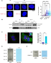
YBX1 interacts with PARP1, facilitating ADP-ribosylation of ysRNA. (A) PLA of YBX1 and γH2AX in HeLa cells, with and without IR treatment (10 Gy, +IR, and -IR, respectively), including single antibody control assays. Left panel shows representative images, and right panel shows quantification carried out in CellProfiler. Statistical significance was determined using Kruskall–Wallis test with Dunn’s multiple comparison, ****P ≤ 0.0001. (B) Detection of YBX1 in co-immunoprecipitation of YFP-PARP1 from mock-transfected or YFP-PARP1-transfected wild type (WT) and YBX1 knockout (YBX1-/-) HEK293T cells, following subjection to 10 Gy IR (+) or left untreated (-). (C) Representative images (left) and quantification (right) of PLA between YBX1 and PARP1 upon IR treatment (10 Gy, 10 min) of U2OS cells, combined with transfection of AlexaFluor488-labelled synthetic ysRNA oligonucleotides (Y1 3′ and Y3 5′). Quantification represented as percentage of cells with overlapping green and red foci per frame, N ≥ 6 frames. Statistical significance determined by Mann–Whitney test. **P ≤ 0.01. (D) Slot blot for ADP-ribosylation (ADPr) modification (left) and total RNA stain (right) following pulldown of biotinylated Y3 5′ ysRNA oligonucleotide or beads only control after incubation with cytoplasmic (Cyt) and nuclear (Nuc) fractions from IR-treated (10 Gy, 10 min, +IR) and untreated (-IR) HEK293T cells. Numbers represent signal intensity of ADPr in Y3 5′ pulldown, normalised to corresponding total RNA signal. Quantified using ImageJ. (E) Slot blot for ADPr modification (left) and total RNA stain (right) following pulldown of biotinylated Y3 5′ ysRNA oligonucleotide, after incubation with nuclear fractions from IR-treated (10 Gy, 10 min, +IR) wild-type (WT) or PARP1 knockout (PARP1-/-) HEK293T cells. Numbers represent signal intensity of ADPr in Y3 5′ pulldown, normalised to corresponding total RNA signal. Quantified using ImageJ.
Figure 6.
Figure 6.
YBX1/ysRNA promote PARP1 residency at DSBs by affecting its auto-modification. (A) Western blot for pan-ADP-ribosylation signal in untreated or IR-treated wild-type (WT) or YBX1 knockout (YBX1-/-) HEK293T cells harvested at indicated time points following IR treatment (10 Gy). Bands corresponding to PARP1 auto-modification have been marked with an arrow. (B) Western blot for pan-ADP-ribosylation signal in untreated or IR-treated wild-type (WT) or PARP1 knockout (PARP1-/-) HEK293T cells harvested at indicated time points following IR treatment (10 Gy). Bands corresponding to PARP1 auto-modification have been marked with an arrow. (C) Western blot for pan-ADP-ribosylation signal in HEK293T cells treated with Y3 or control (Luc) ASO, harvested at indicated time points following IR treatment (10 Gy). Bands corresponding to PARP1 auto-modification have been marked with an arrow. (D) PLA of PARP1 and γH2AX in wild type (WT) or YBX1 knockout (YBX1-/-) HEK293T cells, with and without IR treatment (10 Gy, +IR, and -IR, respectively), fixed at indicated time points, including single antibody control assays. Left panel shows representative images, right panel shows quantification carried out in CellProfiler. Statistical significance was determined using Kruskal–Wallis test with Dunn’s multiple comparison, ****P ≤ 0.0001. (E) Laser induced DNA damage of wild type (WT) and YBX1-/- HEK 293T cells transiently transfected with YFP-PARP1 plasmid. Representative spinning disc confocal microscopy images before and after the laser induced damage at indicated time points. (F) Quantification of relative YFP signal from panel E (N ≥ 10) along the time considered (0–264 s). Mean ± SEM. Statistical significance was determined by two-way ANOVA with multiple comparison test (P ≤ 0.0001). (G) PLA of PARP1 and γH2AX upon IR treatment (10 Gy, 10 min) of Y3 or control (Luc) ASO-treated HEK293T cells (48 h). Left panel shows representative images, and right panel shows quantification carried out in ImageJ. Mean ± SEM. Statistical significance was determined using Student’s t-test, **P ≤ 0.01.
Figure 7.
Figure 7.
Proposed model of YBX1-ysRNA mediated radioprotective bystander phenotype. YsRNA fragments are loaded into exosomes by YBX1 and can be taken up by recipient cells, where they modulate PARP1 residency on chromatin by serving as its substrate and preventing PARP1 auto-modification. Generated in Biorender.

References

    1. Jackson SP, Bartek J The DNA-damage response in human biology and disease. Nature. 2009; 461:1071–8. 10.1038/nature08467. - DOI - PMC - PubMed
    1. Groelly FJ, Fawkes M, Dagg RA et al. Targeting DNA damage response pathways in cancer. Nat Rev Cancer. 2023; 23:78–94. 10.1038/s41568-022-00535-5. - DOI - PubMed
    1. Bader AS, Hawley BR, Wilczynska A et al. The roles of RNA in DNA double-strand break repair. Br J Cancer. 2020; 122:613–23. 10.1038/s41416-019-0624-1. - DOI - PMC - PubMed
    1. Zhang J, Li S, Li L et al. Exosome and exosomal microRNA: trafficking, sorting, and function. Genom Proteom Bioinform. 2015; 13:17–24. 10.1016/j.gpb.2015.02.001. - DOI - PMC - PubMed
    1. Mutschelknaus L, Azimzadeh O, Heider T et al. Radiation alters the cargo of exosomes released from squamous head and neck cancer cells to promote migration of recipient cells. Sci Rep. 2017; 7:12423. 10.1038/s41598-017-12403-6. - DOI - PMC - PubMed

MeSH terms