. 2025 Jul 22;99(7):e0074125.
doi: 10.1128/jvi.00741-25. Epub 2025 Jun 18.

UNC0638 inhibits SARS-CoV-2 entry by blocking cathepsin L maturation

Affiliations

UNC0638 inhibits SARS-CoV-2 entry by blocking cathepsin L maturation

Yongjun Chen et al. J Virol. .

Abstract

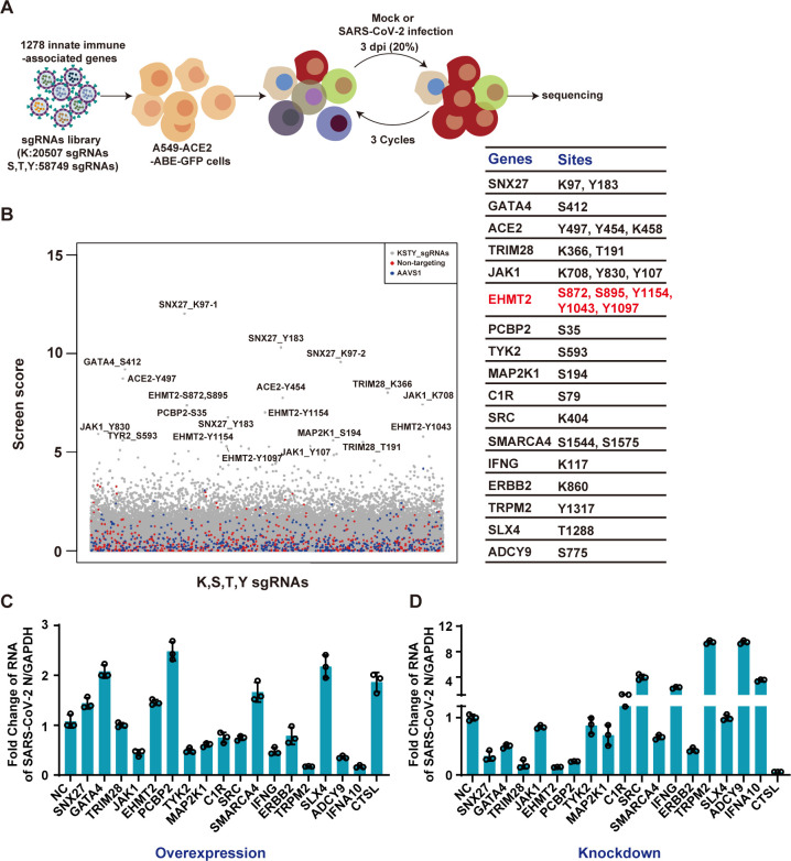
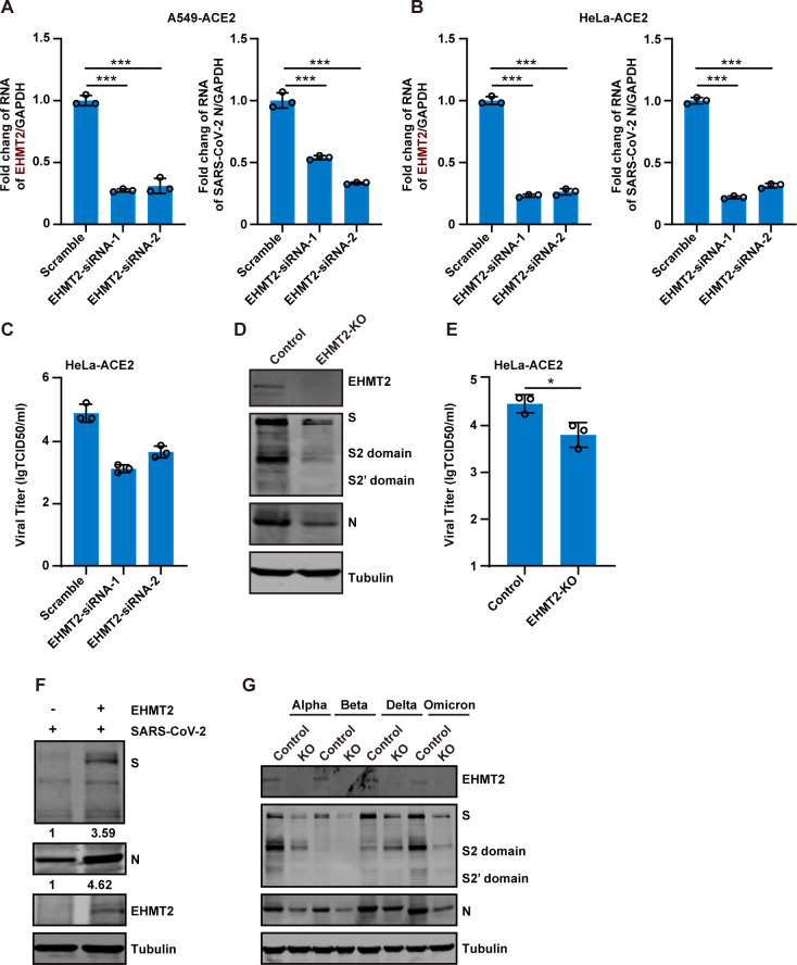
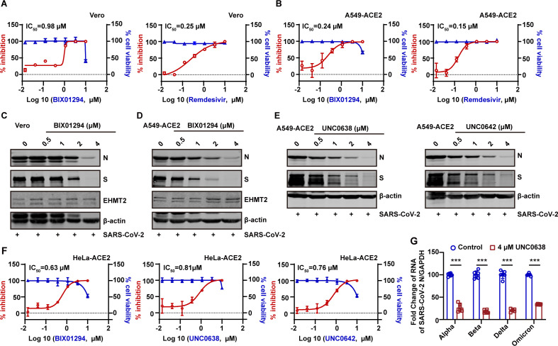
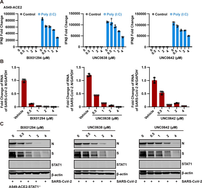
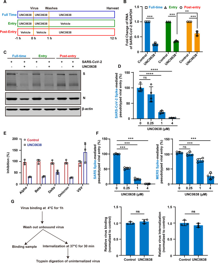
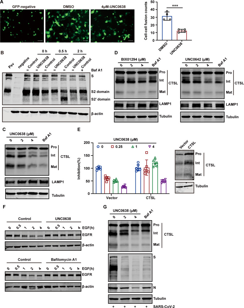
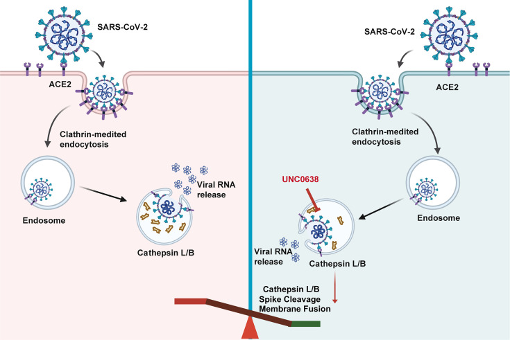
Since the outbreak of SARS-CoV-2, viral mutations have posed significant challenges in identifying therapeutic targets and developing broad-spectrum antiviral drugs. Post-translational modifications of genes involved in interferon production and signaling pathways play a crucial role in regulating interferon responses. In this study, we employed CRISPR-Cas9 screening based on adenine base editors to investigate functional amino acids in 1,278 innate immune-related genes. This approach, which converts A-T base pairs into G-C base pairs to probe the functional importance of specific amino acids, allowed us to identify 17 vital factors involved in SARS-CoV-2 infection. Among the candidate genes, genetic knockdown of EHMT2 exhibited the strongest antiviral effect. Further analysis revealed that UNC0638, a selective inhibitor of EHMT2, significantly reduced the endosomal entry of SARS-CoV-2 in pseudovirus assays. The observed inhibitory effect was consistently observed across multiple SARS-CoV-2 variants, including Alpha, Beta, Delta, and Omicron. Mechanistically, UNC0638 reduced mature cathepsin L (CTSL) levels, impairing the proteolytic cleavage of SARS-CoV-2 spike protein and subsequent membrane fusion, a critical step for viral entry. Our findings uncover EHMT2 as a host dependency factor and reveal the antiviral mechanism of EHMT2 inhibitors through CTSL maturation blockade. These results advance the understanding of host factors in SARS-CoV-2 infection and provide a strategic framework for developing host-targeted antiviral therapies.IMPORTANCEIn this study, we demonstrated that knockdown or knockout of EHMT2 inhibited SARS-CoV-2 infection, and inhibitors of EHMT2, including UNC0638, UNC0642, and BIX01294 showed similar restrictive effects. Mechanistically, the EHMT2 inhibitor UNC0638 restricts spike-mediated cell entry by inhibiting the maturation of CTSL, a critical protease required for SARS-CoV-2 entry via the endosomal pathway. Importantly, CTSL is not only essential for SARS-CoV-2 but also plays a key role in the entry of other coronaviruses that utilize similar pathways. Therefore, EHMT2 inhibitors could have broader applications as pan-coronavirus therapeutic agents.

Keywords: EHMT2; SARS-CoV-2; UNC0638; cathepsin L; viral entry.


Conflict of interest statement

The authors declare no conflict of interest.

Figures

Fig 1
Fig 1
ABE system screens identify host factors for SARS-CoV-2 infection. (A) schematic diagram of the screening process. ACE2 and ABE-GFP expressing A549 cells transduced with a lentiviral sgRNA library targeting innate immune-related molecules. The cells were infected with SARS-CoV-2 at an MOI of 0.2. DNA was extracted from surviving cells after three rounds of infection and analyzed for sgRNA enrichment. (B) Gene enrichment from ABE screen of SARS-CoV-2 infection. (Left) Bubble plot showing data from SARS-CoV-2 screen. (Right) List of differentially enriched genes and their corresponding mutation sites. (C) Overexpression verification of candidate genes. Candidate genes were overexpressed in HEK 293T-ACE2, which were then infected with SARS-CoV-2 at an MOI of 0.1 for 24 h. Intracellular SARS-CoV-2 RNA levels were quantified with RT-qPCR, using GAPDH as an internal control. (D) Loss-of-function screen to identify functional host factors. Candidate genes were silenced using siRNA in A549-ACE2 cells, followed by infection with SARS-CoV-2 at an MOI of 0.1 for 24 h. Intracellular SARS-CoV-2 RNA levels were quantified by RT-qPCR, with GAPDH as the internal control.
Fig 2
Fig 2
EHMT2 acts as a proviral factor in SARS-CoV-2 infection. (A and B) A549-ACE2 cells (A) and HeLa-ACE2 cells (B) were transfected with two independent siRNA targeting EHMT2, followed by infection with SARS-CoV-2 at an MOI of 0.1 for 24 h. Intracellular SARS-CoV-2 RNA levels were quantified by RT-qPCR, normalized to GAPDH. (C) Virus supernatants as in panel B were collected, and the virus titers were determined by TCID50 assay. (D) HeLa-ACE2 and HeLa-ACE2-EHMT2-KO cells were infected with SARS-CoV-2 at an MOI of 0.1 for 24 h. Cell lysates were collected and analyzed by western blot. (E) Virus supernatants as in panel D were collected, and the virus titers were determined by TCID50 assay. (F) HeLa-ACE2-EHMT2-KO cells were transfected with control plasmid or plasmid expressing EHMT2. After 24 h of transfection, cells were infected with SARS-CoV-2 at an MOI of 0.1 for 24 h. Cell lysates were evaluated using western blotting. (G) HeLa-ACE2-EHMT2-KO cells were infected with mutant strains of SARS-CoV-2 at an MOI of 0.1 for 24 h. All data are means ± SD. These experiments were repeated at least twice. ***P < 0.001, **P < 0.01, *P < 0.05, ns P > 0.05.
Fig 3
Fig 3
The antiviral activity of EHMT2 inhibitors. (A and B) Vero cells (A) and A549-ACE2 cells (B) were infected with SARS-CoV-2 at an MOI of 0.1 after treating with indicated doses of BIX01294 or Remdesivir. The SARS-CoV-2 RNA levels in supernatant were assessed through RT-qPCR. In parallel, their effects on cell viability were measured by CCK8 assay. The left Y-axis of the graphs represents % inhibition of the infection (red dots), and the right Y-axis of the graphs presents % cell viability (blue triangles). (C and D) Western blot detected the inhibition efficiency of BIX01294 in Vero cells (C) and A549-ACE2 cells (D). (E) Antiviral activity of EHMT2 inhibitors, UNC0638 (left) and UNC0642 (right), against SARS-CoV-2. A549-ACE2 cells were pretreated with inhibitors for 1 h and subsequently infected with SARS-CoV-2 at an MOI of 0.1 for 24 h. (F) % inhibition and % cell viability of BIX01294, UNC0638, and UNC0642 in HeLa-ACE2 cells. (G) Following either treatment or no treatment with UNC0638, HeLa-ACE2 cells were exposed to SARS-CoV-2 variants, including Alpha, Beta, Delta, and Omicron, at an MOI of 0.1 for 24 h. Intracellular SARS-CoV-2 RNA levels were quantified with RT-qPCR, using GAPDH as an internal control. Data are means ± SD. These experiments were repeated at least twice. ***P < 0.001, **P < 0.01, *P < 0.05, ns P > 0.05.
Fig 4
Fig 4
The antiviral activity of EHMT2 inhibitors is independent of the interferon pathway. (A) The effects of EHMT2 inhibitors, BIX01294 (left), UNC0638 (middle), and UNC0642 (right), on the interferon responses induced by high-molecular weight poly(I:C) were investigated. A549-ACE2 cells were transfected with poly(I:C) for 6 h after treating with varying concentrations of the inhibitors. The change of IFN-β was detected with RT-qPCR using GAPDH as an internal control. (B and C) A549-ACE2-STAT1−/− cells were treated with EHMT2 inhibitors at indicated doses and then were infected with SARS-CoV-2 at an MOI of 0.1 for 24 h. Intracellular SARS-CoV-2 RNA levels were quantified with RT-qPCR, using GAPDH as the internal control (B). Intracellular SARS-CoV-2 protein levels were quantified with western blotting (C). Data are means ± SD. These experiments were repeated at least twice.
Fig 5
Fig 5
UNC0638 targets an early post-entry step primarily blocking endosomal entry of SARS-CoV-2. (A, B, and C) Schematic diagram of the time-of-drug-addition assay (A). HeLa-ACE2 cells were treated with UNC0638 at the different stages of the viral life cycle and categorized into full-time, entry, and post-entry groups. These cells were infected with SARS-CoV-2 at an MOI of 0.1 for 12 h. Intracellular SARS-CoV-2 RNA levels were quantified with RT-qPCR, using GAPDH as an internal control (B). Intracellular SARS-CoV-2 protein levels were quantified with western blotting (C). (D) Following the treatment with UNC0638, HeLa-ACE2 cells were infected with pseudovirions, and luciferase activity was assessed after 48 h post-infection. (E) After the treatment with UNC0638, HeLa-ACE2 cells were infected with pseudovirions that carry the S protein from various strains of SARS-CoV-2, including Alpha, Beta, Delta, and Omicron variants. The luciferase activity was assessed after 48 h post-infection. (F) Following UNC0638 treatment, HeLa-ACE2 (left) or HeLa-DPP4 (right) cells were infected with SARS-CoV-1 or MERS-CoV pseudovirion, respectively. Luciferase activity was assessed after 48 h post-infection. (G) Viral binding and internalization in HeLa-ACE2 cells with or without UNC0638 treatment. Intracellular SARS-CoV-2 RNA levels were quantified with RT-qPCR, using GAPDH as an internal control. Data are means ± SD. These experiments were repeated at least twice. ***P < 0.001, **P < 0.01, *P < 0.05, ns P > 0.05.
Fig 6
Fig 6
UNC0638 suppresses SARS-CoV-2 endocytosis by inhibiting the maturation of CTSL. (A) Left, representative images showing syncytia formation after incubating UNC0638-treated or UNC0638-untreated HeLa-ACE2 with HEK 293T expressing SARS-CoV-2 S and EGFP. Scale bars, 100 µm. Right, quantitative analysis of syncytia with ImageJ. (B) The cleavage of the SARS-CoV-2 S protein was examined in HeLa-ACE2 cells, both treated and untreated with UNC0638, following infection with pseudovirions over specified time intervals. Cell treated with bafilomycin A1 (Baf A1) (200 nM) served as a negative control. The level of S2′ domain was detected by western blotting. (C) Western blot analysis of CTSL protein levels in HeLa-ACE2 cells with or without UNC0638 treatment. Cell treated with Baf A1 (200 nM) served as a negative control. (D) The effects of BIX0194 and UNC0642 on CTSL levels in HeLa-ACE2 cells. (E) HeLa-ACE2 cells were transfected with control plasmid or plasmid expressing CTSL. After 24 h of transfection, cells were infected with pseudovirions after treatment with indicated doses of UNC0638. Left, luciferase activity was assessed after 48 hours post-infection. Right, detection of expression efficiency of CTSL in HeLa-ACE2 with western blotting. (F) HeLa-ACE2 cells were stimulated with 40 ng/mL of EGF for different time after treatment with UNC0638. Cells treated with bafilomycin A1 (200 nM) served as a negative control. The level of EGFR was detected by western blotting. (G) HeLa-ACE2 cells were pretreated with UNC0638 and infected with SARS-CoV-2 at an MOI of 0.1. Intracellular SARS-CoV-2 S, N, and CTSL protein levels were evaluated using western blotting. Data are means ± SD. These experiments were repeated at least twice. ***P < 0.001, **P < 0.01, *P < 0.05, ns P > 0.05.
Fig 7
Fig 7
Schematic diagram illustrating the proposed mechanism by which UNC0638 antagonizes SARS-CoV-2 infection.

Similar articles

References

    1. Ren L-L, Wang Y-M, Wu Z-Q, Xiang Z-C, Guo L, Xu T, Jiang Y-Z, Xiong Y, Li Y-J, Li X-W, et al. 2020. Identification of a novel coronavirus causing severe pneumonia in human: a descriptive study. Chin Med J 133:1015–1024. doi: 10.1097/CM9.0000000000000722 - DOI - PMC - PubMed
    1. Lu R, Zhao X, Li J, Niu P, Yang B, Wu H, Wang W, Song H, Huang B, Zhu N, et al. 2020. Genomic characterisation and epidemiology of 2019 novel coronavirus: implications for virus origins and receptor binding. Lancet 395:565–574. doi: 10.1016/S0140-6736(20)30251-8 - DOI - PMC - PubMed
    1. Coronaviridae Study Group of the International Committee on Taxonomy of Viruses . 2020. The species Severe acute respiratory syndrome-related coronavirus: classifying 2019-nCoV and naming it SARS-CoV-2. Nat Microbiol 5:536–544. doi: 10.1038/s41564-020-0695-z - DOI - PMC - PubMed
    1. Drosten C, Günther S, Preiser W, van der Werf S, Brodt H-R, Becker S, Rabenau H, Panning M, Kolesnikova L, Fouchier RAM, et al. 2003. Identification of a novel coronavirus in patients with severe acute respiratory syndrome. N Engl J Med 348:1967–1976. doi: 10.1056/NEJMoa030747 - DOI - PubMed
    1. Guery B, Poissy J, el Mansouf L, Séjourné C, Ettahar N, Lemaire X, Vuotto F, Goffard A, Behillil S, Enouf V, Caro V, Mailles A, Che D, Manuguerra J-C, Mathieu D, Fontanet A, van der Werf S, MERS-CoV study group . 2013. Clinical features and viral diagnosis of two cases of infection with middle east respiratory syndrome coronavirus: a report of nosocomial transmission. Lancet 381:2265–2272. doi: 10.1016/S0140-6736(13)60982-4 - DOI - PMC - PubMed

LinkOut - more resources