. 2025 Jul 23;91(7):e0101925.
doi: 10.1128/aem.01019-25. Epub 2025 Jun 18.

Chlorobaculum tepidum outer membrane vesicles may transport biogenic elemental sulfur

Affiliations

Chlorobaculum tepidum outer membrane vesicles may transport biogenic elemental sulfur

Vadesse L Noundou et al. Appl Environ Microbiol. .

Abstract

Outer membrane-derived vesicles (OMVs) have been studied in different phyla of gram-negative bacteria, most extensively in the Pseudomonadota, where they have been shown to participate in diverse biological and environmental processes. To date, the production of OMVs has not been reported in the Chlorobiaceae within the phylum Chlorobiota. Chlorobaculum tepidum is the model organism for the Chlorobiaceae that synthesizes and consumes insoluble extracellular sulfur (S(0)) globules by an unknown mechanism. Here, we report evidence implicating outer membrane vesicles in biogenic S(0) globule synthesis. We demonstrate that Cba. tepidum secretes OMVs in the extracellular milieu and that OMV concentration and size vary with growth conditions, particularly sulfide concentration. A core of 31 proteins involved in diverse biological processes such as cell wall biogenesis, inorganic ion transport, and metabolism was found to be shared between OMVs, extracellular S(0) globules, and Cba. tepidum-intact cells. Multiple analytical methods indicated that OMVs contain S(0) and that OMVs and biogenic S(0) globules share protein and polysaccharide signatures, including lipooligosaccharides. Together, these lines of evidence indicate that Cba. tepidum's OMVs are one component of sulfur transport between cells and extracellular sulfur globules.IMPORTANCEAll living cells must exchange material with their environment while maintaining cellular integrity. This is a particular challenge for materials that are not water-soluble; however, many bacteria utilize insoluble materials for energy conservation and as nutrients for growth. Here, we show that Cba. tepidum makes outer membrane vesicles, and these vesicles are likely involved in the exchange of material with extracellular elemental sulfur globules formed and consumed by Cba. tepidum as part of its energy metabolism based on oxidizing reduced sulfur compounds like hydrogen sulfide. These data expand our basic understanding of Cba. tepidum's metabolism. As elemental sulfur is an industrial by-product with a limited number of uses, the information here may help enable the use of additional sulfur compounds by Cba. tepidum to drive the synthesis of biomass and/or specialty biochemicals from waste elemental sulfur by this autotrophic bacterium.

Keywords: Chlorobaculum tepidum; Chlorobiaceae; NanoIR spectroscopy; Raman spectroscopy; outer membrane vesicles; proteomics; sulfur globules.


Conflict of interest statement

Y.Y. is a named inventor on a patent application (PCT/US2023/020215) for the E3 technology, which has been licensed exclusively to CDS Analytical LLC (Oxford, PA).

Figures

Fig 1
Fig 1
The effect of Cba. tepidum growth phase on extracellular particle concentration in cultures and protein concentration in purified particle preparations. See Fig. S1A for details on purification. (A) Extracellular particle concentration from different growth phases. (B) Total protein concentration of particles purified at different growth phases. Error bars represent the standard error, n = 3 with * indication P < 0.05 and ** indicating P < 0.01 (t-test, two-tailed, unequal variance).
Fig 2
Fig 2
Transmission electron microscopy (TEM) images of Cba. tepidum’s OMVs. (A and B) TEM images show spherical OMVs budding out of rod-shaped Cba. tepidum after 8 h and 14 h of growth, respectively (white arrows). (C and D) TEM analysis of OMVs purified via serial ultracentrifugation from a 14 h old Cba. tepidum culture.
Fig 3
Fig 3
Analysis of Cba. tepidum extracellular particles for hallmark characteristics of OMVs including size (A), protein (B), LPS or LOS (C), and KDO (D).
Fig 4
Fig 4
Comparative proteome analysis of Cba. tepidum whole cells, OMVs from cultures grown on sulfide (HS-OMV) or sulfide + thiosulfate (PF7-OMV), and biogenic sulfur globules (S(0) glob.). (A) Venn diagram indicating the distribution of 1,447 proteins across sample groups (n = 3 samples per group). (B) Principal component analysis score plot of samples based on label-free quantification. The first and the second principal components (PC) account for 80.6% and 7.3% of the variability in the data, respectively. Loadings for PC1 and PC2 are displayed (blue dashed arrows) for proteins grouped by PSORTb predicted localization: Cytoplasmic, Unknown, Inner Membrane (IM), Outer Membrane (OM), Periplasmic (Peri), and Extracellular (ExC).
Fig 5
Fig 5
OMV concentration and size vary with increased sulfide (HS) concentration. (A) OMV concentrations were measured after 14 h of growth with different concentrations of sulfide as the sole electron donor. (B) MRSP-based size distribution of particles purified from the cultures in panel A (see Fig. S1A for purification details). (C and D) TEM analysis of OMVs purified from 4 mM and 7 mM sulfide cultures in panel A corresponds to the green and black traces in panel B.
Fig 6
Fig 6
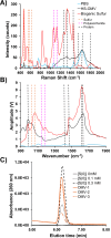
OMVs and S(0) share multiple characteristics. Characteristic Raman scattering (A) or IR absorption (B) spectra for purified OMVs (black), biogenic S(0) (red), and PBS (blue). Scattering (A) or absorption (B) wavenumbers are indicated by vertical dashed lines for sulfur (orange), polysaccharide (pink), and protein (black). (C) Chromatograms of elemental sulfur standards (0–0.3 mM S(0), black traces) and OMVs purified from three independent 7 mM sulfide Cba. tepidum cultures (orange traces). The standards indicate the peak from OMV samples co-migrates with authentic S(0), and the replicates are within the range of detection.
Fig 7
Fig 7
Summary of the proposed OMV S(0) transport mechanism. (A) Cba. tepidum produces both OMVs and polysulfides during growth that contribute to the growth of extracellular S(0) globules. (B) Enlarged view of the periplasm and OM with increased detail. Sulfide oxidation may induce unfolded protein stress that leads to OMV production. The dashed arrows for sulfide and polysulfide indicate possible routes to enter and exit either OMVs or S(0) globules. SQR = sulfide:quinone oxidoreductase (CT0117 and CT1087), Psr = putative polysulfide oxidoreductase complex (CT0496-CT0494). (C) Enlarged view of a Cba. tepidum OMV with selected cargo molecules detected by proteomics analyses denoted at the right. Created in BioRender (https://BioRender.com/bzod9yf).

Update of

Similar articles

References

    1. Hanson TE, Bonsu E, Tuerk A, Marnocha CL, Powell DH, Chan CS. 2016. Chlorobaculum tepidum growth on biogenic S(0) as the sole photosynthetic electron donor. Environ Microbiol 18:2856–2867. doi: 10.1111/1462-2920.12995 - DOI - PubMed
    1. Gralnick JA, Bond DR. 2023. Electron transfer beyond the outer membrane: putting electrons to rest. Annu Rev Microbiol 77:517–539. doi: 10.1146/annurev-micro-032221-023725 - DOI - PubMed
    1. Beblawy S, Bursac T, Paquete C, Louro R, Clarke TA, Gescher J. 2018. Extracellular reduction of solid electron acceptors by Shewanella oneidensis. Mol Microbiol 109:571–583. doi: 10.1111/mmi.14067 - DOI - PubMed
    1. Gadd GM. 2010. Metals, minerals and microbes: geomicrobiology and bioremediation. Microbiology (Reading) 156:609–643. doi: 10.1099/mic.0.037143-0 - DOI - PubMed
    1. Schertzer JW, Whiteley M. 2013. Bacterial outer membrane vesicles in trafficking, communication and the host-pathogen interaction. J Mol Microbiol Biotechnol 23:118–130. doi: 10.1159/000346770 - DOI - PubMed

MeSH terms

LinkOut - more resources