. 2025 Jun 18;91(6):e0021825.
doi: 10.1128/aem.00218-25. Epub 2025 May 7.

ARTP mutagenesis for genome-wide identification of genes important for biofilm regulation in spoilage bacterium Pseudomonas fluorescens PF08

Affiliations

ARTP mutagenesis for genome-wide identification of genes important for biofilm regulation in spoilage bacterium Pseudomonas fluorescens PF08

Feifei Wang et al. Appl Environ Microbiol. .

Abstract

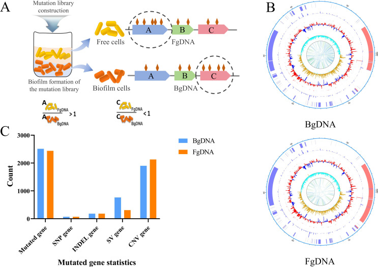
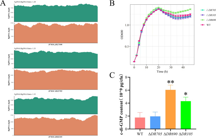
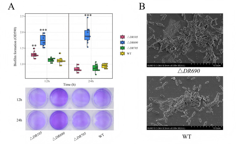
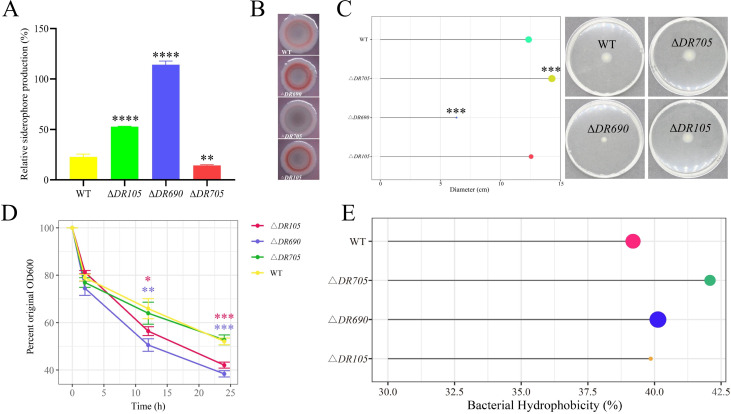
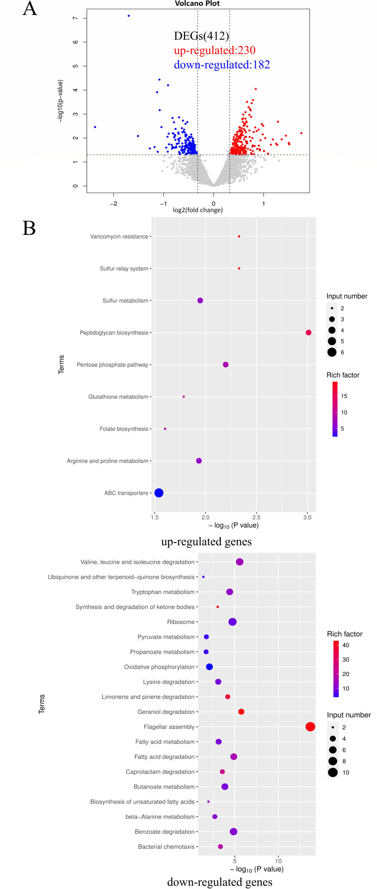
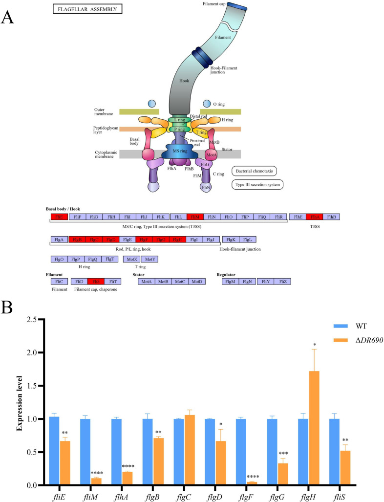
Pseudomonas fluorescens is a vital food spoilage bacterium and commonly spoils foods in the form of biofilms. Yet its biofilm regulation strategies have not been fully revealed. Here, we conducted a genome-wide screen of genes important for biofilm regulation using atmospheric and room temperature plasma mutagenesis together with the whole-genome resequencing technology. Three genes (D7M10_RS02105, D7M10_RS27690, and D7M10_RS25705) encoding GGDEF-EAL domain-containing proteins were found to have different mutation manifestations between biofilm cells and free cells. On direct testing, null mutants of D7M10_RS02105 and especially D7M10_RS27690 exhibited significantly elevated cyclic di-GMP (c-di-GMP) levels. Further studies indicated that a higher level of c-di-GMP caused by the null mutant of D7M10_RS27690 triggered cell growth, the production of siderophore and exopolysaccharide as well as autoaggregation, and hindered cell motility, all of which together promote biofilm formation. RNA-sequencing analysis revealed the transcription profile regulated by D7M10_RS27690, mostly including flagellar assembly and peptidoglycan biosynthesis pathways. Therein, the downregulated genes enriched in flagellar assembly were verified by qRT-PCR; the result of which was in agreement with the decreased cell motility.IMPORTANCEBiofilms formed by spoilage bacterium Pseudomonas fluorescens will bring about food quality and safety issues. In this study, we present the establishment of a genetic method and verified its reliability and efficiency for identifying genes associated with biofilm regulation. The genes we discovered offer new perspectives on the mechanisms of biofilm regulation in spoilage bacterium P. fluorescens. Moreover, the gene screen method based on atmospheric and room temperature plasma mutagenesis and whole-genome resequencing-coupled technology overcomes the labor-intensive issues caused by traditional methods and should generally be suitable for identifying genes associated with biofilm formation or dispersion in other bacteria.

Keywords: ARTP mutagenesis; Pseudomonas fluorescens; biofilm; c-di-GMP; siderophore.


Conflict of interest statement

The authors declare no conflict of interest.

Figures

Fig 1
Fig 1
Genome-wide screen for genes involved in biofilm regulation using ARTP mutagenesis combined with WGR technology. (A) Schematic diagram of the strategy for identifying genes involved in biofilm regulation. Cells of the mutant library were grown statically in the Petri dish at 28°C and separated into free cells and biofilm cell fractions. DNA was extracted from free cells and biofilm cells, named FgDNA and BgDNA, respectively. After whole-genome resequencing and data analysis, the FgDNA:BgDNA ratio of total mutation counts (including SNP, INDEL, SV, and CNV) for each gene was calculated. As shown in the hypothetical example on the right, gene A and gene C have more and fewer mutations in FgDNA than in BgDNA, and are therefore potentially required for biofilm formation or dispersion. Therefore, we focused on genes in categories A and C. (B) The mutation profile of biofilm cells and free cells. The outermost circle represents the genome size. The second circle represents CNV, with red indicating duplication and blue indicating deletion. The third circle represents INDEL, depicted as a standard histogram showing the frequency of insertion/deletion variations in the region. The fourth circle represents SNP, shown as a standard histogram indicating the frequency of SNP variations in the region. The fifth circle represents GC content, with the outward red part indicating GC content higher than the genome-wide average and the inward blue part indicating GC content lower than the genome-wide average. The sixth circle represents the GC skew value, which is positive when the forward strand tends to transcribe CDS (coding sequences) and negative when the reverse strand tends to transcribe CDS. The innermost circle indicates SV links, with forward SVs in blue and reverse SVs in green. (C) Statistics of mutant genes in different types of biofilm cells and free cells, including SNP, INDEL, SV, and CNV.
Fig 2
Fig 2
Effects of three EAL domain-containing proteins on c-di-GMP metabolism. (A) Sequencing depth spectrum of D7M10_RS02105, D7M10_RS27690, and D7M10_RS25705 in FgDNA and BgDNA. (B) Growth curves of WT and mutant strains. (C) Intracellular c-di-GMP levels of WT and mutant strains. Data were presented as the mean ± standard deviation (n = 3, *P < 0.05, **P < 0.01).
Fig 3
Fig 3
Roles of three EAL domain-containing proteins in biofilm formation. (A) Biofilm biomass quantification using the crystal violet method. (B) SEM observation of biofilms for WT and ΔDR690. Scale bars, 10 µm. Data were presented as the mean ± standard deviation (n = 10, **P < 0.01, ***P < 0.001).
Fig 4
Fig 4
Exploring the biofilm-associated biochemical process regulated by three EAL domain-containing proteins. (A) Siderophore relative production of WT and mutant strains. (B) Bacterial colony morphology on Congo red agar plates. (C) The swimming motility and the swimming zone formed by different strains. (D) The cell autoaggregation of WT and mutant strains. (E) The cell hydrophobicity of WT and mutant strains. Data were presented as the mean ± standard deviation (n = 3, *P < 0.05, **P < 0.01, ***P < 0.001, ****P < 0.0001).
Fig 5
Fig 5
Transcriptomic analysis of ΔDR690 compared with WT using RNA sequencing. (A) The volcano plot of DEGs in ΔDR690 compared to WT. Red circles are upregulated genes, and blue circles are downregulated genes. (B) Scatter diagram of KEGG enrichment pathways for upregulated and downregulated genes of ΔDR690, respectively. |log2 fold change| > 0.32 and an adjusted P-value <0.05 were used to judge the statistical significance of gene expression differences. KEGG enrichment analysis for DEGs was implemented by KOBAS software (version: 2.1.1) with a P-value cutoff of 0.05 to judge statistically significant enrichment.
Fig 6
Fig 6
qRT-PCR verification of flagellar assembly-related genes obtained from transcription analysis. (A) The positions of enriched genes in the flagellar assembly pathway. (B) qRT-PCR verification of the transcription of genes enriched in the flagellar assembly pathway. Data were presented as the mean ± standard deviation (n = 3, *P < 0.05, **P < 0.01, ***P < 0.001, ****P < 0.0001).

Similar articles

References

    1. Gallique M, Decoin V, Barbey C, Rosay T, Feuilloley MGJ, Orange N, Merieau A. 2017. Contribution of the Pseudomonas fluorescens MFE01 Type VI secretion system to biofilm formation. PLoS One 12:e0170770. doi: 10.1371/journal.pone.0170770 - DOI - PMC - PubMed
    1. Rumbaugh KP, Sauer K. 2020. Biofilm dispersion. Nat Rev Microbiol 18:571–586. doi: 10.1038/s41579-020-0385-0 - DOI - PMC - PubMed
    1. Aswathanarayan JB, Vittal RR. 2014. Attachment and biofilm formation of Pseudomonas fluorescens PSD4 isolated from a dairy processing line. Food Sci Biotechnol 23:1903–1910. doi: 10.1007/s10068-014-0260-8 - DOI
    1. Xie J, Zhang Z, Yang S-P, Cheng Y, Qian Y-F. 2018. Study on the spoilage potential of Pseudomonas fluorescens on salmon stored at different temperatures. J Food Sci Technol 55:217–225. doi: 10.1007/s13197-017-2916-x - DOI - PMC - PubMed
    1. Mlipano CL, Alistair G, Michael L. 2018. Detection of proteolysis in milk by Pseudomonas fluorescens using urea PAGE method. J Food Stud 7:14. doi: 10.5296/jfs.v7i1.12019 - DOI

MeSH terms

LinkOut - more resources