. 2025 Jun;29(12):e70042.
doi: 10.1111/jcmm.70042.

HEY1 promotes the development and metastasis of osteosarcoma through CD44/EGFR/FAK pathway

Affiliations

HEY1 promotes the development and metastasis of osteosarcoma through CD44/EGFR/FAK pathway

Yuhang Liu et al. J Cell Mol Med. 2025 Jun.

Abstract

Osteosarcoma (OS) is a highly prevalent and deadly malignant tumour primarily affecting adolescents. However, the identification of new therapeutic targets remains an urgent need. The advent of bioinformatics technology has offered us a novel approach to screen key genes from diverse OS-related databases, thereby providing valuable insights into the mechanistic understanding of OS prognosis. In this study, we comprehensively integrated multiple databases to identify the crucial oncogene, HEY1, which exerts a significant impact on OS prognosis. Subsequently, we conducted a experimental validations to explore influence of HEY1 knockdown on OS cells. HEY1 exhibited significant overexpression in OS tissues and cells and its silencing resulted in a significant inhibition of proliferation. The interaction between HEY1 and CD44 was identified through transcriptome sequencing and mass spectrometry analysis. Additionally, our findings suggested that HEY1 could potentially influence the EGFR-FAK pathway. Further experiments established that HEY1 regulates the EGFR-FAK pathway via CD44, thereby influencing the biological phenotype of OS cells. These findings were subsequently validated using in vivo animal models. In summary, HEY1 demonstrated significant overexpression in both OS tissues and cells, exerting a substantial impact on the prognosis of OS.

Keywords: Bioinformatic; CD44; HEY1; focal adhesion pathway; osteosarcoma.


Conflict of interest statement

The authors declare no competing interests.

Figures
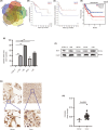
FIGURE 1
FIGURE 1
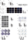
HEY1 exhibits significant upregulation in both osteosarcoma tissues and cells, correlating with patient prognosis. (A) Identification of commonly differentially expressed genes across five osteosarcoma databases. (B, C) Analysis of the association between HEY1 expression levels in the R2 database and overall survival as well as metastasis‐free survival in osteosarcoma patients. (D) Investigation of the relationship between HEY1 expression levels in the GEPIA database and overall survival in patients with soft tissue sarcoma. (E, F) Quantitative analysis of HEY1 expression using PCR and protein imprinting detection in osteosarcoma cells and normal osteoblasts. (G, H) Assessment of HEY1 expression levels in tumour tissues and normal tissues in osteosarcoma chips. *p < 0.05, **p < 0.01, ***p < 0.001.
FIGURE 2
FIGURE 2
HEY1 plays a pivotal role in promoting the progression of osteosarcoma in vitro. (A–C) We assessed the levels of RNA and protein expression after suppressing HEY1. (D) Cell viability was evaluated using the CCK‐8 assay. (E) The EdU cell proliferation assay demonstrated the inhibitory effects of HEY1 knockdown on osteosarcoma cell proliferation. (F, G) Findings from the cell scratch assay and transwell assay indicate that HEY1 knockdown suppresses the migration and invasion of osteosarcoma cells. (H) The colony formation assay confirms the inhibitory effect of HEY1 knockdown on the formation of osteosarcoma cell colonies. (I) Flow cytometry apoptosis assay results highlight the promotion of osteosarcoma cell apoptosis achieved through HEY1 knockdown. *p < 0.05, **p < 0.01, ***p < 0.001.
FIGURE 3
FIGURE 3
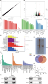
HEY1 exhibits an interaction with CD44. (A, B) Differential gene distribution and volcano plot obtained from high‐throughput transcriptome sequencing. (C, D) GO functional enrichment analysis and KEGG pathway enrichment analysis based on transcriptome sequencing. (E) Results of silver staining on immunoprecipitation gel. (F) GO functional enrichment analysis of proteins identified through protein mass spectrometry. (G) Shared proteins in the cell–cell adhesion and focal adhesion pathways. (H) Correlation analysis of HEY1 and CD44 expression levels in osteosarcoma databases. (I) CO‐IP results confirmed the interaction between HEY1 and CD44. *p < 0.05, **p < 0.01, ***p < 0.001.
FIGURE 4
FIGURE 4
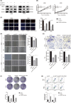
HEY1 promotes osteosarcoma cell invasion by activating CD44. (A) The protein level of CD44 was altered following HEY1 knockdown. (B, C) CD44 reversed the inhibitory effect of HEY1 knockdown on cell proliferation. (D, E) CD44 reversed the inhibitory effect of HEY1 knockdown on cell migration and invasion. (F) CD44 reversed the inhibitory effect of HEY1 knockdown on cell colony formation. (G) CD44 reversed the apoptosis‐promoting effect of HEY1 knockdown. *p < 0.05, **p < 0.01, ***p < 0.001.
FIGURE 5
FIGURE 5
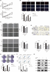
HEY1 promotes osteosarcoma progression through the CD44/EGFR/FAK pathway. (A, B) LeY reversed the inhibitory effect of HEY1 knockdown on osteosarcoma cell proliferation. (C, D) LeY reversed the inhibitory effect of HEY1 knockdown on osteosarcoma cell migration and invasion. (E) LeY reversed the inhibitory effect of HEY1 knockdown on osteosarcoma cell colony formation. F. LeY reversed the pro‐apoptotic effect of HEY1 knockdown. (G) Knockdown of HEY1 resulted in reduced expression levels of EGFR, FAK, Akt, and ERK, which were restored by LeY. *p < 0.05, **p < 0.01, ***p < 0.001.
FIGURE 6
FIGURE 6
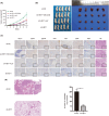
Knockdown of HEY1 suppresses the growth and metastasis of osteosarcoma in vivo. (A) Tumour growth curve. (B) Comparison of tumour volume after 18 days. (C) Immunohistochemical analysis and HE/Ki67 staining of subcutaneous tumours. (D) Knockdown of HEY1 inhibited pulmonary metastasis of osteosarcoma. *p < 0.05, **p < 0.01, ***p < 0.001.

References

    1. Kansara M, Teng MW, Smyth MJ, Thomas DM. Translational biology of osteosarcoma. Nat Rev Cancer. 2014;14(11):722‐735. - PubMed
    1. Ritter J, Bielack SS. Osteosarcoma Annals of oncology. Med Oncol. 2010;21(7):320‐325. - PubMed
    1. Meltzer PS, Helman LJ. New horizons in the treatment of osteosarcoma. N Engl J Med. 2021;385(22):2066‐2076. - PubMed
    1. Gorlick R, Janeway K, Lessnick S, Randall RL, Marina N, COG Bone Tumor Committee . Children's oncology Group's 2013 blueprint for research: bone tumors. Pediatr Blood Cancer. 2013;60(6):1009‐1015. - PMC - PubMed
    1. Zils K, Bielack S, Wilhelm M, et al. Osteosarcoma of the mobile spine. Ann Oncol. 2013;24(8):2190‐2195. - PubMed

MeSH terms