. 2025 Jun 18;21(6):e1012582.
doi: 10.1371/journal.ppat.1012582. eCollection 2025 Jun.

Microglia-specific NF-κB signaling is a critical regulator of prion-induced glial inflammation and neuronal loss

Affiliations

Microglia-specific NF-κB signaling is a critical regulator of prion-induced glial inflammation and neuronal loss

Arielle J D Hay et al. PLoS Pathog. .

Abstract

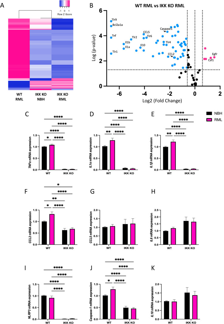
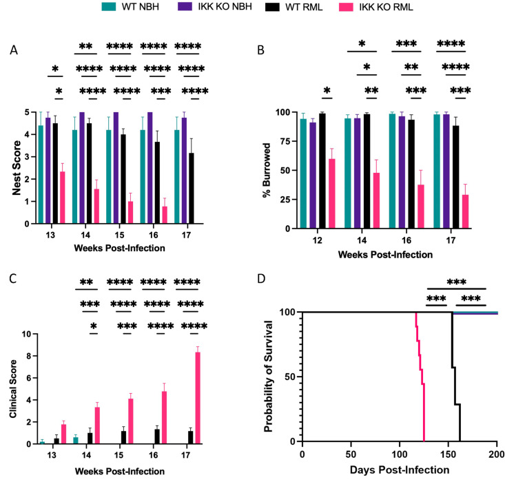
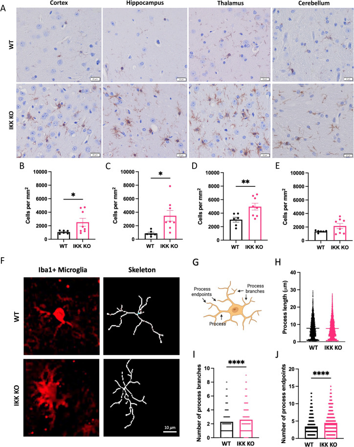
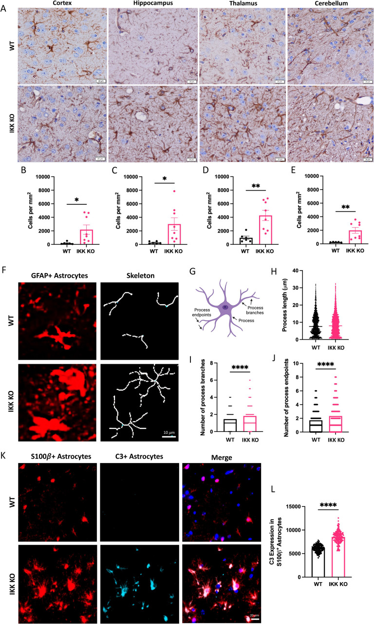
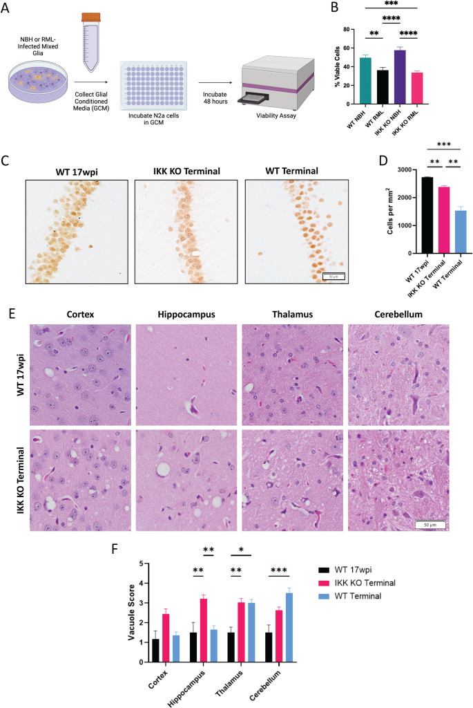
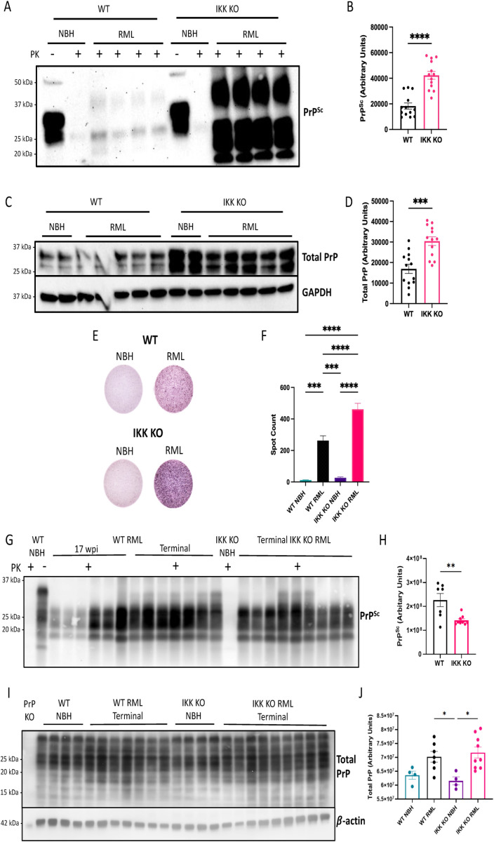
Prion diseases are a group of rare and fatal neurodegenerative diseases caused by the cellular prion protein, PrPC, misfolding into the infectious form, PrPSc, which forms aggregates in the brain. This leads to activation of glial cells, neuroinflammation, and irreversible neuronal loss, however, the role of glial cells in prion disease pathogenesis and neurotoxicity is poorly understood. Microglia can phagocytose PrPSc, leading to the release of inflammatory signaling molecules, which subsequently induce astrocyte reactivity. Animal models show highly upregulated inflammatory molecules that are a product of the Nuclear Factor-kappa B (NF-κB) signaling pathway, suggesting that this is a key regulator of inflammation in the prion-infected brain. The activation of the IκB kinase complex (IKK) by cellular stress signals is critical for NF-κB-induced transcription of a variety of genes, including pro-inflammatory cytokines and chemokines, and regulators of protein homeostasis and cell survival. However, the contribution of microglial IKK and NF-κB signaling in the prion-infected brain has not been evaluated. Here, we characterize a primary mixed glial cell model containing wild-type (WT) astrocytes and IKK knock-out (KO) microglia. These cultures show a near ablation of microglia compared to WT mixed glial cultures, highlighting the role of IKK in microglial survival and proliferation. We show that, when exposed to prion-infected brain homogenates, NF-κB-associated genes are significantly downregulated, but prion accumulation is significantly increased, in mixed glial cultures containing minimal microglia. Mice with IKK KO microglia show rapid disease progression when intracranially infected with prions, characterized by an increased density of activated microglia and reactive astrocytes, development of spongiosis, and accelerated loss of hippocampal neurons and associated behavioral deficits. These animals display clinical signs of prion disease early and have a 22% shorter life expectancy compared to infected wild-type mice. Intriguingly, PrPSc accumulation was significantly lower in the brains of terminal animals with IKK KO microglia compared to terminal WT mice, suggesting that accelerated disease is independent of PrPSc accumulation, highlighting a glial-specific pathology. Together, these findings present a critical role for microglial IKK and NF-κB signaling in host protection against prion disease.


Conflict of interest statement

The authors have declared that no competing interests exist.

Figures

Fig 1
Fig 1. NF-κB-associated genes are significantly downregulated in mixed glial cultures derived from mice with IKK KO microglia.
mRNA expression of the NF-κB-associated cytokines and chemokines A TNFα, B IL1α, C IL1β, D CCL2, E CCL5 and F IL6 were assessed. The NLRP3-associated genes G NLRP3, H Caspase-1 and I IL18 were assessed. Analysis of 3-5 biological replicates, each with 3 technical replicates. One-way ANOVA and post-hoc Tukey test, error bars = SEM, *p < 0.05, **p < 0.01, ****p < 0.0001.
Fig 2
Fig 2. Mice with IKK KO microglia present accelerated prion disease.
Mice were infected intracranially with RML mouse-adapted scrapie and monitored for changes in A nesting behavior, B burrowing behavior, and C clinical scores. D Animals displaying clinical scores of 10 were euthanized. NBH-treated groups contained 4 IKK KO and 5 WT mice, and RML-infected groups contained 9 IKK KO and 6 WT mice. For behavioral and clinical analyses, a two-way repeated measures ANOVA was used. For survival curve, a Log-rank (Mantel-Cox) test was performed. *p < 0.05, **p < 0.01, ***p < 0.001, ****p < 0.0001.
Fig 3
Fig 3. Removal of IKK in microglia induces changes in microglia number and morphology during prion infection.
Terminal mice with IKK KO microglia were compared to wpi-matched WT infected mice. A Brains were stained for Iba1 + microglia, which were counted and compared in the B cortex, C hippocampus, D thalamus and E cerebellum for 9 animals with IKK KO microglia, 6 wpi-matched WT and 3 terminal WT animals. Scale bars = 20·m. F Representative images of Iba1 + hippocampal microglia morphological skeletons between groups. Scale bar = 10·m. G Microglia cartoon depicting features analyzed via skeletonization, created with Biorender.com. H Process length, I number of process branches and J number of process endpoints was compared between hippocampal microglia from WT mice and those with IKK KO microglia. Skeletonization and C3 analysis were performed on 3 randomly selected animals per group. Welch’s t-test, error bars = SEM, **p < 0.01, ****p < 0.0001.
Fig 4
Fig 4. Brains with microglial IKK KO have increased GFAP expression and activated astrocytes during prion infection.
Terminal mice with IKK KO microglia were compared to wpi-matched WT infected mice. A Brains were stained for GFAP+ astrocytes, which were counted and compared in the B cortex, C hippocampus, D thalamus and E cerebellum for 9 animals with IKK KO microglia, 6 wpi-matched WT and 3 terminal WT animals. Scale bars = 20·m. F Representative images of GFAP+ hippocampal astrocyte morphological skeletons between groups. Scale bar = 10·m. G Astrocyte cartoon depicting features analyzed via skeletonization, created with Biorender.com. H Process length, I number of process branches and J number of process endpoints was compared between hippocampal astrocytes from WT mice and those with IKK KO microglia. K Hippocampal astrocytes were co-stained for the pan-astrocytic marker S100β and the reactive astrocyte marker C3. Scale bar = 50·m. L Mean grey intensity of C3 was compared in S100β+ astrocytes (arbitrary units). Skeletonization and C3 analysis were performed on 3 randomly selected animals per group. Welch’s t-test, error bars = SEM, *p < 0.05, **p < 0.01, *** p < 0.001, ****p < 0.0001.
Fig 5
Fig 5. Removal of IKK in microglia does not protect against prion-induced neuronal death in vitro or in vivo.
A Mixed glial cultures were treated for 7 days with NBH or RML homogenates. Glial conditioned media (GCM) was isolated and plated on N2as in 96-well plates and chamber slides for 48 hours. Image created with Biorender.com. B GCM-treated N2as were analyzed for percent viability using a Presto Blue cell viability assay. C Representative images of NeuN+ cells in the CA1 region of the hippocampus for terminal mice with IKK KO microglia (n = 9), infected wpi-matched WT mice (n = 6) and terminal WT mice (n = 7) and D CA1 NeuN+ neuronal counts. The severity of vacuolization for infected mice was scored from 1-5 by three blinded pathologists in the cortex, hippocampus, thalamus and cerebellum. E Representative images of vacuoles for terminal mice with IKK KO microglia and infected wpi-matched WT mice. F Bar graph showing mean vacuole severity for all infected mice. For the viability assay, a two-way ANOVA and post-hoc Tukey test was used. For NeuN counts, a Brown-Forsythe and Welch ANOVA was used. For vacuole counts, a two-way ANOVA and post-hoc Tukey test was used. Error bars = SEM, *p < 0.05, **p < 0.01, *** p < 0.001, ****p < 0.0001. Scale bars = 50·m.
Fig 6
Fig 6. IKK KO in microglia leads to increased accumulation of PK-resistant PrP in vitro but decreased accumulation in vivo.
Mixed glial cells containing WT astrocytes and WT or IKK KO microglia were treated with NBH or infected with RML for 7 days. A Cells were lysed and digested with PK for PrPSc observation, undigested (Total) PrP and GAPDH as a control. B protease-resistant protein signal for PrPSc and C protein signal for total PrP was measured. D Infected cells were trypsinized and transferred to an ELISpot plate for a scrapie cell assay to E count the number of infected cells (normalized). Prion western blot analysis and spot counts are combined data from three separate experiments. F Homogenized brains from terminally infected mice with IKK KO microglia and both wpi-matched and terminally infected WT mice were digested with PK and G protease-resistant protein signal was measured using Sha31 antibody comparing terminal groups. H Total PrP was assessed using Sha31 antibody for both terminally infected and mock-infected mice and I compared between groups. For western blot quantification of PrPSc, an unpaired t-test was performed. For scrapie cell assay, a two-way ANOVA and post-hoc Tukey test was used. For western blot analysis of total PrP, a one-way ANOVA and post-hoc Tukey test was used. Error bars = SEM, *p < 0.05, **p < 0.01, *** p < 0.001, ****p < 0.0001.

Similar articles

References

    1. Wang C, Fan L, Khawaja RR, Liu B, Zhan L, Kodama L. Microglial NF-kappaB drives tau spreading and toxicity in a mouse model of tauopathy. Nat Commun. 2022;13(1):1969. - PMC - PubMed
    1. Frakes AE, Ferraiuolo L, Haidet-Phillips AM, Schmelzer L, Braun L, Miranda CJ, et al. Microglia induce motor neuron death via the classical NF-κB pathway in amyotrophic lateral sclerosis. Neuron. 2014;81(5):1009–23. doi: 10.1016/j.neuron.2014.01.013 - DOI - PMC - PubMed
    1. Rocha SM, Kirkley KS, Chatterjee D, Aboellail TA, Smeyne RJ, Tjalkens RB. Microglia-specific knock-out of NF-kappaB/IKK2 increases the accumulation of misfolded alpha-synuclein through the inhibition of p62/sequestosome-1-dependent autophagy in the rotenone model of Parkinson’s disease. Glia. 2023. - PMC - PubMed
    1. Carroll JA, Striebel JF, Race B, Phillips K, Chesebro B. Prion infection of mouse brain reveals multiple new upregulated genes involved in neuroinflammation or signal transduction. J Virol. 2015;89(4):2388–404. doi: 10.1128/JVI.02952-14 - DOI - PMC - PubMed
    1. Hartmann K, Sepulveda-Falla D, Rose IVL, Madore C, Muth C, Matschke J. Complement 3( )-astrocytes are highly abundant in prion diseases, but their abolishment led to an accelerated disease course and early dysregulation of microglia. Acta Neuropathol Commun. 2019;7(1):83. - PMC - PubMed

MeSH terms

LinkOut - more resources