Review
. 2025 Jun 18;15(1):86.
doi: 10.1038/s41408-025-01286-w.

Guidelines for the testing and reporting of cytogenetic results for risk stratification of multiple myeloma: a report of the Cancer Genomics Consortium Plasma Cell Neoplasm Working Group

Affiliations
Review

Guidelines for the testing and reporting of cytogenetic results for risk stratification of multiple myeloma: a report of the Cancer Genomics Consortium Plasma Cell Neoplasm Working Group

Xinyan Lu et al. Blood Cancer J. .

Abstract

Fluorescence in situ hybridization (FISH) remains the gold-standard clinical assay to detect genetic abnormalities in multiple myeloma (MM). However, FISH panel design, use of conventional chromosome banding analysis and reporting practices have been reported to vary among laboratories. Therefore, standardization in FISH testing and reporting practices is needed to improve report clarity and avoid misinterpretation. The recommendations in this paper represent a consensus of our Cancer Genomics Consortium Plasma Cell Neoplasm Working Group, comprising a joint panel of cytogenetic laboratory directors and clinical investigators with expertise in the diagnosis, risk stratification, and treatment of multiple myeloma. Prior to developing these consensus recommendations, we performed a full literature review and conducted a survey of 102 oncologists to assess current variations and challenges in MM cytogenetic/FISH testing and reporting. Our guidelines establish best practices for the optimization of FISH panel selection, and recommendations for standardized reporting of cytogenetic results to align with the 2025 International Myeloma Society (IMS)/International Myeloma Working Group (IMWG) Updated Risk Stratification.


Conflict of interest statement

Competing interests: EA reports consultancy AbbVie. RB reports consultancy Adaptive Biotech, BMS, Caribou Biosciences, Genentech, Janssen, Karyopharm, Legend Biotech, Pfizer, Sanofi, SparkCures; Research: Novartis, Pack Health. RF reports consultancy for AbbVie, Adaptive Biotechnologies, AMGEN, AZeneca, Bayer, Binding Site, BMS (Celgene), Millenium Takeda, Jansen, Juno, Kite, Merck, Pfizer, Pharmacyclics, Regeneron, Sanofi; scientific advisory boards for Adaptive Biotechnologies, Caris Life Sciences, Oncotracker; board of directors for Antegene, AZBio; and patents for FISH in myeloma. SK reports consultancy from BMS/Celgene, Takeda and Janssen and research funding from BMS (Celgene), Takeda, Novartis, AbbVie, Janssen and Amgen. LBB reports consultancy Genentech. The remaining authors have no interests to disclose.

Figures

Fig. 1
Fig. 1. Previous FISH panels proposed by various expert groups.
FISH panels incorporated into the major risk stratification guidelines (NCCN, IMWG and mSMART). * While not on the recommended diagnostic minimal panel, the NCCN lists MYC-r as a factor considered high-risk for progression/relapse. ** Hyperdiploidy defined by multiple trisomies can be detected by FISH or an equivalent method like flow cytometry. *** Consider implementing a reflex strategy. Many laboratories use an IGH BAP probe to determine rearrangement status. If an IGH BAP signal pattern is detected, laboratories will perform a reflex panel including pertinent IGH partners. However, rare IGH translocations may occur resulting in a “false-negative” using IGH BA; therefore, the decision to perform sequential testing is at the discretion of the individual laboratory. If a TP53 deletion is identified, reflex to evaluate for TP53 mutation can be considered, but is not required.
Fig. 2
Fig. 2. Minimum FISH panel proposed to conform to the 2025 IMS/IMWG risk stratification.
Consensus recommendation for minimum FISH panel design includes detection of TP53 deletion using a probe targeting 17p13, 1q gain or amplification using a probe targeting 1q21 (typically CKS1B), 1p deletion using a probe tageting 1p32 (typically CDKN2C) and IGH-r for all NDMM. * If an IGH-r is identified, reflex to DC-DF probes targeting t(4;14), t(14;16), t(14;20) and t(11;14) should be performed. Detection of an IGH-r occurs when the IGH break-apart (BAP) FISH probe is abnormal. For RRMM, a minimal FISH panel that includes assessment for TP53 deletion, 1p deletion, 1q gain or amplification, and at least one probe targeting the primary abnormality observed at diagnosis is recommended. Inclusion of a previous primary abnormality serves as a positive control for detection of the patient’s previously described MM clone.
Fig. 3
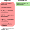
Fig. 3. Cytogenetic risk assessment and reporting for MM according to the 2025 IMS/IMWG risk stratification.
Following FISH detection, laboratories should provide a report using clear language that interprets the FISH results with accurate classification into standard-risk and high-risk based on the 2025 IMS/IMWG Risk Stratification guidelines. Cytogenetic high-risk includes the presence of at least one of the following cytogenetic abnormalities: TP53 deletion or TP53 mutation, 1p biallelic deletion, t(4;14) plus 1q gain or amplification or 1p deletion, t(14;16) plus 1q gain or amplification or 1p deletion, t(14;20) plus 1q gain or amplification or 1p deletion, 1q gain or amplification plus 1p deletion. 17p deletion should have clonality of ≥20%. Standard-risk includes the absense of a high-risk cytogenetic abnormality and can include isolated hyperdiploidy, t(11;14) or t(6;14). Other features such as β2 microglobulin and creatinine also impact the overall IMS/IMWG risk stratification-please refer to Avet-Loiseau, et al. [5] for a complete risk stratification incorporating these variables along with the cytogenetics.
Fig. 4
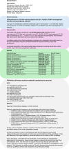
Fig. 4. Recommended format and components of sample report to conform to the new 2025 IMS/IMWG Risk Stratification of Myeloma.
Sample of a FISH report reflecting the critical results highlighted in bold at the top of the report under “result summary” (example in blue shading). In this case, there is a t(11;14)/IGH::CCND1 rearrangement and hyperdiploidy detected. Also indicated are the results for cytogenetic abnormalities not detected in this sample. An interpretation written in plain and standardized language is provided which indicates that FISH analysis was performed on enriched plasma cells and identifies specifically which abnormalities were detected and not detected (example in purple shading). A comment about whether the FISH findings represent a standard or high-risk abnormality according to the 2025 IMS/IMWG risk stratification is also indicated. A table of results is provided (example in green shading), which indicates the FISH probes being tested, the laboratory’s established cut-off values for each probe and the abnormality being detected or not detected. Finally, additional technical details including previous FISH results if available, the ISCN nomenclature and the methods are also included (example in grey shading).

References

    1. Kumar SK, Rajkumar SV. The multiple myelomas - current concepts in cytogenetic classification and therapy. Nat Rev Clin Oncol. 2018;15:409–21. - PubMed
    1. Saxe D, Seo EJ, Bergeron MB, Han JY. Recent advances in cytogenetic characterization of multiple myeloma. Int J Lab Hematol. 2019;41:5–14. - PubMed
    1. Abdallah N, Rajkumar SV, Greipp P, Kapoor P, Gertz MA, Dispenzieri A, et al. Cytogenetic abnormalities in multiple myeloma: association with disease characteristics and treatment response. Blood Cancer J. 2020;10:82. - PMC - PubMed
    1. Rajan AM, Rajkumar SV. Interpretation of cytogenetic results in multiple myeloma for clinical practice. Blood Cancer J. 2015;5:e365. - PMC - PubMed
    1. Avet-Loiseau H, Davies FE, Samur MK, Corre J, D’Agostino M, Kaiser MF, et al. International Myeloma Society/International Myeloma Working Group Consensus Recommendations on the Definition of High-Risk Multiple Myeloma. J Clin Oncol. 2025; In Press. https://healthtree.org/myeloma/community/articles/ims-2024-high-risk-mm-.... - PubMed

MeSH terms

LinkOut - more resources