. 2025 Jun 18;8(1):937.
doi: 10.1038/s42003-025-08359-3.

Optimised nanobody-based quenchbodies for enhanced protein detection

Affiliations

Optimised nanobody-based quenchbodies for enhanced protein detection

Jordan H Cater et al. Commun Biol. .

Abstract

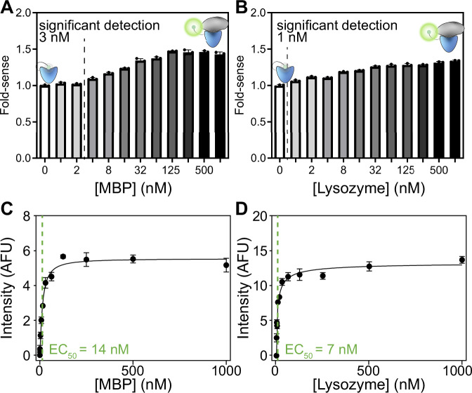
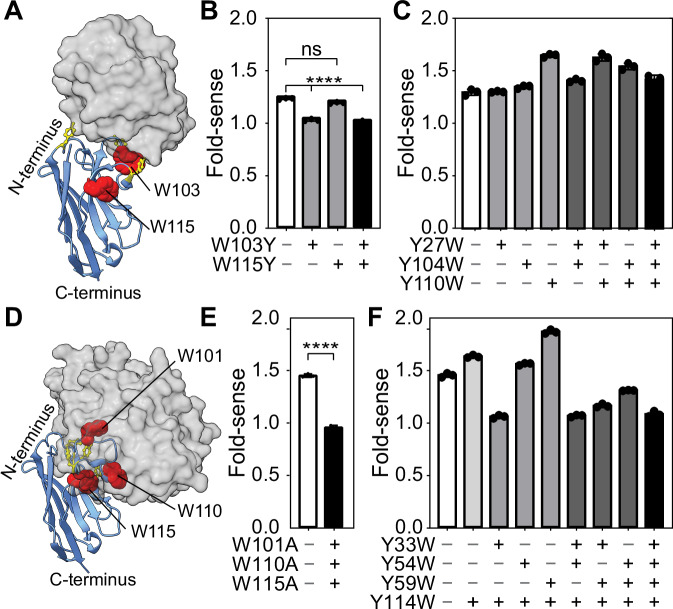
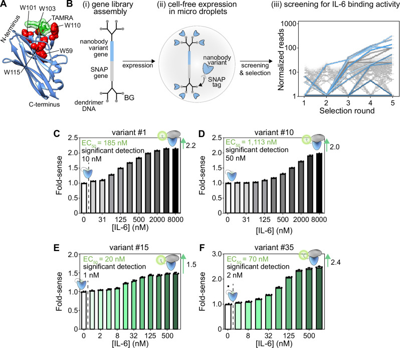
Quenchbodies, antibodies labelled with fluorophores that increase in intensity upon antigen binding, offer great promise for biosensor development. Nanobody-based quenchbodies are particularly attractive due to their small size, ease of expression, high stability, rapid evolvability, and amenability to protein engineering. However, existing designs for protein detection show limited dynamic range, with fluorescence increases of only 1.1-1.4 fold. Here we identify the tryptophan residues in the nanobody complementarity-determining regions (CDRs) that are critical to quenchbody performance. Using a combination of rational design and molecular dynamics simulations, we developed an optimised nanobody scaffold with tryptophans introduced at key positions. We used this scaffold in an in vitro directed-evolution screen against human inflammatory cytokine interleukin-6 (IL-6). This yielded quenchbodies with 1.5-2.4-fold fluorescence increases, enabling IL-6 detection down to 1-2 nM. Our scaffold provides a valuable platform for developing biosensors for diverse protein targets, with applications in research, diagnostics, and environmental monitoring.


Conflict of interest statement

Competing interests: This work was financially supported by Quantum-Si. Marco Ribezzi-Crivellari and Sebastian Hutchinson are employed by Quantum-Si. Antoine van Oijen and Andrew Griffiths were members of the Quantum-Si Scientific Advisory Committee.

Figures

Fig. 1
Fig. 1. Design and in silico modelling of de novo quenchbodies.
A Schematic representation of the working principle of a quenchbody. In the absence of the antigen (left), the fluorophore is quenched by tryptophans in the antibody. In the antigen-bound state (right,) the fluorophore is displaced, resulting in dequenching and an increase in fluorescence intensity. B Proposed mechanism of the MBP-binding quenchbody (blue) modelled from PDB ID: 5M14, with N-terminal covalently conjugated fluorophore (green) undergoing quenching due to interaction with the intrinsic CDR-based tryptophans (red spheres). Upon binding to the MBP antigen (grey surface model), the fluorophore is sterically occluded from tryptophans (W101, W110 and W115), which is associated with increased fluorescence intensity. C Average normalised distribution histogram of the distance of the fluorophore to any of the three tryptophans (W101, W110 and W115) derived from MD simulations in the absence (blue) or presence (green) of antigen for the MBP-binding nanobody. The fluorophore is considered quenched by tryptophan at distances ≤10 Å (hatched zone). Individual plots for W101, W110 and W115 are provided (Fig. S1A). D Average normalised distribution histogram of the distance of the fluorophore to either of the two tryptophans (W103 and W115) derived from MD simulations in the absence (blue) or presence (green) of antigen for the lysozyme-binding nanobody. Individual plots for W103 and W115 are provided (Fig. S1B).
Fig. 2
Fig. 2. Fluorescence intensity changes in quenchbodies upon antigen binding.
A MBP or (B) lysozyme quenchbodies were incubated (60 min, 25 °C) in the presence of increasing concentrations of their cognate antigens. Data are mean ± SD of the fluorescence fold-increase relative to the 0 nM antigen native sample (excitation = 535 ± 20 nm and emission = 585 ± 30 nm). The lowest point of statistically significant detection is indicated (Ordinary one-way ANOVA, Tukey’s multiple comparisons, p = 0.002), with concentrations listed to the left of the hashed line considered to be non-significant (α = 0.05). Fluorescence intensity responses of (C) MBP or (D) lysozyme quenchbodies to their cognate antigens were quantified and fitted to an equation describing a single site-specific binding mode to derive an EC50 as a proxy measure for quenchbody binding affinity (KD). Data are mean ± SD normalised fluorescence intensity.
Fig. 3
Fig. 3. Tryptophan-mediated quenching in lysozyme and MBP quenchbodies.
A Model of the lysozyme quenchbody (blue) highlighting all intrinsic tyrosines (yellow sticks) and tryptophans (red spheres, as labelled), in complex with lysozyme (grey surface model), based on PDB ID: 1ZVH. B Fold-sense for WT, W103Y, W115Y and W103Y/W115Y lysozyme quenchbody variants in the presence of 500 nM lysozyme. C Fold-sense for WT, Y27W, Y104W, Y110W, single-, double- and triple-mutant lysozyme quenchbody variants. D Model of the MBP quenchbody (blue) highlighting all intrinsic tyrosines (yellow sticks) and tryptophans (red spheres, as labelled), in complex with MBP (grey surface model), based on PDB ID: 5M14. E Fold-sense for WT, vs W101A/W110A/W115A triple mutant in the presence of 500 nM MBP. F Fold-sense for WT, Y33W, Y54W, Y59W and Y114W, double-, triple-, and quadruple-mutant MBP quenchbody variants. Data are mean ± SD (excitation = 535 ± 20 nm and emission = 585 ± 30 nm). Ordinary one-way ANOVA with Tukey’s multiple comparisons shows significant (**** = p < 0.0001) or non-significant (ns) differences.
Fig. 4
Fig. 4. Evolution of novel CDR-tryptophan quenchbodies for binding to interleukin-6.
A Model of our tryptophan-optimised nanobody scaffold. B Schematic representation of the directed-evolution workflow. (i) A gene library consisting of variant genes (blue) linked to SNAP genes (grey) is assembled and ligated to dendrimer-like DNA conjugated to a benzylguanine moiety (BG). (ii) Gene constructs are encapsulated in individual microdroplets with cell-free expression reagent. Expressed SNAP-tagged variants bind to the gene construct, resulting in phenotype–genotype linkage. (iii) Variants are screened and sequenced to measure the number of reads for each variant. Lines show normalised read counts over rounds 1–5, selecting for IL-6 binding over 2 technical replicates. Each hit is indicated as a separate colour. C The fold-sense for quenchbody variant #1 and D variant #10 in the presence of increasing concentrations of IL-6. The lowest point of statistically significant detection is indicated (Ordinary one-way ANOVA, Tukey’s multiple comparisons, p < 0.0001), with concentrations listed to the left of the hashed line considered to be non-significant (α = 0.05). E The fold-sense for affinity-matured quenchbody variant #15 in the presence of increasing concentrations of IL-6 shows an EC50 = 20 nM, and significant detection down to 1 nM. F The fold-sense for affinity-matured quenchbody variant #35 in the presence of increasing concentrations of IL-6 shows a fold-sense of 2.4 and significant detection down to 2 nM.

Similar articles

References

    1. Hristova, V. A. & Chan, D. W. Cancer biomarker discovery and translation: proteomics and beyond. Expert Rev. Proteom.16, 93–103 (2019). - PMC - PubMed
    1. Crutchfield, C. A. et al. Advances in mass spectrometry-based clinical biomarker discovery. Clin. Proteomic13, 1 (2016). - PMC - PubMed
    1. Geyer, P. E. et al. Revisiting biomarker discovery by plasma proteomics. Mol. Syst. Biol.13, 942 (2017). - PMC - PubMed
    1. Hawkridge, A. M. & Muddiman, D. C. Mass spectrometry-based biomarker discovery: Toward a global proteome index of individuality. Annu Rev. Anal. Chem.2, 265–277 (2009). - PMC - PubMed
    1. Hage, D. S. Immunoassays. Anal. Chem.71, 294R–304R (1999). - PubMed

MeSH terms

LinkOut - more resources