. 2025 Aug;644(8075):205-213.
doi: 10.1038/s41586-025-09166-w. Epub 2025 Jun 18.

R9AP is a common receptor for EBV infection in epithelial cells and B cells

Affiliations

R9AP is a common receptor for EBV infection in epithelial cells and B cells

Yan Li et al. Nature. 2025 Aug.

Abstract

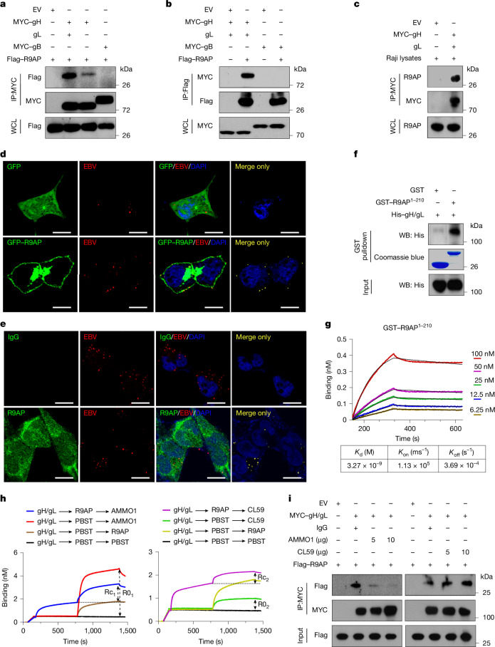
Epstein-Barr virus (EBV) persistently infects more than 90% of the human population, causing infectious mononucleosis1, susceptibility to autoimmune diseases2 and multiple malignancies of epithelial or B cell-origin3. EBV infects epithelial cells and B cells through interaction between viral glycoproteins and different host receptors4, but it has remained unknown whether a common receptor mediates infection of its two major host cell targets. Here, we establish R9AP as a crucial EBV receptor for entry into epithelial and B cells. R9AP silencing or knockout, R9AP-derived peptide and R9AP monoclonal antibody each significantly inhibit, whereas R9AP overexpression promotes, EBV uptake into both cell types. R9AP binds directly to the EBV glycoprotein gH/gL complex to initiate gH/gL-gB-mediated membrane fusion. Notably, the interaction of R9AP with gH/gL is inhibited by the highly competitive gH/gL-neutralizing antibody AMMO1, which blocks EBV epithelial and B cell entry. Moreover, R9AP mediates viral and cellular membrane fusion in cooperation with EBV gp42-human leukocyte antigen class II or gH/gL-EPHA2 complexes in B cells or epithelial cells, respectively. We propose R9AP as the crucial common receptor of B cells and epithelial cells and a potential prophylactic and vaccine target for EBV.


Conflict of interest statement

Competing interests: Patent applications related to the R9AP monoclonal antibody 5E9 have been submitted to the China National Intellectual Property Administration (patent application number 2024119195522; M.-S.Z., C.S. and C.X. are named inventors). The other authors declare no competing interests.

Figures

Fig. 1
Fig. 1. Inhibition of R9AP impairs EBV infection in epithelial and B cells.
a,b, HNE1 cells were transfected with R9AP siRNA (siR9AP 1 or siR9AP 2) or control siRNA (siCtrl), then co-incubated with EBV. a, GFP expression was quantified by flow cytometry analysis and R9AP expression was analysed by western blot. b, Representative fluorescence microscopy images with EBV-positive cells shown in green. Scale bars, 100 μm. c,d, R9AP protein expression in CRISPR-edited HNE1 cells using control (sgVector) or independent R9AP sgRNA (sgR9AP 1 and sgR9AP 2) (c) and R9AP-reconstituted R9AP-knockout cells (d). EBV was added to the R9AP-knockout or reconstituted cells and EBV infection efficiency was analysed by flow cytometry. e,f, EBV infection in control and two R9AP-knockout single-cell HNE1 clones was measured by flow cytometry (f) and quantified (e, top). e, Bottom, R9AP protein expression in control cells and two R9AP-knockout HNE1 single-cell clones. g, R9AP protein expression in AGS cells expressing sgVector or R9AP sgRNA. EBV was added to AGS cells and infection efficiency was analysed by flow cytometry. h,i, EBV infection in control and two R9AP-knockout single-cell AGS clones was measured by flow cytometry (i) and quantified (h, top). h, Bottom, R9AP protein expression in control cells and two R9AP-knockout AGS single-cell clones. j, R9AP protein in control versus in R9AP-knockout Raji cells. EBV infection efficiency was analysed by flow cytometry. k,l, EBV infection in control and two R9AP-knockout single-cell Raji clones was measured by flow cytometry (l) and quantified (k, top). k, Bottom, R9AP protein expression in control cells and two R9AP-knockout Raji single-cell clones. Three independent experiments in triplicates (n = 9); data are mean ± s.e.m.; one-way ANOVA with Tukey’s correction for multiple comparisons (a,ce,g,h,j,k). FSC, forward scatter. Source data
Fig. 2
Fig. 2. R9AP directly interacts with gH/gL.
a,b, HEK-293T cells were co-transfected with Flag–R9AP together with empty vector (EV), MYC–gH and gL, MYC–gH or MYC–gB, lysed and immunoprecipitated (IP) with antibodies against MYC (a) or Flag (b). WCL, whole-cell lysate. c, HEK-293T cells were transfected with empty vector or MYC–gH and gL, lysed and incubated with Raji cell lysates. Anti-MYC immunoprecipitation was then performed with the mixed lysates, followed by western blot (WB) analysis. d,e, HEK-293T or HNE1 cells expressing GFP or GFP–R9AP were incubated with EBV at 4 °C for 1 h, then incubated at 37 °C for 30 min. GFP or GFP–R9AP (d) and R9AP immunofluorescence (e) are shown in green. EBV was detected with Alexa Fluor 594-conjugated gp350 antibody (72A1) and is shown in red. Nuclei were stained with DAPI (blue). The far right panel shows co-localization of green and red signals. Scale bars, 10 μm. f, His–gH/gL (containing gH amino acids 19–682 and gL amino acids 23–137) proteins were precipitated with GST or GST–R9AP1–210 and detected by western blot. GST fusion proteins were detected by Coomassie blue staining. g, His–gH/gL was captured on Ni-NTA biosensors and assayed for binding to GST–R9AP1–210. Kd, on rate (Kon) and off rate (Koff) calculated from the fit model for binding curves are shown below the graph. h, BLI gH/gL competition assay, with gH/gL binding to GST–R9AP1–210 or the monoclonal antibodies AMMO1 or CL59 added in the indicated order. PBST, PBS Tween-20 vehicle. i, HEK-293T cells were lysed after transfection with empty vector, MYC–gH/gL or Flag–R9AP. Lysates containing MYC–gH/gL were pre-incubated with the indicated amount of antibody or IgG control and then incubated with lysates containing Flag–R9AP overnight. Mixtures were then immunoprecipitated with antibodies against MYC and western blot was performed. af, Data are from three independent experiments. gi, Data are from two independent experiments. Source data
Fig. 3
Fig. 3. R9AP receptor function depends on its N-terminal residues and TMDs.
a,b, Protein levels and EBV infection efficiency of ectopically expressed wild-type (WT) and truncated R9AP constructs (a) or WT and multiple deletions (b). ce, EBV was pretreated with indicated peptide or control peptide (Ctrl) and added to HNE1 cells (c), Raji cells (d), primary NPECs or primary B cells (e) and EBV infection efficiency was analysed by flow cytometry (c,d) or qPCR (e). ****P <  0.0001. f, His–gH/gL was assayed for binding to Ctrl peptide or R9AP19–30. n = 2 independent experiments. gi, Humanized B-NDG mice were infected with EBV and treated with Ctrl peptide or R9AP19–30 and EBV DNA copy number in peripheral blood was determined by qPCR at the indicated times (g,h). i, Survival curves of mice. n = 5 mice in each group. Data are mean ± s.e.m. (h). jl, Primary B cells, NPECs, Akata, Raji, HNE1, AGS and HEK-293T cells were pretreated with R9AP monoclonal antibody (5E9) or IgG and infected with EBV, and EBV infection efficiency was analysed by counting colony formation using B cell transformation assays (j), qPCR (k) or flow cytometery (l). Data are mean ± s.e.m. from 3 independent experiments (ad), primary B cells from 3 donors (e) performed in triplicate, n = 9 total replicates (a,ce) or n = 8 total replicates (b). Data are mean ± s.e.m. from 1 representative of 2 (j) or 3 (k,l) independent experiments (n = 6 or 3 biological replicates). One-way ANOVA with Tukey’s correction for multiple comparisons (a,b); two-way ANOVA with Sidak’s correction for multiple comparisons (ce,jl); unpaired two-tailed t-test (h) and log-rank test (i). ****P < 0.0001. NS, not significant. Source data
Fig. 4
Fig. 4. R9AP is expressed in human tissues that are susceptible to EBV infection.
a, Representative immunohistochemical images of human lymphoid tissues (n = 5), basal layers of floor of the mouth (n = 5) and tongue (n = 5), stained with haematoxylin and eosin (H&E) (top row) or with anti-R9AP (bottom row). Inset shows 4× magnified view of the outlined region in the main image. Scale bars, 50 μm. b, Representative images of nasopharyngeal carcinoma (n = 5), gastric carcinoma (n = 3) and B cell lymphoma (n = 4) stained with anti-R9AP (top row) or EBV-encoded EBER probe (bottom row). Inset shows 4× magnified view of the outlined region in the main image. Scale bars, 100 μm. c, Western blot analysis of R9AP expression in human tongue, lymph node, gastric mucosa, liver, lung, thyroid, EBV-positive B cell lymphoma (BL), nasopharyngeal carcinoma (NPC) and EBV-positive gastric carcinoma (GC). HNE1 cells expressing Cas9 and sgR9AP 1 or sgVector were used as negative and positive controls, respectively. Data are representative of three independent experiments. d, Unified model of EBV entry. In B cells, gp42 binding to HLAII allows gH/gL to bind R9AP, which induces activation of gB-mediated viral and host membrane fusion. In epithelial cells, gH/gL simultaneously binds R9AP and EPHA2, which induces gB-mediated viral and host membrane fusion. Part d created in BioRender. Yang, T. (2025) https://BioRender.com/b61o826.
Extended Data Fig. 1
Extended Data Fig. 1. Screening identifies R9AP as a potential EBV receptor.
a, Heatmap of genes in SLCs compared to MLCs of NPEC1-Bmi1 and NPEC2-Bmi1 cells. b, siRNA screening. Relative EBV infection efficiency was determined by flow cytometry. 72 siRNA pools were screened in NPEC2-Bmi1 SLCs first, and 10 siRNA pools were selected and re-screened in NPEC1- Bmi1 SLCs. Arrows indicated genes whose knockdown reduced EBV infection efficiency by more than 50% compared to the siCtrl. The dotted pink line indicated 50% of relative EBV infected cells in siCtrl transfected SLCs. Data are mean (n = 2 biological replicates). c, RT-qPCR was used to detect knockdown efficiency of indicated siRNA. Results were quantified relative to the housekeeping gene beta-actin (ACTB). Two independent experiments in either triplicate or duplicate (n = 5) and mean and S.E.M. of those n = 5 were used. (****P <  0.0001) d, WB analysis of the endogenous CNGA1, R9AP, GPR1, and SLC26A9 in indicated EBV susceptible cells. Data are representative of two independent experiments. e, f, EBV infection efficiencies, and ectopic R9AP, CNGA1 protein level of HEK-293T cells after being transfected with CNGA1, R9AP, or EV (e). Three independent experiments in triplicates (n = 9) and the mean and S.E.M. of those n = 9 were used. The percentage of EBV-infected cells was shown (f), data are representative of three independent experiments. g, Schematic summary of screening. Microarray was used to analyze gene expression in NPECs-Bmi1 SLCs compared to MLCs. A siRNA library targeting upregulated transmembrane associated genes in SLCs was transfected into NPECs-Bmi1. Knocking down of CNGA1, R9AP, SLC26A9, or GPR1 reduced EBV infection more than 50%, while only CNGA1 and R9AP were expressed in all indicated cell lines by WB. Finally, ectopic expressing R9AP but not CNGA1 enhanced EBV infection in HEK-293T cells. One-way ANOVA was carried out with Tukey’s correction for multiple comparisons (c, e). Source data
Extended Data Fig. 2
Extended Data Fig. 2. R9AP mediates EBV infection in nasopharyngeal epithelial cells.
a, RT-qPCR was used to quantify the mRNA level of R9AP in HNE1, AGS, and Raji cells. The mRNA without reverse transcription was used as a negative control. Three independent experiments in either triplicate or duplicate (n = 8) and mean and S.E.M. of those n = 8 were used. b, Raji, HNE1, and AGS cells were stained with antibodies against R9AP and analyzed by flow cytometry. c, HNE1 cells were transfected with R9AP siRNAs (siR9AP 1 or siR9AP 2) or siCtrl, then infected with EBV, and the percentage of infected cells was determined using flow cytometry. d, e, HNE1 cells were knockout of R9AP using sgR9AP 1 and sgR9AP 2 (d). Then R9AP knockout cells were reconstituted with an R9AP expression vector (sgR9AP 1 +R9AP) (e). EBV was added to the R9AP knockout and reconstituted HNE1 cells, and EBV infection efficiency was analyzed by flow cytometry. f, g, Representative images of R9AP knockout HNE1 single-cell clones or sgVector HNE1 cells infected with EBV then hybridized with an EBV EBERs probe (f). Scale bar=50 μm. The proportion of EBERs-positive cells was independently evaluated by three pathologists (g). h, Single-cell clones of R9AP knockout HNE1 cells were infected with EBV and analyzed by RT-qPCR to quantify the mRNA level of indicated EBV genes. i, j, The R9AP protein in R9AP knockout single-cell clone 1 HNE1 cells without or with reconstitution with R9AP (i). EBV was added to these cells, and the EBV infection efficiency was analyzed by flow cytometry (j) and quantified (i). Data are representative of three independent experiments (b-f, j). Three independent experiments by three individual evaluation (n = 9; g) or in triplicates (n = 9; h, i) and mean and S.E.M. of those n = 9 were used, and one-way ANOVA was carried out with Tukey’s correction for multiple comparisons (g-i). Source data
Extended Data Fig. 3
Extended Data Fig. 3. R9AP mediates EBV infection in gastric epithelial and B cells.
a, EBV infection was determined in R9AP knockout AGS cells using flow cytometry. b, c, The R9AP protein in MKN74 cells knockout of R9AP using sgR9AP 1 and sgR9AP 2 (b). EBV infection was determined using flow cytometry (c), and the quantification data was shown in (b). d, e, The R9AP protein level in R9AP knockout single-cell clone 1 AGS cells without or with reconstitution with R9AP (d). EBV was added to these cells, and the EBV infection efficiency was analyzed by flow cytometry (e) and quantified (d). f, EBV infection was determined in R9AP knockout Raji cells using flow cytometry. g, h, The R9AP protein level in R9AP knockout single-cell clone 1 Raji cells without or with reconstitution with R9AP (g). EBV was added to these cells, and the EBV infection efficiency was analyzed by flow cytometry (h) and quantified (g). i, sgVector, sgR9AP 1, or sgR9AP 2 was delivered into primary B cells by a lentivirus package system. The R9AP protein level was analyzed by WB. EBV infection was analyzed by qPCR. Three independent experiments in triplicates (n = 9) and mean and S.E.M. of those n = 9 were used (b, d, g), Two independent experiments from two donors’ primary B cells experiments in triplicate (n = 6) and mean and S.E.M. of those n = 6 were used (i). One-way ANOVA was carried out with Tukey’s correction for multiple comparisons (b, d, g, i). Data are representative of three independent experiments (a, c, e, f, h). Source data
Extended Data Fig. 4
Extended Data Fig. 4. R9AP mediates EBV entry and fusion.
a-d, Ectopic R9AP protein and EBV infection efficiency in CNE1 and AGS cells (a, b) or EBV-negative Akata cells (c, d). e, EBV binding in R9AP, CR2, or EV transiently transfected HEK-293T cells. f, g, EBV binding in R9AP or EV transiently transfected CNE1 cells or stably delivered EBV-negative Akata cells (f), in R9AP knockout HNE1 cells and EBV-negative Akata cells (g). h, i, EBV entry in R9AP or EV transiently transfected CNE1 cells or stably delivered EBV-negative Akata cells (h), in R9AP knockout HNE1 cells or EBV-negative Akata cells (i). j-l, Cell-based EBV fusion assay by co-culture of R9AP knockdown HEK-293T cells and HEK-293T cells transfected with gB and gH/gL (j), by co-culture of R9AP knockout Daudi cells, HEK-293T cells transfected with gB and gH/gL and His-gp42 protein at 0.5 μg/mL (k), by co-culture of R9AP overexpressed HEK-293T and HEK-293T cells transfected with gB, gH/gL, or gH/gL/gB (l). Three independent experiments in triplicates (n = 9) and mean and S.E.M. of those n = 9 were used (a, c, j, k, e), two independent experiments in triplicates (n = 6) and mean and S.E.M. of those n = 6 were used (f-i), or three independent experiments in quadruplicates (n = 12) and mean and S.E.M. of those n = 12 were used (l). Two-tailed unpaired Student’s t-test (a, c, f-k), one-way ANOVA was carried out with Tukey’s correction for multiple comparisons (e), or two-way ANOVA was carried out with Sidak’s correction for multiple comparisons (l). Data are representative of three independent experiments (b, d). NS, not significant. Source data
Extended Data Fig. 5
Extended Data Fig. 5. R9AP interacts with gH and gH/gL and cooperates with gp42-receptor HLA II.
a, HEK-293T cells were transfected with FLAG-R9AP, Myc-gH, and Myc-gL, Myc-gH alone, or Myc-gB and then lysed. The cell lysate containing FLAG-R9AP was incubated with the cell lysate containing Myc-gH and Myc-gL, Myc-gH alone, or Myc-gB, then immunoprecipitated with an antibody against Myc followed by WB analysis with the indicated antibody. b, HEK-293T cells were co-transfected with R9AP and EV, Myc-gH/gL, Myc-gH/gL/gp42, or Myc-gH/gL/gp42, and FLAG-HLA II (HLA-DR1). Cells were lysed and immunoprecipitated with antibodies against Myc, followed by WB analysis with the indicated antibodies. c, Cell-based EBV fusion assay by co-culture of R9AP, HLA II (HLA-DR1), or R9AP/HLA II (HLA-DR1) overexpressed HEK-293T cells, HEK-293T cells transfected with gB/gH/gL, and His-gp42 protein. Data are mean ±  S.E.M. (n = 3 biological replicates). Two-way ANOVA was carried out with Sidak’s correction for multiple comparisons. (P <  0.0001) Data are representative of two independent experiments (a-c). Source data
Extended Data Fig. 6
Extended Data Fig. 6. R9AP, EPHA2, and NRP1 are not redundant for EBV infection of epithelial cells.
a-c, Cell-based EBV fusion assay by co-culture of sgVector transfected HNE1 cells, EPHA2 knockout HNE1 cells, R9AP overexpressed while EPHA2 knockout HNE1 cells or EPHA2 rescued EPHA2 knockout HNE1 cells, and HEK-293T cells transfected with gH/gL/gB (a). EBV infection efficiency was analyzed by flow cytometry (c) and quantified (b) in the above-indicated HNE1 cells. d-f, Cell-based EBV fusion assay by co-culture of sgVector transfected HNE1 cells, R9AP knockout HNE1 cells, EPHA2 overexpressed while R9AP knockout HNE1 cells or R9AP rescued R9AP knockout HNE1 cells, and HEK-293T cells transfected with gH/gL/gB (d). EBV infection efficiency was analyzed by flow cytometry (f) and quantified (e) in the above-indicated HNE1 cells. g, HEK-293T cells were transfected with Myc-R9AP, EPHA2 together with FLAG-gH/gL or EV, lysed, and immunoprecipitated with antibody against FLAG as indicated IPx1:FLAG. The immunoprecipitated proteins were eluted by FLAG peptide and re-immunoprecipitated with antibody against Myc as indicated IPx2: Myc, followed by WB analysis with indicated antibodies. Data are representative of two independent experiments. h-j, Cell-based EBV fusion assay by co-culture of sgVector transfected HNE1 cells, R9AP knockout HNE1 cells, NRP1 overexpressed while R9AP knockout HNE1 cells or R9AP rescued R9AP knockout HNE1 cells, and HEK-293T cells transfected with gH/gL/gB (h). EBV infection efficiency was analyzed by flow cytometry (j) and quantified (i) in the above-indicated HNE1 cells. Two independent experiments in triplicates (n = 6) and mean and S.E.M. of those n = 6 were used (a, d, h, i), or three independent experiments in triplicates (n = 9) and mean and S.E.M. of those n = 9 were used (b, e). One-way ANOVA was carried out with Tukey’s correction for multiple comparisons. Data are representative of three (c, f) or two (j) independent experiments. Source data
Extended Data Fig. 7
Extended Data Fig. 7. Functional domains of R9AP.
a, Schematic representation of R9AP wild-type (WT), R9AP1-210, R9AP1-231, R9AP mutants deleted of amino acids 1-50 (∆1-50), 51-100 (∆51-100), 101-152 (∆101-152), or 153-200 (∆153-200). b, c, EBV infection efficiency was analyzed in EV, R9AP1-210, R9AP1-231, or WT transiently transfected HEK-293T cells (b), as well as EV, ∆1-50, ∆51-100, ∆101-152, ∆153-200 or WT transiently transfected HEK-293T cells (c). d, EV, N-terminus FLAG-tagged WT or ∆1-50 R9AP mutant was transiently transfected into HEK-293T cells. Cells were stained with FITC-FLAG antibody and analyzed by flow cytometry. Data are representative of three independent experiments (b-d).
Extended Data Fig. 8
Extended Data Fig. 8. N-terminus of R9AP locates at cell surface.
a, b, Prediction of R9AP localization by using TMHMM (a) or InterPro (b). c, HK1 cells were fixed and then incubated with antibody targeting N-terminal of R9AP and Alexa Flour 594-labelled goat antibody (red), nuclei were stained with DAPI (blue). d, HNE1 cells transfected with Myc-R9AP1-210 or Myc-R9APFL expression plasmid for 24 h, fixed and treated with or without Triton X-100, followed by incubation with the antibody specific for Myc tag and Alexa Flour 594-labelled goat antibody (red), nuclei were stained with DAPI (blue). e, f, PreScission protease (PSP) cleavage site was inserted between the FLAG tag and the N-terminal end of R9AP1-210 (FLAG-psp-R9AP1-210) or R9APFL (FLAG-psp-R9APFL) expressing plasmid. Cells transfected with indicated plasmid were cultured for 24 h followed by PSP treatment to remove FLAG located at outside of cells. The schematic was used to show that FLAG tag is removed by the PSP when it is located outside the cell whereas it is retained when located in the cytoplasm (e). WB was used to detect the R9AP or FLAG tag after adding PSP enzyme (f). Data are representative of three (c) two (d, f) independent experiments.
Extended Data Fig. 9
Extended Data Fig. 9. R9AP peptide inhibits EBV infection in vitro and in vivo.
a, b, EBV was pretreated with R9AP1-12, R9AP13-24, R9AP19-30, R9AP30-41, R9AP35-46, or Ctrl peptide, then added to HNE1 cells (a), R9AP19-30 or Ctrl pretreated EBV was added to Raji cells (b). EBV infection efficiency was analyzed by flow cytometry. c, EBV was pretreated with R9AP19-30 at a concentration of 0, 50, 100, 200, or 400 μg mL−1, then added to HNE1 or Raji cells. EBV infection efficiency was analyzed by flow cytometry, and the EC50 of R9AP19-30 was determined. d, Schematic representation of in vivo EBV infection using humanized B-NDG mice. e, The body weight of humanized B-NDG mice treated with R9AP19-30 or Ctrl peptide at indicated time points (Ctrl, n = 5 mice; R9AP19-30, n = 5 mice). f, Representative images of spleen tissue sections of humanized B-NDG mice infected with EBV and treated with R9AP19-30 or Ctrl peptide stained with hematoxylin-eosin staining (H& E), anti-human CD20, or hybridized with EBV EBERs probe. Scale bar=50 μm. The samples’ representative images were detected (Ctrl, n = 5 mice; R9AP19-30, n = 5 mice). g, The proportion of EBERs-positive cells in the spleen of EBV-infected and R9AP19-30 or Ctrl peptide-treated humanized B-NDG mice were independently evaluated by three pathologists. Three independent evaluation of each mouse (n = 15) and mean and S.E.M. of those n = 15 were used, two-tailed unpaired Student’s t-test (Ctrl, n = 5 mice; R9AP19-30, n = 5 mice). (P <  0.0001) Data are representative of three (a, b) two (c) independent experiments. Source data
Extended Data Fig. 10
Extended Data Fig. 10. Characterization of the anti-R9AP monoclonal antibody 5E9 and its effects on blocking EBV infection.
a, Complementarity determining regions (CDR) details for the anti-R9AP monoclonal antibody 5E9 antibody. b, ELISA binding for 5E9 and control antibody to R9AP1-210. The A450 signals were from triplicate wells and the mean signal were presented. c, BLI binding assay for 5E9 to R9AP1-210. 5E9 were captured onto ProA biosensors and assayed for binding to R9AP1-210. d, WB for 5E9 binding to R9AP in comparison to commercial antibody. HEK-293T cells were transfected with FLAG-R9AP. Cells were lysed and immunoprecipitated with 5E9 or anti-R9AP antibody (Cat #HPA049791, Sigma). e, Primary B cells were pretreated with anti-R9AP monoclonal antibody (5E9), then infected with EBV. The proportion of EBERs-positive cells were independently evaluated by three pathologists. Each pathologist counted 3 representative high-power fields (×40 objective) per sample, with approximately 100 cells/field, and obtained a mean value. Bars represent proportion of EBERs-positive cells. Data are mean ±  S.E.M. and representative of two independent experiments (n = 3). f-j, Akata, Raji, HNE1, AGS and HEK-293T cells were pretreated with anti-R9AP monoclonal antibody (5E9), then infected with EBV. EBV infection efficiency was analyzed by flow cytometry. Data are representative of two (b-d) or three independent experiments (f-j). Source data
Extended Data Fig. 11
Extended Data Fig. 11. R9AP expression in human tonsil and blood tissues.
a, RT-qPCR was used to quantify the mRNA level of the R9AP in tonsil epithelial cells of 2 donors and memory B cells and naïve B cells from 2 donor’s blood and tonsil tissues. Data are mean ±  S.E.M. (n = 3 biological replicates) and are from 2 donors. b, Blood PBMC were co-stained with CD19/R9AP antibody and analyzed by flow cytometry. Results are representative of 10 donors. c, Cells from tonsil tissues were co-stained with EpCAM/R9AP, CD19/CD27/R9AP, or CD19/CD10/R9AP antibody and analyzed by flow cytometry. Results are representative of 3 donors. d, e, Epithelium, and lymphoid tissues from tonsil were stained with hematoxylin-eosin staining (H&E), co-stained with R9AP (imaged as green) antibody and EpCAM (imaged as red) antibody, or co-stained with R9AP (imaged as green), CD19 (imaged as red) and CD27 (imaged as gray) antibodies, nuclei were stained with DAPI (blue), and analyzed by immunofluorescence. Epithelial cells were delineated by the dotted line (d). The white dotted areas indicated CD19+CD27+R9AP+ cells and were magnified 3 times, and the white solid areas indicated CD19+CD27R9AP+ cells (e). Results are representative of 2 donors. Source data
Extended Data Fig. 12
Extended Data Fig. 12. R9AP expression in human lymphoid, tongue, floor of mouth, nasopharynx epithelium and gastric mucosa tissues.
a, Human lymphoid tissues (n = 3) were stained with hematoxylin-eosin (H&E) (left), R9AP antibody (middle), and CR2 antibody (right). Images of insets were magnified 4 times. GC: germinal center; MCZ: mantle cell zone; MZ: marginal zone. Scale bars: 100 μm. b, The keratinocytes and spinous cell layer of human tongue (n = 5) and floor of the mouth tissues (n = 5) were stained with hematoxylin-eosin (H&E) (left) and R9AP antibody (right). Images of insets were magnified 4 times. Scale bars: 50 μm. c, Tissues of human normal (n = 5) and dysplastic nasopharynx epithelium (n = 4) and normal gastric mucosa (n = 5) were stained with H& E (top row) and R9AP antibody (bottom row). Images of insets were magnified 4 times. Scale bar=50 μm. Representative images from the samples detected (a-c).

References

    1. Luzuriaga, K. & Sullivan, J. L. Infectious mononucleosis. N. Engl. J. Med.362, 1993–2000 (2010). - PubMed
    1. Bjornevik, K. et al. Longitudinal analysis reveals high prevalence of Epstein–Barr virus associated with multiple sclerosis. Science375, 296–301 (2022). - PubMed
    1. Shannon-Lowe, C. & Rickinson, A. The global landscape of EBV-associated tumors. Front. Oncol.9, 713 (2019). - PMC - PubMed
    1. Hutt-Fletcher, L. M. Epstein–Barr virus entry. J. Virol.81, 7825–7832 (2007). - PMC - PubMed
    1. Zhong, L. Y. et al. Research landmarks on the 60th anniversary of Epstein–Barr virus. Sci. China Life Sci.68, 354–380 (2025). - PubMed

MeSH terms

LinkOut - more resources