. 2025 Aug;644(8075):214-220.
doi: 10.1038/s41586-025-09177-7. Epub 2025 Jun 18.

Targeting de novo purine biosynthesis for tuberculosis treatment

Affiliations

Targeting de novo purine biosynthesis for tuberculosis treatment

Dirk A Lamprecht et al. Nature. 2025 Aug.

Abstract

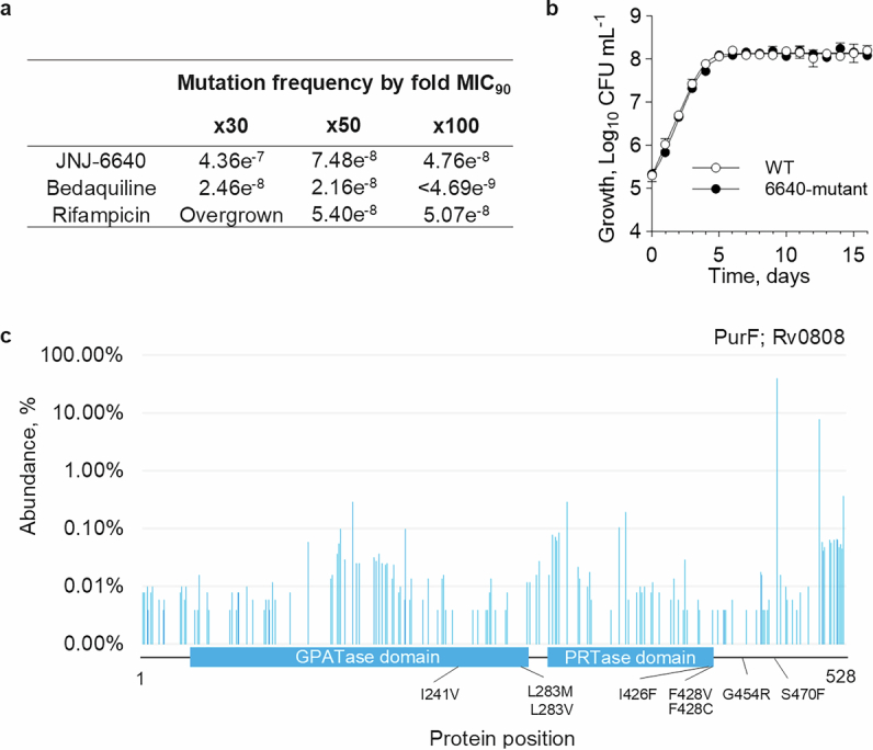
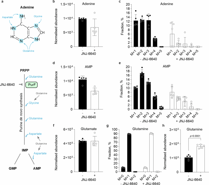
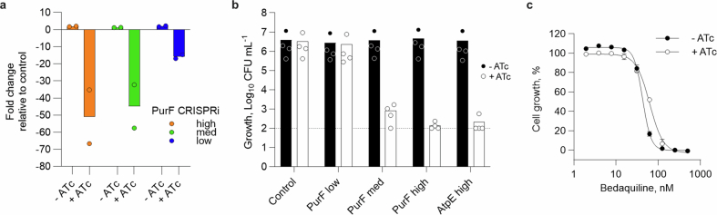
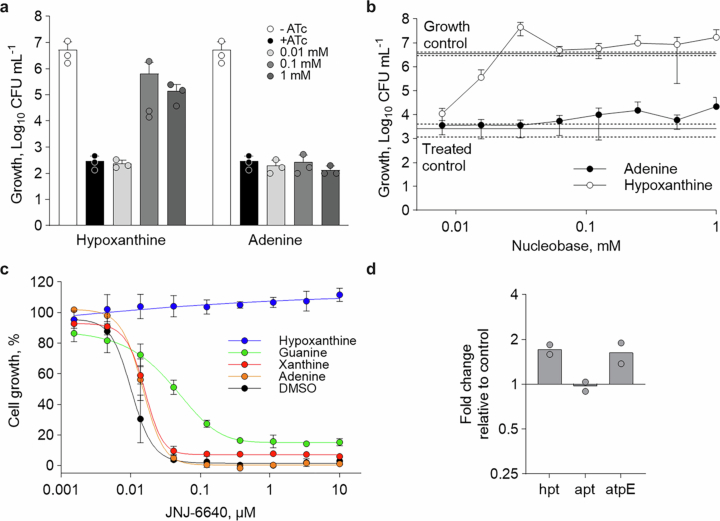
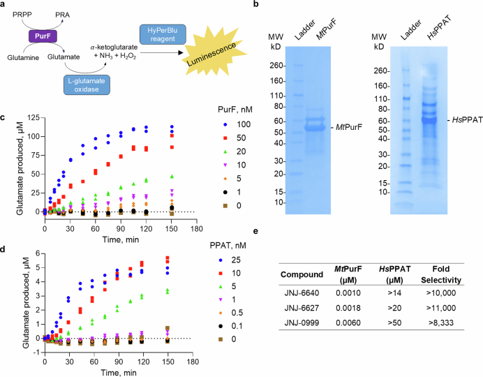
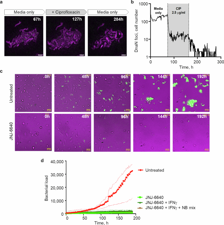
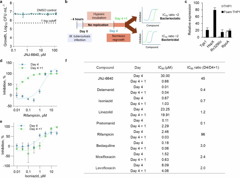
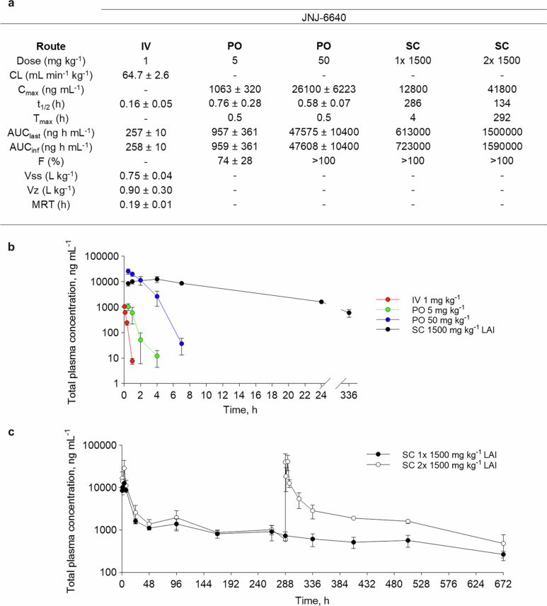
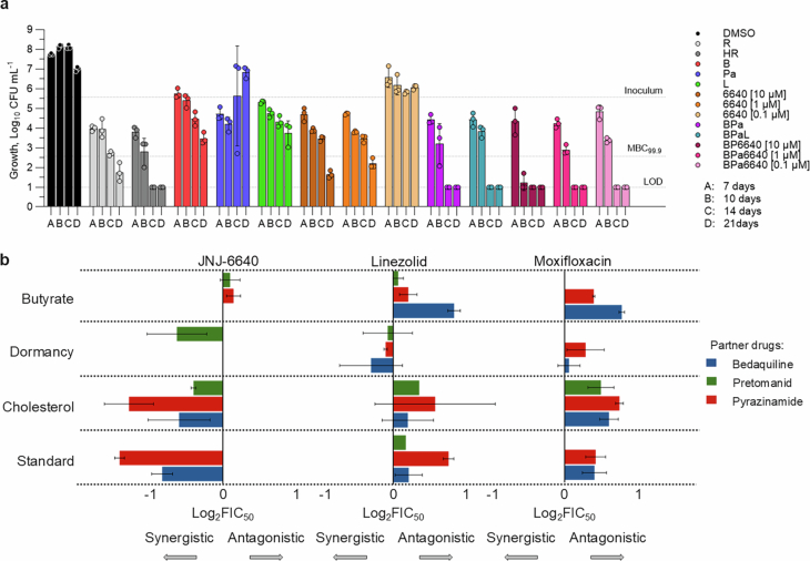
Tuberculosis remains the leading cause of death from an infectious disease1,2. Here we report the discovery of a first-in-class small-molecule inhibitor targeting PurF, the first enzyme in the mycobacterial de novo purine biosynthesis pathway. The lead candidate, JNJ-6640, exhibited nanomolar bactericidal activity in vitro. Comprehensive genetic and biochemical approaches confirmed that JNJ-6640 was highly selective for mycobacterial PurF. Single-cell-level microscopy demonstrated a downstream effect on DNA replication. We determined the physiologically relevant concentrations of nucleobases in human and mouse lung tissue, showing that these levels were insufficient to salvage PurF inhibition. Indeed, proof-of-concept studies using a long-acting injectable formulation demonstrated the in vivo efficacy of the compound. Finally, we show that inclusion of JNJ-6640 could have a crucial role in improving current treatment regimens for drug-resistant tuberculosis. Together, we demonstrate that JNJ-6640 is a promising chemical lead and that targeting de novo purine biosynthesis represents a novel strategy for tuberculosis drug development.


Conflict of interest statement

Competing interests: D.A.L., A.L., J.S., P.F., J.W., S.D., S. Saylock, M.D.R., A.H., T.G., A.V., K.W.C., P.S., M.E., M.M., J.E., R.D.A., H.F., A.S.P., B.S., C.A.-P., P.J. and A.K. were or are all full-time employees of Janssen Pharmaceutica, a Johnson & Johnson company, and/or potential stockholders of Johnson & Johnson. J.S. and P.F. were or are employees of Charles River Laboratories, a contract research organization. A.R., C.R., N.C. and S. Sans were or are all full-time employees of Evotec. R.J.W., C.D., W.P., E.D., S.L., M.G., J.D., S.J.W., G.L.-M., N.D. and A.K. received funding from Janssen Pharmaceutica to perform contract research. The other authors declare no competing interests.

Figures

Fig. 1
Fig. 1. Discovery of JNJ-6640.
a, Series evolution from the hit compound JNJ-7310 to the series lead JNJ-6640. MIC90 values against whole-cell M. tuberculosis are shown. R*, R*-enantiomer; S*, S*-enantiomer. b, Dose–response curve depicting the bactericidal activity (measured as CFU) of JNJ-6640. MBC99.9 = 140 ± 63 nM. n = 4 biological replicates. Data shown are mean ± s.d. LoD, limit of detection. Source data
Fig. 2
Fig. 2. JNJ-6440 target identification and de-risking.
a, Susceptibility of JNJ-6640-resistant clones (R1–R5). MIC90 values are shown in Extended Data Table 2. b, Induced fit docking model of JNJ-6640 (cyan) in the MtPurF-binding pocket (grey). Phe428 (pink), key interactions and resistance residues are annotated. c, Percentage 15N-stable isotope incorporation following 100 nM JNJ-6640 treatment. Control metabolite (glutamine) was the most abundant nitrogen-containing metabolite. n = 5 biological replicates, representative of two independent experiments. Significance was calculated with two-sided (Bonferroni–Dunn) Student’s t-test with Welsh correction. d, Growth kinetics of CRISPRi-mediated purF transcript knockdown strains (low, medium and high) compared with ‘empty’ vector (control) and kill (atpE high) controls after ATc induction. n = 3 independent experiments. e, CRISPRi-mediated PurF ‘low’ knockdown increases susceptibility to JNJ-6640 (MIC50 = 6.4 nM for −ATc and MIC50 = 0.8 nM for +ATc). n = 3 technical replicates, representative of two independent experiments. f, Nucleobase rescue assay with CRISPRi-mediated high, medium and no (control) purF transcript knockdown in the presence of 10 µM nucleobase or nucleoside with (+ATc) and without (−ATc) induction. Starting inoculum was approximately 1 × 105 CFU ml−1. n = 3 independent experiments. g, MtPurF (IC50 = 1 nM) and Homo sapiens PPAT (HsPPAT; IC50 = 14 µM) enzymatic assays with JNJ-6640. n = 2 biological replicates. h, Cell proliferation assay with JNJ-6640 and three known cell proliferation inhibitors using 93 different cancer cell lines derived from different tissue types. The average pIC50 shown for each tissue type: pIC50 ~ 4 (IC50 ~ 100 µM) was considered non-active. Aza, azathioprine; MMPR, 6-methyl-mercaptopurine riboside; MPA, mycophenolic acid. Representative dataset from two independent experiments. For panels cf, data shown are mean ± s.d. Source data
Fig. 3
Fig. 3. M. tuberculosis single-cell analysis of PurF inhibition.
a, M. tuberculosis expressing TdTomato, cultured in a microfluidic device and imaged over 312 h at 1-h intervals, exposed to 0.6 µM JNJ-6640 (95–238 h). NB mix, nucleobase mix (239–311 h). Snapshot images of representative microcolony are shown in magenta (TdTomato fluorescence). Scale bars, 3 µm. b, Fraction of intact cells upon exposure to 0.6 µM JNJ-6640 (grey shading) and after washout and supplementation with 1× NB mix (blue shading), from the single-cell imaging experiments. The lines represent independent xy frames imaged. n = 1,195 cells, across 15 fields. A representative dataset from three independent experiments. c, Representative snapshots of M. tuberculosis replisome reporter strain (MTB::gfp-dnaN, green channel) expressing cytoplasmic TdTomato (magenta channel), before, during and after exposure to 0.6 µM JNJ-6640. Bacteria undergoing DNA replication identified by green foci representing active replisome complex. Scale bars, 3 µm. d, Number of bacteria with a GFP–DnaN foci when exposed to 0.6 µM JNJ-6640 (grey shading) and after washout and supplementation with 1× NB mix (blue shading). The lines (n = 8) represent individual xy positions imaged over time. A representative dataset from two independent experiments. Source data
Fig. 4
Fig. 4. Translation of JNJ-6640 activity.
a, Susceptibility of JNJ-6640 in a foamy macrophage assay. IC50 comparison at day 4 (30 µM) and day 4 + 1 (0.66 µM); ratio of 45 (bactericidal). n = 2 individual experiments with 4 technical replicates. b, JNJ-6640 (1,500 mg kg−1 LAI; subcutaneous) efficacy in an acute mouse model for TB administered once fortnightly (6640 1/14, 7 days post-infection and day 1 treatment phase) or once weekly (6640 2/14, 7 and 14 days post-infection, and day 1 and day 7 treatment phase). 21 Days post-infection data are shown. n = 5 animals. Representative of two independent experiments. BDQ, 25 mg kg−1 bedaquiline oral administration (PO) once daily (qd). c, JNJ-6640 (1,500 mg kg−1 LAI; subcutaneous) efficacy in the chronic mouse model for TB. 84 Days post-infection are shown. n = 6 animals. 6640 8/56, 8× doses, once weekly of JNJ-6640. d, In vitro combination studies replacing linezolid with JNJ-6640. Day 10 data are shown. 6640, 10 µM JNJ-6640; B, 0.5 µM bedaquiline; HR, 5.8 µM isoniazid and 14.58 µM rifampicin; L, 6 µM linezolid; Pa, 7 µM pretomanid. Bedaquiline, pretomanid and linezolid concentrations reflect the mouse equivalent human dose based on efficacious exposure. The full dataset is shown in Extended Data Fig. 9. n = 3 biological replicates, representative of two independent experiments. Dashed line indicates the starting inoculum. e, In vivo combination study demonstrating that JNJ-6640 can replace linezolid in a high acute model. Two week treatment. 24 Days post-infection are shown. 6,640, 1,500 mg kg−1 JNJ-6640 LAI (subcutaneous; once weekly); B, 25 mg kg−1 bedaquiline (PO qd); L, 100 mg kg−1 linezolid (PO qd); Pa, 40 mg kg−1 pretomanid (PO qd); SoT, start of treatment (10 days post-infection). n = 5 (SoT) or 6 (treatment groups) animals. For all panels, data shown are mean ± s.d. Significance was calculated with one-way analysis of variance (ANOVA) with Dunnett’s (b), Tukey’s multiple comparisons (d,e) or two-sided unpaired t-test (c). Source data
Extended Data Fig. 1
Extended Data Fig. 1. JNJ-6640 resistance profiling.
a, Mutation frequency of JNJ-6640. b, Growth of a JNJ-6640-resistant strain harbouring a I241V mutation had comparable growth to the parental WT strain based on CFU counts. n = 3 biological replicates. Data shown are mean ± SD. c, Genetic variance in 51,183 clinical TB isolates. Protein domains predicted using InterPro domain search (ebi.ac.uk/interpro). Highlighted mutations identified from resistance generation experiments with PurF-targeting inhibitors including JNJ-6640. Sequencing of additional clones, not further profiled, also identified additional mutations in PurF. Twelve clinical isolates (from a total of 49,982) contained an amino acid change within 5 angstrom distance of JNJ-6640 binding that also maintained a genetic variation. None of these variations have been linked to JNJ-6640 resistance.
Extended Data Fig. 2
Extended Data Fig. 2. The effect of JNJ-6640 on de novo purine biosynthesis.
15N-stable isotope incorporation following co-treatment with 100 nM JNJ-6640 and 2 mM 15N- amide labelled glutamine for 4 h showing inhibition of de novo purine biosynthesis. a, Schematic of the structure of adenine, indicating the potential sources of 15N atoms, and de novo purine biosynthetic pathway showing (cyan) the amino acid donors of the purine nitrogen atoms. Note, 15N atoms may be incorporated from glutamine indirectly (grey). The total metabolite abundances, regardless of labelling, are shown for (b) adenine, (d) AMP and (f) glutamate. For the metabolites with multiple nitrogen atoms, the percentage of the total abundance made up by each labelled isotopologue are shown; (c) adenine, (e) AMP and (g) glutamine. The different isotopologue species are indicated on the x-axes by M + (x), where x= the number of 15N atoms (after accounting for the natural abundance of 15N). For (c) adenine and (e) AMP, the remaining percent is made up of the unlabelled M + 0 isotopologue species. n = 5 replicates. PRPP: Phosphoribosyl pyrophosphate; GMP: guanosine monophosphate; AMP: adenosine monophosphate. h, Measurement of total glutamine abundance (without labelling) following 24-h treatment with 1000 nM JNJ-6640. n = 5. For all panels, data shown are mean ± SD.
Extended Data Fig. 3
Extended Data Fig. 3. Validation of purF CRISPRi-mediated knockdown strains.
a, qRT-PCR demonstrating transcript knockdown for each strain (low, med and high transcript reduction) ± induction with 100 ng mL−1 anhydrotetracycline (ATc). Individual sample Ct values were normalised against the reference gene, SigA (Rv2703), followed by normalisation against the transcript levels of a control strain containing the CRISPRi machinery without specific guide RNA. n = 2 independent experiments. b, Growth defects observed from purF transcript knockdown are ATc inducible. No growth defect observed for strains grown in the absence of ATc (induction) after 9 days. Starting inoculum ~1 ×10⁵ CFU mL−1. n = 4 biological replicates. c, Dose-response curves of bedaquiline in the presence ( + ATc; EC50 = 61 nM) and absence (- ATc; EC50 = 44 nM) of CRISPRi-mediated ‘low’ transcript knockdown of PurF. n = 3 technical replicates. Representative of two independent experiments. For all panels, data shown are mean ± SD.
Extended Data Fig. 4
Extended Data Fig. 4. Impact of the purine salvage pathway on PurF inhibition.
a, CRISPRi-mediated PurF‘high’ transcript knockdown in the presence of 0.01 - 1 mM hypoxanthine or adenine after 14 days. n = 3 independent experiments. b, Nucleobase rescue assay with 1 µM JNJ-6640 in the presence and absence of hypoxanthine and adenine. CFU count collected after 17 days treatment. n = 3 biological replicates. c, Nucleobase rescue assay with dose-response of JNJ-6640 in the presence and absence of a fixed concentration of 500 µM nucleobase. Hypoxanthine, but not other nucleobases, rescues JNJ-6640 activity. n = 3 technical replicates. For a-c, data shown are mean ± SD. d, Salvage pathway gene expression in response to treatment with 25 nM JNJ-6640 (3x MIC90) for 24 h. Transcript levels were normalised against a reference gene, sigA, then against WT relative transcript levels. Data shown are mean.
Extended Data Fig. 5
Extended Data Fig. 5. Validation of the MtPurF and HsPPAT enzymatic assays.
a, Schematic for enzymatic assays. b, SDS-PAGE gels showing MtPurF and HsPPAT protein purification. c, PurF reaction progress curves in HyPerBlu assay with 70 mM glutamine and 300 µM PRPP. n = 2 biological replicates. d, PPAT reaction progress curves in HyPerBlu assay with 200 µM glutamine and 30 µM PRPP. n = 2 biological replicates. e, IC50 values and fold selectivity from MtPurF and HsPPAT enzymatic assays. n = 2 biological replicates. Data shown are mean.
Extended Data Fig. 6
Extended Data Fig. 6. Single-cell analysis of M. tuberculosis.
a, Representative snapshots of M. tuberculosis replisome reporter strain (MTB::gfp-dnaN, green channel) expressing cytoplasmic TdTomato (magenta channel), before (67 h), during (127 h) and after (284 h) exposure to 2.5 µg mL−1 ciprofloxacin. Bacteria undergoing DNA replication are identified by the green foci which represents the active replisome complex. The bacteria cultured in microfluidic device were exposed to the compound between 73–163 h. Scale bar represents 3 µm. b, Plot depicting the number of bacteria with a GFP-DnaN foci before, during exposure to 2.5 µg mL−1 ciprofloxacin (CIP; gray area) and after washout. Cumulative number of foci observed across 13 independent xy fields. n = 242 cells. c, Mouse bone marrow-derived macrophages were infected with M. tuberculosis expressing TdTomato and imaged by timelapse microscopy at 2 h intervals for 190 h. Representative time series snapshots showing macrophages infected with fluorescent bacteria (green) over time, untreated (upper panels); treated with 3 µM JNJ-6640. Numbers on top right indicate hours elapsed. Scale bar represents 25 µm. d, Plot depicting the bacterial load in infected macrophages over time under the different treatment conditions: no treatment; 3 µM JNJ-6640; 3 µM JNJ-6640 plus 100 ng mL−1 IFNγ; 3 µM JNJ-6640 plus 100 ng mL−1 IFNγ plus 1x nucleobases mix (NB mix). Bacterial load was determined from the fluorescence area in each time frame. Data shown are median ± interquartile range. n = 11 independent xy fields for each condition. Representative dataset from two independent experiments.
Extended Data Fig. 7
Extended Data Fig. 7. Activity of JNJ-6640 under non-replicating conditions.
a, Bactericidal activity of JNJ-6640 in the ex vivo rabbit caseum assay. Bacterial burden and JNJ-6640 concentration are expressed on log scales. n = 3 biological replicates. b, Schematic outlining the foamy macrophage assay adapted from previous work. THP-1 cells were differentiated and incubated with M. tuberculosis expressing LuxABCDE-based bioluminescence for 4 h. Infected cells were then incubated with various concentrations of compound for 4 days under hypoxic conditions. After 4 days, infected macrophages were incubated in normoxic conditions for 1 day. IC50 values were determined through measurement of bacterial bioluminescence, before (day 4) and after (day 4 + 1) regrowth in normoxic conditions. Bactericidality was measured based on ratio of these IC50 values. c, Relative expression of known dormancy genes Tgs1, HspX and Rv3290c in macrophages grown in dormancy conditions (Foam THP-1) compared to normoxic conditions (THP-1). No difference in the expression of the control gene, RpoA (Rv3457c), was observed. Relative expression compared to 16S-RSS shown. n = 3 biological replicates. Dose-response curves of control compounds d, rifampicin (bactericidal) and e, isoniazid (bacteriostatic) in a foamy macrophage assay. n = 2 individual experiments with 4 technical replicates. For a, c-e, data shown are mean ± SD. f, Foamy macrophage IC50 ratios to determine bactericidality in hypoxic conditions. IC50 values were determined based on measurement of bacterial bioluminescence before (day 4; D4) and after (day 4 + 1; D4+1) regrowth in normoxic conditions. IC50 ratios of >2 classed as bactericidal. n = 2 independent experiments containing 4 technical replicates.
Extended Data Fig. 8
Extended Data Fig. 8. Murine pharmacokinetic (PK) parameters for JNJ-6640.
a, Results are expressed as the mean ± SD. n = 3 animals were dosed for the IV and PO arms and n = 3 animals by alternating sampling (from a total of 6 dosed) for SC arms (LAI), alternating sampling per time point was performed to limit the number of times blood was collected per animal, therefore we were unable to calculate error for these treatments. CL: clearance; Cmax = maximum concentration reached; t1/2: half-life; AUC: area under the curve; MRT: Mean residence time; F: bioavailability; Vss: volume of distribution; Vz: terminal elimination phase. b, Comparison of total plasma concentration profiles of JNJ-6640 administered as IV (1 mg kg−1), PO (5 mg kg−1 and 50 mg kg−1) or SC (1500 mg kg−1; LAI). n = 3 mice ± SD (IV and PO) or n = 3 mice (from a total of 6) for LAI PK studies, alternating sampling in 3 mice per time point. c, PK profiles following one or two doses of 1500 mg kg−1 JNJ-6640 LAI over 4 weeks. n = 3 mice (from a total of 6) alternating sampling in 3 mice per time point. Data shown are mean ± SD.
Extended Data Fig. 9
Extended Data Fig. 9. Contribution of JNJ-6640 to relevant TB treatment regimens.
a, In vitro combination studies over 21 days focused on replacing linezolid with JNJ-6640. Inoculum: starting inoculum: 5 ×10⁵ CFU; LOD: Limit of detection. R: 0.1 µM rifampicin; HR: 5.8 µM isoniazid and 14.58 µM rifampicin; B: 0.5 µM bedaquiline; Pa: 7 µM pretomanid; L: 6 µM linezolid; 6640: 0.1–10 µM JNJ-6640. n = 3 biological replicates. Representative of two independent experiments. Drug interaction profiles of partner drugs with JNJ-6640 compared to moxifloxacin. Data shown are mean ± SD. b, Drug interactions are quantified with log2 FIC50 values for JNJ-6640, linezolid and moxifloxacin in pairwise combination with bedaquiline, pretomanid, and pyrazinamide. Partner drugs were selected based on their inclusion in a Phase 2C clinical trial,. Lower log2 FIC50 values (towards the left) are indicative of more synergistic drug interactions. Missing bars depict pairs for which specific pairwise drug combinations did not meet quality control metrics. Drug interactions with linezolid and moxifloxacin were measured previously and the data replotted here for comparison. n = 3 independent experiments. Data shown is mean ± SEM.

Similar articles

References

    1. TB alliance. Get the facts. TB Alliancehttps://www.tballiance.org/why-new-tb-drugs/global-pandemic (2023).
    1. World Health Organization. Global tuberculosis report 2023. WHOhttps://www.who.int/teams/global-tuberculosis-programme/tb-reports/globa... (2023).
    1. Bald, D., Villellas, C., Lu, P. & Koul, A. Targeting energy metabolism in Mycobacterium tuberculosis, a new paradigm in antimycobacterial drug discovery. mBio8, e00272-17 (2017). - PMC - PubMed
    1. Bosch, B. et al. Genome-wide gene expression tuning reveals diverse vulnerabilities of M. tuberculosis. Cell184, 4579–4592.e24 (2021). - PMC - PubMed
    1. Block, A. M., Wiegert, P. C., Namugenyi, S. B. & Tischler, A. D. Transposon sequencing reveals metabolic pathways essential for Mycobacterium tuberculosis infection. PLoS Pathog.20, e1011663 (2024). - PMC - PubMed

MeSH terms

LinkOut - more resources